– Short Communication –
Nonivamide, a natural analog of capsaicin, affects intracellular Ca
2+level in
rat thymic lymphocytes
Shohei Saitoh, Eri Fukunaga, Sari Honda, Kaori Kanemaru, Masaya Satoh, Yasuo Oyama
Laboratory of Cellular Signaling, Graduate School of Integrated Arts and Sciences,
The University of Tokushima, Tokushima 770-8502, Japan
Corresponding author: Yasuo Oyama, Ph.D. E-mail: [email protected]Abstract
Effect of nonivamide, a natural analog of capsaicin, on intracellular Ca2+ level of rat thymocytes was examined using a flow-cytometric technique with appropriate fluorescent probes in order to further characterize the cytotoxicity because nonivamide can be used as an active intergradient of antifouling paints. Nonivamide at concentrations ranging from 30 µM to 300 µM significantly increased the intensity of Fluo-3 fluorescence. The potency of 100 µM nonivamide to increase the fluorescence was similar to that of 100 µM capsaicin. The increase in Fluo-3 fluorescence by 100 µM nonivamide was attenuated under an external Ca2+-free condition.
Nonivamide at 100 µM also increased the intensity of Fluo-3 fluorescence in the continued presence of 100 µM capsaicin. It is suggested that nonivamide at high micromolar concentrations increases intracellular Ca2+ level
via the activation of vanilloid receptors. Nonivamide concentrations (30 µM or more) that increase intracellular Ca2+ level in rat thymocytes are comparable to those in algal cells. However, it is something hard
to argue the implications in environmental science because nonivamide doesn’t seem to be released into environment in such a high concentration, and because bioaccumulation of nonivamide has not been reported. Keywords: nonivamide; intracellular Ca2+; lymphocyte; capsaicin
Introduction
Nonivamide is a natural analog of capsaicin that is an active component of chili peppers (Constant et al., 1996). This irritant substance is used as incapacitant spray, food additive, and ointment (Wang et al., 2001; Manirakiza et al., 2003; Haber et al., 2007). It is recently proposed that capsaicin analogs, including nonivamide, are used as an active ingredient in antifouling paints that are applied to the bottom of a ship or boat that floats on the water to inhibit mussel attachment to the hull (Angarano et al., 2007). Thus, capsaicin analogs are toxic to organisms living around ships or boats (Angarano et al., 2007). If the active ingredient is released from the paints to waters, the agent would be an environmental pollutant. It is the cases for organotins (Hoch, 2001). Therefore, it is necessary to further characterize the toxicity of nonivamide.
Capsaicin increases intracellular Ca2+ levels in several types of cells. The increase in intracellular Ca2+
level is one of common features in the cytotoxicity of chemical compounds (Kass and Orrenius, 1999). In this study, the effect of nonivamide on intracellular Ca2+ levels of thymic lymphocytes was examined by
a flow cytometric technique with fluo-3, a fluorescent indicator of intracellular Ca2+.
Methods and Materials
This study was approved by the committee for animal experiments in our university (Registered No. 05279).
All methods employed in this study were described in our previous papers (Chikahisa and Oyama, 1992; Matsui et al., 2008). In brief, thymus glands dissected from ether-anesthetized Wistar rats were sliced with a blade under ice-cold conditions.
The slices were triturated by gently shaking in chilled Tyrode’s solution to dissociate the thymocytes. Thereafter, the beaker containing the cell suspension was incubated in a water bath at 36–37°C for 1 h before the experiment.
Fluo-3-AM was used to monitor changes in the intracellular Ca2+ level (Kao et al., 1989). The cells were incubated with 500 nM Fluo-3-AM and 5 µM propidium iodide for 60 min prior to any fluorescence measurements. Fluo-3 fluorescence was measured in the living cells that were not stained with 5 µM propidium iodide. The excitation wavelength used for Fluo-3 and propidium was 488 nm, and the emission was detected at 530 ± 15 nm for fluo-3 fluorescence and at 600 ± 20 nm for propidium fluorescence. The fluorescence was analyzed by JASCO software (Version 3.06; JASCO, Tokyo, Japan). Statistical analysis was performed with Tukey’s multivariate analysis. A P value of <0.05 was considered significant. Values (columns and bars in figures) are expressed as the mean and standard deviation, respectively.
Results
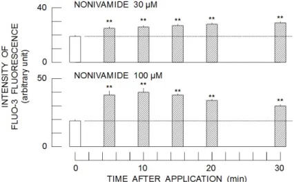
Nonivamide at 30 µM increased the intensity of Fluo-3 fluorescence in a time-dependent manner after the start of application (Upper panel of Fig. 1). Increase in the concentration of nonivamide (100 µM) produced further augmentation of Fluo-3
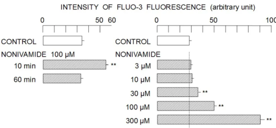
fluorescence (Lower panel of Fig. 1). The peak intensity was observed at 10 min after the start of drug application, thereafter, it gradually decreased. The intensity of Fluo-3 fluorescence monitored at 10 min after the start of drug application (100 µM nonivamide) was significantly higher than that at 60 min (Left panel of Fig. 2). Therefore, the concentration-dependent change in the intensity of Fluo-3 fluorescence by nonivamide was examined at 10 min after the start of drug application. As shown in the right panel of Fig. 2, nonivamide at the concentrations of 30 µM and more (up to 300 µM) significantly increased the intensity of Fluo-3 fluorescence. In order to reveal the source of Ca2+
for nonivamide-induced increase in Fluo-3 fluorescence, the effect of nonivamide was examined under an external Ca2+-free condition. As shown in the left panel of Fig. 3, nonivamide at 100 µM did not affect the intensity of Fluo-3 fluorescence under external Ca2+-free conditions.
Capsaicin is reported to activate vanilloid receptor (Caterina et al., 1997), resulting in the increase in intracellular Ca2+ level (Inoue et al., 2002). Our experiment also showed that capsaicin at 100 µM equally increased the intensity of Fluo-3 fluorescence (Right panel of Fig. 3). To test the possibility of the cross desensitization between nonivamide and capsaicin, the effect of capsaicin on Fluo-3 fluorescence was examined at 60 min after the start of nonivamide application and vice versa.
Figure 1. Time-dependent changes in the intensity of Fluo-3 fluorescence in the continued presence of nonivamide. Column and bar respectively indicate average intensity and standard deviation of four samples. Asterisks (**) show significant difference (P < 0.01) between the groups treated without and with nonivamide.
Figure 2. Concentration-dependent change in the intensity of Fluo-3 fluorescence by nonivamide. Left panel; the comparison of Fluo-3 fluorescence intensity at 10 min and 60 min after the start of drug application. Right panel; the concentration-dependent changes in Fluo-3 fluorescence intensity by 3–300 µM nonivamide. Column and bar respectively indicate average intensity and standard deviation of four samples. Asterisks (**) show significant difference (P < 0.01) between the groups treated without and with nonivamide.
Figure 3. Effect of 100 µM nonivamide under normal Ca2+ and Ca2+-free conditions and the comparison with
100 µM capsaicin. Left panel; attenuation of nonivamide-induced increase in the intensity of Fluo-3 fluorescence by removal of external Ca2+. Right panel; comparison with capsaicin-induced increase in Fluo-3
fluorescence intensity. Column and bar respectively indicate average intensity and standard deviation of four samples. Asterisks (**) show significant difference (P < 0.01) between the groups treated without and with nonivamide.
Figure 4. Effect of 100 µM nonivamide on Fluo-3 fluorescence in the continued presence of 100 µM capsaicin and vice versa. Column and bar respectively indicate average intensity and standard deviation of four samples. Asterisks (**) show significant difference (P < 0.01) between the group treated with nonivamide or capsaicin for 60 min and the group treated with capsaicin or nonivamide for 10 min in the continued presence of nonivamide or capsaicin, respectively.
As shown in Fig. 4, 100 µM capsaicin significantly increased the intensity of Fluo-3 fluorescence in the continued presence of 100 µM nonivamide. It was also true for 100 µM nonivamide in the continued presence of 100 µM capsaicin.
Discussion
As shown in Figs. 1–4, nonivamide at concentrations ranging from 30 µM to 300 µM significantly increased the intensity of Fluo-3 fluorescence, indicating the nonivamide-induced increase in intracellular Ca2+ level. The potency of
nonivamide to increase intracellular Ca2+ level seems to be similar to that of capsaicin when the comparison is made at 100 µM (Fig. 3). It is likely that the increase in intracellular Ca2+ level by nonivamide is
dependent on external Ca2+ because the removal of external Ca2+ attenuated the nonivamide-induced
increase in intracellular Ca2+ level (Fig. 3). Capsaicinoids activate vanilloid receptors coupled with nonselective cation channels, resulting in the increase in intracellular Ca2+ level (Inoue et al., 2002).
Thymocytes possess vanilloid receptors that induce Ca2+ influx (Amantini et al., 2004). In fact, both
nonivamide and capsaicin increased the intensity of Fluo-3 fluorescence, indicating the increase in intracellular Ca2+ level (Fig. 3). Desensitization of vanilloid receptors is dependent on the elevation of intracellular Ca2+ (Vyklicky et al., 2008). As shown in Figs. 2 and 4, the intensity of Fluo-3 fluorescence returns to control level during the prolonged incubation (60 min) with 100 µM nonivamide. Interestingly, capsaicin significantly increased the
intensity of Fluo-3 fluorescence in the continued presence of 100 µM nonivamide and vice versa (Fig. 4). The result may implicate that nonivamide and capsaicin function to the vanilloid receptors in an independent manner. Further analysis on the desensitization of vanilloid receptors in rat thymocytes will be necessary to elucidate the mechanism for the nonivamide-induced increase in intracellular Ca2+ level in the continued presence of capsaicin.
As described above, nonivamide at high micromolar concentrations increases intracellular Ca2+ levels. It may be likely that nonivamide affects cellular functions because cellular Ca2+ signaling
plays important roles in thymocyte functions (Lewis, 2001; Hogan et al., 2010). Nonivamide at concentrations ranging from 5 mg/L (17 µM) to 15 mg/L (51 µM) increased cytoplasmic Ca2+ level of
algal cells (Zhou et al., 2013). Therefore, nonivamide may exert cytotoxic action on mammalian cells at the concentrations similar to those on algal cells. Furthermore, there is no report that nonivamide is accumulated in wild animals. At present, it is hard to argue the implication of nonivamide cytotoxicity in environmental sciences. Conflict of interest
We have no conflicts of interests to declare. Acknowledgment
Experiments were performed with annual educational and research expenditure.
References
Amantini, C., Mosca, M., Lucciarini, R., Perfumi, M., Morrone, S., Piccoli, M., Santoni, G., 2004. Distinct thymocyte subsets express the vanilloid receptor VR1 that mediates capsaicin-induced apoptotic cell death. Cell Death Differ. 11, 1342–1356.
Angarano, M.B., McMahon, R.F., Hawkins, D.L., Schetz, J.A., 2007. Exploration of structure–antifouling relationships of capsaicin-like compounds that inhibit zebra
Biofouling 23, 295–305.
Caterina, M.J., Schumacher, M.A., Tominaga, M., Rosen, T.A., Levine, J.D., Julius, D., 1997. The capsaicin receptor: a heat-activated ion channel in the pain pathway. Nature 389, 816–824.
Chikahisa, L., Oyama, Y., 1992. Tri-n-butyltin increases intracellular Ca2+ in mouse thymocytes: a flow-cytometric study using fluorescent dyes for membrane potential and intracellular Ca2+. Pharmacol. Toxicol. 71, 190–195.
J. Nat. Prod. 59, 425–426.
Haber, L., Nance, P., Maier, A., Price, P., Olajos, E., Bickford, L., McConnell, M., Klauenberg, B.J., 2007. Human effectiveness and risk characterization of oleoresin capsicum (OC) and pelargonic acid vanillylamide (PAVA or nonivamide) hand-held devices (PP. 1–137). Toxicology excellence for risk assessment, Cincinnati OH.
Hoch, M., 2001. Organotin compounds in the environment—an overview. Appl. Geochem. 16, 719-743.
Hogan, P.G., Lewis, R.S., Rao, A., 2010. Molecular basis of calcium signaling in lymphocytes: STIM and ORAI. Annu. Rev. Immunol. 28, 491–533. Inoue, K., Koizumi, S., Fuziwara, S., Denda, S.,
Inoue, K., Denda, M., 2002. Functional vanilloid receptors in cultured normal human epidermal keratinocytes. Biochem. Biophys. Res. Comm. 291, 124–129.
Kao, J.P., Harootunian, A.T., Tsien, R.Y., 1989. Photochemically generated cytosolic calcium pulses and their detection by fluo-3. J. Biol. Chem. 264, 8179–8184.
Kass, G.E., Orrenius, S., 1999. Calcium signaling and cytotoxicity. Environ. Health Perspect. 107(Suppl 1), 25–35.
Lewis, R.S., 2001. Calcium signaling mechanisms in
T lymphocytes. Annu. Rev. Immunol. 19, 497–521.
Manirakiza, P., Covaci, A., Schepens, P., 2003. Pungency principles in Capsicum—Analytical determinations and toxicology (pp. 71–86). A.K. De (Ed.). Taylor & Francis: London, UK.
Matsui, H., Sakanashi, Y., Oyama, T.M., Oyama, Y., Yokota, S., Ishida, S., Okano, Y., Oyama, T.B., Nishimura, Y., 2008. Imidazole antifungals, but not triazole antifungals, increase membrane Zn2+
permeability in rat thymocytes: Possible contribution to their cytotoxicity. Toxicol. 248, 142–150.
Vyklicky, L., Novakova-Tousova, K., Benedikt, J., Samad, A., Touska, F., Vlachová, V., 2008. Calcium-dependent desensitization of vanilloid receptor TRPV1: a mechanism possibly involved in analgesia induced by topical application of capsaicin. Physiol. Res. 57, S59–S68.
Wang, Y.Y., Hong, C.T., Chiu, W.T., Fang, J.Y., 2001. In vitro and in vivo evaluations of topically applied capsaicin and nonivamide from hydrogels. Int. J. Pharmaceutics 224, 89–104. Zhou, J., Yang, C., Wang, J., Sun, P., Fan, P., Tian,
K., Liu, S., Xia, C., 2013. Toxic effects of environment-friendly antifoulant nonivamide on Phaeodactylum tricornutum. Environ. Toxicol. Chem. 32, 802–809.
Article History
Received MS June 13, 2014
Received Revised MS July 17, 2014 Accepted MS July 29, 2014