INTRODUCTION
Myxopapillary ependymoma is defined as a slow-growing, glial-derived tumor that arises from epen-dymal cells within the central nervous system. Its preferential manifestation is in young adults with an annual incidence of less than 0.01 per 100,000 (1-4). The majority of myxopapillary ependymomas are found in the conus medullaris or filum terminale (5, 6) and tend to have an insidious presentation with a gradual onset of symptoms and prolonged history (7, 8). Therefore, it is rare for myxopapillary ependymoma to present with acute neurological de-terioration secondary to rupture and hemorrhage. To the best of our knowledge, only 12 reports of acute neurological deterioration caused by ruptured
myxopapillary ependymoma with accompanying hemorrhage have been published in the English literature.
In this report, we present a case of acute onset cauda equina syndrome caused by a ruptured myxo-papillary ependymoma with accompanying hemor-rhage.
CASE REPORT
A 26-year-old healthy woman with no history of major trauma or coagulation disorders presented with acute low back pain. During the following 3 days, she developed muscle weakness and sensory disturbances in her bilateral lower extremities. A diagnosis of paraparesis secondary to cauda equina tumor was made by a local physician prior to her referral to our department for surgical treatment. Muscle strength of the right iliopsoas, quadriceps femoris, tibialis anterior (TA), extensor halluces longus (EHL), and triceps surae (TS) was reduced
CASE REPORT
Spontaneous Rupture and Hemorrhage of Myxopapillary
Ependymoma of the Filum Terminale : A Case Report
and Literature Review
Ichiro Tonogai, Toshinori Sakai, Fumitake Tezuka, Yuichiro Goda, Yoichiro Takata,
Kosaku Higashino, and Koichi Sairyo
Department of Orthopedics, Institute of Health Biosciences, the University of Tokushima Graduate School, Tokushima, Japan
Abstract : We present a rare case of acute onset cauda equina syndrome caused by a rup-tured myxopapillary ependymoma with accompanying hemorrhage. A 26-year-old healthy woman developed muscle weakness and sensory disturbances in her bilateral lower ex-tremities. Magnetic resonance imaging showed a huge mass from the L1 body to the L2-3 disc level. She was able to ambulate with crutches after the tumor was successfully re-moved. To prevent recurrence, she received whole brain and spinal cord radiation. No sing of recurrence were detected at the 8 month follow up. J. Med. Invest. 61 : 430-435, Au-gust, 2014
Keywords :spinal myxopapillary ependymoma, hemorrhage, rupture, acute neurological deterioration, magnetic resonance imaging
Received for publication May 9, 2014 ; accepted June 10, 2014. Address correspondence and reprint requests to Koichi Sairyo, M.D., Ph. D., Department of Orthopedics, Institute of Health Biosciences, University of Tokushima Graduate School, 3 - 18 - 15 Kuramoto, Tokushima 770 8503, Japan and Fax : +81 88 633 -0178.
A B
C
D
to 0/5 on manual muscle testing (MMT), and that of the left was reduced to 1/5. Loss of pin-prick and vibration sensation, severe reduction of light touch sensation, and urinary dysfunction were found. Anal tone was lax.
Magnetic resonance imaging (MRI) revealed an intradural tumorous mass extending from the L1 body to the L2-3 disc level, severely compressing the epiconus and conus level of the spinal cord (Fig. 1A-D). These findings were suggestive of bleeding from the tumorous mass secondary to capsule rup-ture. Notably, the hemorrhage extended from the cranial end of the tumor to the T10-11 disc level, and a finding similar to drop coagulation in the bot-tom of the dural sac was detected.
Surgery was performed via the dorsal midline
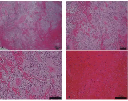
approach. After L1-2 laminectomy with durotomy, xanthochromic fluid was observed in the subarach-noid space. Blood clots and hematoma protruded from the ruptured capsule (Fig. 2A). After removing the blood clots and hematoma, a reddish-gray, soft, pliable tumor with attachment to the filum terminale was found among the cauda equina (Fig. 2B). The conus and cauda equina were compressed by the tumor to the ventral side. The granular tumor aris-ing from the filum terminale was excised (Fig. 2C). Histological examination of the tumor revealed typical features of a myxopapillary ependymoma with fresh blood cells : marked mucous changes of the stroma and papillary arrangement of tumor cells (Fig. 3A-C). The tumor cells were also seen within the hematoma (Fig. 3D). The glial cells were
Fig. 2. Intraoperative findings
(A) Opening of the dura reveals an extensive blood clot intermingled with cauda equine (arrow : hematoma). (B) The mass consists of solid blood clots and reddish- gray tumor (arrow : tumor).
(C) Tumor originating from the filum terminale (arrow : tumor, arrowhead : filum terminale). Fig. 1. Preoperative magnetic resonance imaging (MRI)
(A, B) Sagittal images showing a huge mass from the L1 body to the L2 - 3 disc level (arrows). The lesion was heterogeneous isoin-tense on T1 - weighted images and had low intensity on T2 - weighted images. The hemorrhage extends from the cranial end of the tumor to the T10 - 11 level, and drop coagulation in the bottom of the dural sac is visible (arrowheads).
A B strongly positive for glial fibrillary acidic protein,
which was consistent with a diagnosis of myxopa-pillary ependymoma. The Ki67 labeling index was around 1%.
Immediately after the surgery, the patient’s low back pain, bladder dysfunction, and sensory distur-bance of the bilateral lower extremities resolved. Her bilateral lower extremity strength on the MMT returned to 5/5, except for the right TA (1/5) and
right EHL (1/5), and she was able to ambulate with crutches.
To prevent recurrence, she received whole brain and spinal cord radiation (45 Gy). Postoperative MRI at the 8 month follow up revealed complete re-section of the tumor and hematoma, as well as the disappearance of the drop coagulation in the bottom of the dural sac (Fig. 4A, B).
Fig. 3. Pathological analysis
(A- C) Pathological evaluation after resection of the mass resulted in a diagnosis of hemorrhage from a mxyopapillary ependymoma. Cuboidal to elongated tumor cells are radially arranged in a papillary manner or like pseudorosettes around vascularized stromal cores with no mitotic activity. Hematoxylin and eosin staining ; magnification for A, B, and C :
!
4,!
10, and!
20, respectively.(D) Tumor cells are apparent within the hematoma. Hematoxylin and eosin staining ; Magnification :
!
20.Fig. 4. Postoperative MRI
(A, B) Sagittal images showing disappearance of drop coagulation in the bottom of the dural sac (arrowheads), and no signs of tumor recurrence 8 months after the operation (arrows).
DISCUSSION
Myxopapillary ependymoma is defined as a slow-growing glioma and is considered a distinct variant of ependymomas classified as World Health Organi-zation grade 1 (1-4). More than 50% of myxopapil-lary ependymomas are located in the conus medul-laris or filum terminale (5, 6), and the majority of myxopapillary ependymomas exhibit an insidious presentation with a gradual onset of symptoms and a prolonged subjective history, although clinical manifestations are frequently nonspecific (7, 8). Therefore, it is rare for a patient with myxopapillary ependymoma to present with acute neurological de-cline after spontaneous hemorrhage, even if the myxopapillary ependymoma has a more vascular architecture than the other types of ependymomas (9-11).
We presented here a rare case of a 26-year-old woman with a ruptured myxopapillary ependymoma and accompanying hemorrhage that expanded from the L1 body to the L2-3 disc level. A review of the English literature revealed only 12 reports describ-ing 15 cases of acute neurological decline caused by hemorrhage from myxopapillary ependymoma of the filum terminale or conus medullaris (Table 1) (7, 8, 12-21). Clear descriptions of the tumors or MRI findings were documented in only 8 of these cases. All 8 and the present case showed heterogeneous
lesions, although the presentation may have been affected by the phase of the hemorrhage from the tumor. Furthermore, drop coagulation in the bottom of the dural sac indicating dissemination of the tu-mor cells was detected in 4 patients.
Myxopapillary ependymoma tends to disseminate and recur despite its benign histologic character (22). The role of radiotherapy is controversial, but some authors proposed that radiation to the whole brain and spinal cord (including local radiation ; total dose, 30-50 Gy) prevented further tumor progres-sion (23, 24). Nakamura et al. suggested that the surgical margins obtained in the initial surgery and the extent and amount of postoperative radiation could be crucial factors in determining the progno-sis of patients with myxopapillary ependymoma be-cause cerebrospinal dissemination can occur once the tumor capsule is violated before or during sur-gery (24). In the present case, radiotherapy was per-formed because tumor cells were detected within the hemorrhage. Although the long-term outcome is unclear as yet, postoperative radiotherapy should be considered in such cases.
CONFLICT OF INTEREST
There are no conflicts of interest to disclose.
Table 1 :Literature review of case reports on spinal myxopapillary ependymomas presenting with hemorrhage as a cause of acute neurological deterioration
Authors and year Age (years) Sex
Level of tumor
localization TI intensity T2 intensity
Tumor homogeneity
Drop coagulation in the dural sac end
Djindjian M et al., 1978 18 M L2 N/A N/A
34 F L3 N/A N/A
Destee A et al., 1984 47 F L5 - S1 N/A N/A
Gomori JM et al., 1985 32 M N/A N/A N/A
Rappaport B et al., 1990 21 M L1 N/A N/A
Admiraal P et al., 1992 23 M L1 - 2 N/A iso N/A N/A
Malbrain ML et al., 1994 65 F L2 - 3 N/A N/A
Argyropoulou PI et al., 2001 15 F L1 - L4 iso iso heterogeneous +
Saadah M et al., 2004 33 M L1 - 3 N/A N/A N/A N/A
48 M T12 - L4 N/A N/A heterogeneous N/A
Heuer GC et al., 2007 31 F L1 - L2 low low heterogeneous +
31 M T11 - L2 N/A N/A heterogeneous N/A
Becco de Souza R et al., 2012 13 F L1 - 3 iso - low iso - high heterogeneous + Martinez - Perez R et al., 2012 32 M L2 - L3 iso low heterogeneous N/A Khalatbari MR et al., 2014 15 M L1 high low - iso heterogeneous
-Present case 26 F L1 - 2 iso low heterogeneous +
AKNOWLEDGEMENTS
This article was not funded by any grant. No benefits have been received or will be received in any form from a commercial party related directly or indirectly to the subject of this article.
REFERENCES
1. Louis DN, Ohgaki H, Wiestler OD, Cavenee WK, Burger PC, Jouvet A, Scheithauer BW, Kleihues P : The 2007 WHO classification of tumours of the central nervous system. Acta Neuropathol 114(2) : 97-109, 2007
2. Duong LM, McCarthy BJ, McLendon RE,
Dolecek TA, Kruchko C, Douglas LL, Ajani UA : Descriptive epidemiology of malignant and nonmalignant primary spinal cord, spinal meninges, and cauda equina tumors, United States, 2004-2007. Cancer 118(17) : 4220-4227, 2012
3. Schild SE, Nisi K, Scheithauer BW, Wong WW, Lyons MK, Schomberg PJ, Shaw EG : The re-sults of radiotherapy for ependymomas : the Mayo Clinic experience. Int J Radiat Oncol Biol Phys 42(5) : 953-958, 1998
4. Wager M, Lapierre F, Blanc JL, Listrat A, Bataille B : Cauda equina tumors : a French multicenter retrospective review of 231 adult cases and review of the literature. Neurosurg Rev 23(3) : 119-129, 2000
5. Celli P, Cervoni L, Morselli E, Ferrante L : Spi-nal ependymomas and papilledema : report of 4 cases and review of the literature. J Neuro-surg Sci 37(2) : 97-102, 1993
6. Schweitzer JS, Batzdorf U : Ependymoma of the cauda equina region : diagnosis, treatment, and outcome in 15 patients. Neurosurgery 30(2) : 202-207, 1992
7. Heuer GG, Stiefel MF, Bailey RL, Schuster JM : Acute paraparesis from hemorrhagic spinal ependymoma : diagnostic dilemma and surgi-cal management. Report of two cases and re-view of the literature. J Neurosurg Spine 7(6) : 652-655, 2007
8. Martinez-Perez R, Hernandez-Lain A, Paredes I, Munarriz PM, Castaño-Leon AM, Lagares A : Acute neurological deterioration as a re-sult of two synchronous hemorrhagic spinal ependymomas. Surg Neurol Int 3 : 33, 2012 9. Lagares A, Rivas JJ, Lobato RD, Ramos A,
Alday R, Boto GR : Spinal cord ependymoma presenting with acute paraplegia due to tu-moral bleeding. Neurisurg Sci 44 : 95-97, 2000 10. Tait MJ, Chelvarajah R, Garvan N, Bavetta S : Spontaneous hemorrhage of a spinal ependy-moma : a rare cause of acute cauda equina syn-drome : a case report. Spine (Phila Pa 1976) 29(21) : E502-505, 2004
11. Ozdemir O, Calisaneller T, Coven I, Altinors N : Posttraumatic intratumoural haemorrhage : an unusual presentation of spinal ependymoma. Eur Spine J 16 Suppl 3 : 293-295, 2007
12. Dijindjian M, Djindjian R, Houdart R, Hurth M : Subarachnoid hemorrhage due to intraspinal tumors. Surg Neurol 9(4) : 223-229, 1978 13. Destée A, Lesoin F, Warot M, Mendolia G,
Devos P, Warot P : Tumoral spinal meningeal hemorrhage during anticoagulant treatment. Rev Neurol (Paris) 140(8-9) : 517-519, 1984 14. Gomori JM, Grossman RI, Bilaniuk LT,
Zimmerman RA, Goldberg HI : High-field MR imaging of superficial siderosis of the central nervous system. J Comput Assist Tomogr 9(5) : 972-975, 1985
15. Rappaport B, Emsellem HA, Shesser R, Millstein E : An unusual case of proctalgia. Ann Emerg Med 19(2) : 201-203, 1990
16. Admiraal P, Hazenberg GJ, Algra PR, Kamphorst W, Wolbers JG : Spinal subarach-noid hemorrhage due to a filum terminale ependymoma. Clin Neurol Neurosurg 94(1) : 69-72, 1992
17. Malbrain ML, Kamper AM, Lambrecht GL, Hermans P, Baeck E, Verhoeven F, Wyffels G, Verbraeken H : Filum terminale ependymoma revealed by acute cauda equina compression syndrome following intratumoral and spinal subarachnoid hemorrhage in a patient on oral anticoagulants. Acta Neurol Belg 94(1) : 35-43, 1994
18. Argyropoulou PI, Argyropoulou MI, Tsampoulas C, Gogos P, Manavis I, Efremidis SC : Myxopa-pillary ependymoma of the conus medullar is with subarachnoid haemorrhage : MRI in two cases. Neuroradiology 43(6) : 489-491, 2001 19. Sa’adah M, Al Shunnar K, Saadah L, Shogan
A, Inshasi J, Afifi H : Atypical presentations of conus medullaris and filum terminale myxopa-pillary ependymomas. J Clin Neurosci 11(3) : 268-272, 2004
20. Becco de Souza R, Brasileiro de Aguiar G, Saade N, Esteves Veiga JC : Cauda equina syndrome
caused by spontaneous bleeding in the filum terminale myxopapillary ependymoma : a rare pediatric case. Pediatr Neurosurg 48(6) : 385-388, 2012
21. Khalatbari MR, Moharamzad Y : Myxopapillary ependymoma of the conus medullar is pre-senting with intratumoral hemorrhage during weight lifting in a teenager. Childs Nerv Syst 30(1) : 181-183, 2014
22. Plans G, Brell M, Cabiol J, Villà S, Torres A, Acebes JJ : Intracranial retrograde dissemination in filum terminale myxopapillary ependymomas.
Acta Neurochir (Wien) 148(3) : 343-346, 2006 23. Chan HS, Becker LE, Hoffman HJ, Humphreys RP, Hendrick EB, Fitz CR, Chuang SH : Myxopapillary ependymoma of the filum ter-minale and cauda equina in childhood : report of seven cases and review of the literature. Neurosurgery 14(2) : 204-210, 1984
24. Nakamura M, Ishii K, Watanabe K, Tsuji, T, Matsumoto M, Toyama Y, Chiba K : Long-term surgical outcomes for myxopapillary ependy-momas of the cauda equina. Spine (Phila Pa 1976) 34(21) : E756-760, 2009