INTRODUCTION
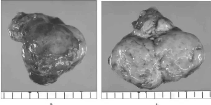
Soft tissue sarcomas are uncommon tumors that represent only 1 to 1.5% of all malignancies (1). Of these tumors, only 15% are located in the retroperi-toneum. We herein describe a case of perinephric liposarcoma which recurred ten years later from the initial operation. At the first operation, histology
of the tumor was well-differentiated type. By con-trast, the recurrent histological type of the tumors showed dedifferention.
CASE REPORT
A 58-year old man was referred to our hospital for treatment of an abdominal mass. As for him, tu-mor resection with right nephrectomy for a 22
!
19!
16 cm well - differentiated right perinephric liposarcoma had been performed ten years ago (Fig. 1). Abdominal computed tomography (CT) examination was performed every year after initialCASE REPORT
A case of perinephric liposarcoma which recurred ten
years later from the initial operation
Shinji Kuratate
1), Motoya Chikakiyo
1), Yuji Kaneda
1), Yukari Harino
1),
Toshiyuki Hirose
1), Toshiyuki Yagi
1), Seiya Saitoh
1), Masayuki Sumitomo
1),
Ryozo Fujino
1), Nobuo Satake
2), and Takahiro Hirose
2) 1)Department of Surgery and2)
Department of Human Pathology, Tokushima Prefectural Central Hospital, Tokushima, Japan
Abstract : A 58-year old man was referred to our hospital for treatment of an abdominal mass. As for him, tumor resection with right nephrectomy had been performed ten years ago for a giant well-differentiated perinephric liposarcoma. CT examination showed a huge tumor shadow in the abdominal cavity. Abdominal MRI examination showed a 15 8 cm tumor with almost high signal intensity on the T2 weighted images. At lapalotomy, a large bulky retroperitoneal tumor pointed out before an operation was found. Surgical extirpation of the tumor was performed. Besides, several tumors of the thumb head size were detected into right retroperitoneal fatty tissue. The right side mesocolon and the tumors were not able to exfoliate, therefore right hemicolectomy was performed. Histo-logical features showed dedifferentiated liposarcoma. The postoperative course was un-eventful. But eight months after surgery, he was admitted again for treatment of a 4 3 cm retroperitoneal tumor. Extirpation of the tumor was performed. Histological finding of this tumor also showed dedifferentiated liposarcoma. Dedifferentiation, occurring in 15%% of the well-differentiated liposarcomas, sometimes may develop later. Long-term de-tailed follow-up is necessary for well-differentiated liposarcoma. J. Med. Invest. 58 : 154-158, February, 2011
Keywords : perinephric liposarcoma, retroperitoneal liposarcoma, dedifferentiated liposarcoma, well-differentiated
liposarcoma
Received for publication October 25, 2010 ; accepted December 28, 2010.
Address correspondence and reprint requests to Dr. Shinji Kuratate, Department of Surgery, Tokushima Prefectural Central Hospital, Kuramoto - cho, Tokushima 770 - 8539, Japan and Fax : + 81 - 88 - 631 - 8354.
surgery. Until 1 year before this admission, CT showed no recurrence sign. Laboratory findings at this admission showed no abnormalities on periph-eral blood and serum examination except slight re-nal function disorder. Abdomire-nal CT examination showed a huge tumor shadow having well-defined margins in the abdominal cavity (Fig. 2). Abdominal
magnetic resonance imaging (MRI) examination showed a 15
!
8 cm tumor with almost low signal intensity on the T1 and with almost high signal in-tensity on the T2 weighted images (Fig. 3). Further-more, the tumor had the part where showed high signal intensity on the T1 and T2 weighted images in MRI. That part regarded as the adipose constitu-ent. The area accompanied with homogeneous low signal intensity on the T1 weighted image in MRIwas regarded as a myxomatous component. Con-sidering the above facts, we presumed that well dif-ferentiated liposarcoma had converted to the other differentiation type. Though the definitive diagnosis was not provided, the tumor was thought to be a recurrence of liposarcoma. At lapalotomy, a bulky tumor was found in the retroperitoneal adipose tis-sue of the left upper abdominal region. The tumor showed expansive growth without any invasion into surrounding structures such as the liver, pancreas, or inferior vena cava. Extirpation of the tumor was performed. The tumor size was 15
!
9!
8 cm (Fig. 4). Besides, several tumors of the thumb head size were detected into right retroperitoneal fatty tissue. The right side mesocolon and the tumors were notFig. 1 :The excised tumor size was 22
!
19!
16 cm (1 - a, 1 - b). Pathological examination revealed a well - differentiated liposar-coma (HE) (1 - c, 1 - d).Fig. 2 :CT demonstrated a huge tumor shadow having well -defined margins in the abdominal cavity (arrow).
Fig. 4 :The excised tumor size was 15
!
9!
8 cm (4 - a). Cut sur-face (4 - b). Besides, several tumors of the thumb head size were detected into right retroperitoneal fatty tissue (4 - c) (arrow).Fig 3. MRI demonstrated a 15
!
8 cm tumor with almost low signal intensity on the T1 and with almost high signal intensity on the T2 weighted images (arrow).able to exfoliate, therefore right hemicolectomy was performed. Histological features of the tumors showed dedifferentiated liposarcoma (DDLPS) (Fig. 5). The postoperative course was uneventful and the
patient was discharged soon. But eight months after surgery, he was admitted again for treatment of a 4
!
3 cm tumor in the retroperitoneal region (Fig. 6). Extirpation of the tumor was performed. Histologi-cal features of this tumor showed loHistologi-cal recurrence of the DDLPS (Fig. 7). He got well immediately and left the hospital.DISCUSSION
Liposarcomas, corresponding to 19% of all soft tis-sue sarcomas, are malignancies of adipose tistis-sue (2). They are the most frequent histopathological variety (41%) of the retroperitoneal sarcoma (3). The World Health Organization classification of soft tis-sue tumors has classified liposarcoma into five main subgroups : well-differentiated, which includes the adipocytic, sclerosing, and inflammatory subtypes, myxoid, round cell, pleomorphic and dedifferen-tiated (4). More than 90% of the retroperitoneal liposarcomas are well-differentiated type (56%) and dedifferentiated type (37%) (5).
Dedifferentiation is defined as the presence of nonlipogenic high grade areas within the well-differ-entiated liposarcoma (WDLPS). Dedifferentiation is rare, occurring in 15% of the WDLPS (6). About 90% of DDLPS arise de novo, while 10% occur in recurrence. According to current literature, WDLPS and DDLPS share the same basic genetic abnormal-ity characterized by a simple genomic profile with a 12q14-15 amplification involving MDM2 gene (7). The risk of dedifferentiation is higher in the retrop-eritoneum and is probably a time-dependent phe-nomenon (8-10). In our present case, initial histo-logical type was a well-differentiation, whereas re-current histological type was a dedifferentiation. It took for a long time, more than about 9 years, till the histological change from well-differentiation to dedifferentiation occurred. At the moment histologi-cal change occurred, the recurrent tumor grew rap-idly.
As for treatment of liposarcoma, surgical resec-tion is considered the mainstay of curative treat-ment, and complete surgical resection with negative margins is required as the goal of therapy for most patients. However, liposarcoma forms a pseudo cap-sule without having a capcap-sule histologically, so that a border is indistinct. Therefore, an aggressive sur-gical technique with en-bloc multiorgan resection is necessary in order to achieve negative margins (5, 11-14). The most frequent organ resected is the kidney like our first resection was so. Even with aggressive surgical approaches, local recurrence remains a common type of failure (13). As to our case, to keep negative margins, we performed right nephrectomy at initial surgery, and right hemicolec-tomy at second surgery. Despite aggressive surgical procedures performed mentioned above, we failed in the local control 2 times, and the third surgery performed against the recurrent tumor. Fortunately,
Fig 5 :Pathological examination revealed dedifferentiated liposar-coma (HE).
Fig. 6 :The excised tumor size was 4
!
3 cm (6 - a). Cut surface (6 - b).Fig. 7 :Pathological examination revealed a dedifferentiated tu-mor (HE).
the patient is alive without recurrence after the third operation. If a resectable local recurrence tumor appears in future, we shall extirpate the tumor.
Some articles report that retroperitoneal WDLPS does not recur after an operation (15-18). It seems WDLPS has favorable prognoses. However, about these cases, postoperative follow-up survey period is a short term. Like our present case, long term follow-up for at least 10!20 years has revealed that WDLPS in the retroperitoneum recur regionally in almost 100% of cases even if the tumor seems to have been completely resected (7). DDLPS in the retroperitoneum also recur in almost 100% of cases and often cause the patient’s death (7). In our case, DDLPS recurred regionally in a short term. The interval of the second operation and the third one was only eight months.
The effectiveness of chemotherapy and radiation therapy for both primary and metastatic liposarco-mas is still controversial (19-22). Survival benefits have not been demonstrated (23). Hence, we are using no chemotherapy or radiation therapy. But if a non-resectable local recurrence lesion appears in future, we will consider radiotherapy such as heavy particle beam for a purpose of the regional control (24).
In conclusion, we have experienced a case of huge perinephric WDLPS which recurred ten years later from the initial operation. Once WDLPS re-curs, recurrence may cause the tumor to evolve into a higher grade of sarcoma or to dedifferentiate. A long-term follow-up after surgery is mandatory due to high rates of recurrence.
REFERENCES
1. Clark MA, Fisher C, Judson I, Thomas JM : Soft-tissue sarcomas in adults. N Engl J Med 353 : 701-711, 2005
2. Juan C. Gutierrez, Eduardo A. Perez, Dido Franceschi, Frederick L. Moffat Jr., Alan S. Livingstone, and Leonidas G. Koniaris. : Out-comes for Soft-Tissue Sarcoma in 8249 Cases from a Large State Cancer Registry. J Surg Res 141 : 105-114, 2007
3. Lewis JJ, Leung D, Woodruff JM, Brennan MF : Retroperitoneal Soft - Tissue Sarcoma Analysis of 500 Patients Treated and Followed at a Single Institution. Ann Surg 228 : 355-365, 1998
4. Fletcher CDM, Unni KK, Mertens F : World
Health Organization Classification of Tumors. Pathology and Genetics of Tumours of soft tissue and bone. IARC Press, Lyon, 2002 5. Singer S, Antonescu CR, Riedel E, Brennan
MF : Histologic subtype and margin of resec-tion predict pattern of recurrence and survival for retroperitoneal liposarcoma. Ann Surg 238 : 358-371, 2003
6. Hasegawa T, Seki K, Hasegawa F, Matsuno Y, Shimodo T, Hirose T, Sano T, Hirohashi S : Dedifferentiated liposarcoma of retroperito-neum and mesentery : varied growth patterns and histological gradesa clinicopathologic study of 32 cases. Hum Pathol 31 : 717-727, 2000 7. Jean-Michel C, Florence P, Alain A :
Well-dif-ferentiated and dedifWell-dif-ferentiated liposarcomas. Virchows Arch 456 : 167-179, 2010
8. McCormick D, Mentzel T, Beham A, Fletcher CD : Dedifferentiated liposarcoma. Clinicopa-thologic analysis of 32 cases suggesting a bet-ter prognostic subgroup among pleomorphic sarcomas. Am J Surg Pathol 18 : 1213-1223, 1994
9. Henricks WH, Chu YC, Goldblum JR, Weiss SW : Dedifferentiated liposarcoma : a clinicopa-thological analysis of 155 cases with a proposal for an expanded definition of dedifferentiation. Am J Surg Pathol 21 : 271-281, 1997
10. Nascimento AG : Dedifferentiated liposarcoma. Semin Diagn Pathol 18 : 263-266, 2001
11. Hassan I, Park SZ, Donohue JH, Nagorney DM, Kay PA, Nasciemento AG, Schleck CD, Ilstrup DM : Operative management of primary retroperitoneal sarcomas. A reappraisal of an institute experience. Ann Surg 239 : 244-50, 2004
12. Neuhans SJ, Barry P, Clark MA, Hayers AJ, Fisher C, Thomas JM : Surgical management of primary and recurrent retroperitoneal liposar-coma. Br J Surg 992 : 246-252, 2005
13. Sato T, Yamaguchi T, Azekura K, Ueno M, Ohyama S, Ohya M, Yamamoto J, Muto T, Ishikawa Y, Kanda H : Repeated resection for intra-abdominal and retroperitoneal liposarco-mas : long-term experience in a single cancer center in Japan. Int Surg 91 : 267-271, 2006 14. Hueman MT, Herman JM, Ahuja N :
Manage-ment of Retroperitoneal Sarcomas. Surg Clin N Am 88 : 583-597, 2008
15. Kim ES, Jang SH, Park HC, Jung EH, Moon GB : Dedifferentiated liposarcoma of the retrop-eritoneum. Cancer Res Treat 42 : 57-60, 2010
16. Fu Q : Huge retroperitoneal liposarcoma : a case report. Chin Med J 120 : 1117-1118, 2007 17. Herrera - Gómez A, Ortega - Gutiérrez C, Betancourt AM, Luna-Ortiz K : Giant retroperi-toneal liposarcoma. World J Surg Oncol 31 : 115, 2008
18. Yildirim O, Namdaroglu OB, Menekse E, Albayrak AL : Giant well-differentiated liposar-coma of retroperitoneum. Bratisl Lek Listy 109 : 418-420, 2008
19. Mussi C, Collini P, Miceli R, Barisella M, Mariani L, Fiore M, Casali PG, Gronchi A : The Prognostic Impact of Dedifferentiation in Retrop-eritoneal Liposarcoma A Series of Surgically Treated Patients at a Single Institution. Cancer 113 : 1657-65, 2008
20. Sepideh G, Charlotte D. J, Daniel S K, Layla M P, Jeffrey A : The value of surgery for retrop-eritoneal sarcoma. Sarcoma 8 : 1-6, 2009 (Epub)
21. Dalal KM, Antonescu CR, Singer S : Diagno-sis and management of lipomatous tumors. J Surg Oncol 97 : 298-313, 2008
22. Jones RL, Fisher C, Al-Muderis O, Judson IR., Ian R J : Differential sensitivity of liposarcoma subtypes to chemotherapy. Eur J Cancer 41 : 2853-2860, 2005
23. Yoshida Y, Inoue K, Ohsaco T, Nagamoto N, Tanaka E, Tsuruzoe S : Weekly paclitaxel ther-apy is curative for patients with retroperitoneal liposarcoma. Gan To Kagaku Ryoho 34 : 465-467, 2007 (in Japanese)
24. Kamada T, Tsujii H, Tsuji H, Yanagi T, Mizoe J, Miyamoto T, Kato H, Yamada S, Morita S, Yoshikawa K, Kandatsu S, Tateishi A and the working group for the bone and soft tissue sar-comas : Efficacy and safety of carbon ion radio-therapy in bone and soft tissue sarcomas. J Clin Oncol 22 : 4472-4477, 2002.