IRUCAA@TDC : 嚥下機能に関連する上咽頭収縮筋の形態学的特徴
9
0
0
全文
(2) 66. 二次出版. 嚥下機能に関連する上咽頭収縮筋の形態学的特徴 津守伸明1). 阿部伸一1). 上松博子1). 橋本正次2). 井出吉信1). 抄録:嚥下運動において重要な役割を担っている上. 上咽頭収縮筋,中咽頭収縮筋,下咽頭収縮筋の3つ. 咽頭収縮筋の形態的特徴を明らかにする目的で上咽. の筋から構成されている1)∼8)。これまで食道と近接. 頭収縮筋の翼突咽頭部,頬咽頭部,顎咽頭部および. する下咽頭収縮筋の解剖学的な形態,筋の特性につ. 舌咽頭部の肉眼解剖学的観察を行った。頬咽頭部の. いて報告されてきた9)10)11)。それらの報告の中で形. 筋の起始形態は,上部から下部まで膜状のもの. 態的にみた下咽頭収縮筋の動きを推測している。一. (A型) ,上部だけが膜状のもの(B型) ,膜が欠如し. 方,口腔に近接した上咽頭収縮筋は,起始部が4. たもの(C型) の3型に分けられた。この3つの型す. つ,停止部が1つという複雑な形態を呈している。. べてにおいて,頬咽頭部の筋は,頬筋から移行的に. この筋は蝶形骨翼突鈎から起始する翼突咽頭部,翼. 起始していた。顎咽頭部の筋の起始形態は,先端が. 突下顎縫線から起始する頬咽頭部,顎舌骨筋線から. 顎舌骨筋線上にみられたもの(A型) と顎舌骨筋線か. 起始する顎咽頭部,舌から起始する舌咽頭部の4部. ら離れていたもの(B型) の2型に分けられた。今回. から構成されている4)7)8)12)。この筋の解剖学的研究. の研究より,上咽頭収縮筋は咀嚼に重要な役割を果. は, Howland と Brodie13)によって報告された。彼ら. たしている頬筋と結合組織で接し,下顎骨および舌. は,頬咽頭部の翼突下顎縫線を観察し,その翼突下. 根部から起始していたことが明らかとなった。これ. 顎 縫 線 を“the sphenoid tendon between the jaws”. らの所見から上咽頭収縮筋は摂食行動において咀嚼. (上下の顎骨間に存在する蝶形骨からの腱) と記載し. から嚥下に至るスムーズな協調運動を発揮するのに. た。また,Brand と Isselhard6)は,この縫線を腱の. 重要な役割を果たしていると推測された。. ようなバンドと報告し,Williams4)は縫線を腱の線 維と述べている。Sicher14)は,縫線を腱様構造物と. 緒 言. してこの中間部を除けば連続的な括約筋様の band. 近年,加齢に伴う摂食・嚥下機能の低下や障害が. を 形 成 し て い る と 報 告 し て い る。し か し,Gau-. 臨床上問題となっている。これに伴い,咀嚼から嚥. ghran15)によればこの翼突下顎縫線は全ての標本で. 下への口腔・咽頭の運動が調べられ,そのメカニズ. 欠如し,従来記載されている縫線は anatomical ar-. ムが解明されてきた。. tifact(解剖学的な剖出の過程で作られた腱様構造. 咽頭収縮筋が嚥下に関与していることはすでに知. 物) であると述べている。このように翼突下顎縫線. られている。咽頭の後壁を形成する咽頭収縮筋は,. の形態についての所見は一致しておらず,結論は明. キーワード:上咽頭収縮筋,頬筋,舌,嚥下,咽頭 東京歯科大学解剖学講座 2) 東京歯科大学法人類学研究室 (2007年10月25日受付) (2007年11月13日受理) 別刷請求先:〒261‐8502 千葉市美浜区真砂1−2−2 東京歯科大学解剖学講座 津守伸明. 本論文は,Springer Science and Business Media, Inc. の 許可を得て,Dysphagia Volume 22:122∼129,2007.に 掲載された論文を和文により二次出版したものである。. 1). ― 66 ―.
(3) 歯科学報. Vol.108,No.1(2008). 67. 察した。. らかとなっていない。 また,上咽頭収縮筋頬咽頭部以外の部に関して. ⑵. 頬咽頭部の筋の起始形態について顔面側から観察. は,舌咽頭部の形態について近年報告されているも 16). 頬咽頭部. のの ,翼突咽頭部と顎咽頭部の形態について詳細. した。. な報告はあまりなく,上咽頭収縮筋の4部の形態を. ⑶. 顎咽頭部 顎咽頭部の筋の起始形態について口腔側から観察. 総合的に観察した研究は,ほとんどみられない。 本研究は,上咽頭収縮筋の形態的特徴を明らかに することを目的とし,上咽頭収縮筋を構成する4部. した。 ⑷. 舌咽頭部 舌咽頭部の筋の起始形態について口腔側から観察. の起始部,停止部,および筋の走行について肉眼解. した。. 剖学的観察を行った。. 2)停止部の形態. 試料と方法. 咽頭収縮筋の停止部である咽頭後壁について顔面. 観察試料は東京歯科大学解剖学講座所蔵の53歳か ら91歳まで(平均73歳) の10%ホルマリン固定された. 側から観察した。 上咽頭収縮筋頬咽頭部の組織学的観察 上咽頭収縮筋頬咽頭部の筋(第4,第6鰓弓由来). 日本人成人実習用屍体(男性24体,女性13体,計37 体) を用いた。. は,発生学的に異なる頬筋(第2鰓弓由来) と接して. 上咽頭収縮筋の剖出. いる。そこで,この両筋の関係を明らかにするため. 頬筋を除く表情筋,咀嚼筋,頬骨弓及び下顎枝の. 肉眼観察に加えて組織学的な観察を行った。肉眼観. 一部を除去した。また後部は頚椎から後方の組織を. 察を行った試料から上咽頭収縮筋頬咽頭部の筋と頬. 離 断 し,上 咽 頭 収 縮 筋 を 顔 面 側 か ら 観 察 し た. 筋を一塊として取り出した。その試料を10%ホルマ. (Fig. 1) 。さらに,頭部を正中矢状断し,口腔粘膜. リンで再固定後,通法に従いパラフィン包埋し,顔. を剥離した後,上咽頭収縮筋を口腔側から観察し. 面側から口腔側にかけて筋線維の走行に平行した8. た。. μm の連続切片を作製した。その後,Azan 染色を. 上咽頭収縮筋の肉眼解剖学的観察. 施し光学顕微鏡にて観察した。. 1)起始部の形態 ⑴. 翼突咽頭部 翼突咽頭部の筋の起始形態について顔面側から観. Fig. 2 The superior pharyngeal constrictor muscle at the pterygopharyngeal part as observed from the facial side. The muscle at the pterygopharyngeal part originates between the pterygoid hamulus of the sphenoid bone and the posterior margin of the medial pterygoid plate of the sphenoid bone. 1 = superior pharyngeal constrictor muscle at the pterygopharyngeal part, S = sphenoid bone.. Fig. 1 Superior pharyngeal constrictor muscle as observed from the facial side. SPC = superior pharyngeal constrictor muscle, B = buccinator muscle, M = mandible, SG = styloglossus muscle. ― 67 ―.
(4) 68. 津守, 他:嚥下機能に関連する上咽頭収縮筋の形態学的特徴. 頭収縮筋の最上部を形成していた。起始直後,この. 結 果. 部の筋は徐々に幅を狭くして咽頭頭底板の側方下部. 本研究において男性24体,女性13体を観察対象と. を走行していた。その後,この筋は咽頭頭底板の後. したが上咽頭収縮筋の4部の形態について性差は認. 方下部を湾曲して走行していた。今回,観察を行っ. められなかった。したがって,結果は,性を区別す. たほぼ全ての試料で同じような起始および走行形態. ることなく集計した。. を呈しており, 大きな違いは見られなかった (Fig. 2) 。. 上咽頭収縮筋の肉眼解剖学的観察. ⑵. 上咽頭収縮筋頬咽頭部の筋は,上方から起始する. 1)起始部の形態 ⑴. 頬咽頭部. 翼突咽頭部の筋および下方から起始する顎咽頭部の. 翼突咽頭部 上咽頭収縮筋翼突咽頭部の筋は,蝶形骨翼突鈎か. 筋と合流し,上咽頭の側壁から後壁を形成していた. ら蝶形骨翼状突起内側板後縁にかけて起始し,上咽. (Fig. 1) 。この頬咽頭部の筋の起始形態は,上部か. Fig. 3 Type A as observed from the facial side. The origin of the superior pharyngeal constrictor muscle at the buccopharyngeal part comprises membranous connective tissue from the superior to inferior areas. 2 = superior pharyngeal constrictor muscle at the buccopharyngeal part, B = buccinator muscle, arrow = membranous.. Fig. 4 Type B as observed from the facial side. Only the superior area of the origin of the superior pharyngeal constrictor muscle at the buccopharyngeal part is membranous. The inferior area of the superior pharyngeal constrictor muscle at the buccopharyngeal part transitionally originates from the buccinator muscle. 2 = superior pharyngeal constrictor muscle at the buccopharyngeal part, B = buccinator muscle, arrow = membranous. ― 68 ―.
(5) 歯科学報. Vol.108,No.1(2008). 69. Fig. 5 Type C as observed from the facial side. The origin of the superior pharyngeal constrictor muscle at the buccopharyngeal part completely lacks membrane from the superior to inferior areas. From the superior to inferior areas of the origin, the superior pharyngeal constrictor muscle at the buccopharyngeal part transitionally originates from the buccinator muscle. 2 = superior pharyngeal constrictor muscle at the buccopharyngeal part, B = buccinator muscle.. ら下部まで結合組織性の膜状を呈していたもの(A. 筋線上にみられたもの(A型) (Fig. 7) と顎舌骨筋線. 型) (Fig. 3) ,上部だけが膜状を呈していたもの(B. から離れていたもの(B型) (Fig. 8) の2型に分けら. 型) (Fig. 4) ,上部から下部まで膜が完全に欠如して. れた。44例中, A型は32例 (72. 7%) , B型は12例 (27. 3. いたもの(C型) (Fig. 5) の3型に分けられた。54例. %) であった。この顎咽頭部の筋は,起始直後レト. 中,A型は7例(13. 0%) ,B型は10例(18. 5%) ,C. ロモラーパッド上縁で上咽頭収縮筋頬咽頭部の筋と. 型は37例(68. 5%) であった。. 合流し,咽頭の側壁から後壁を形成していた。この. ⑶. 顎咽頭部の筋は,上咽頭収縮筋頬咽頭部の筋と合流. 顎咽頭部 上咽頭収縮筋顎咽頭部の筋は,レトロモラーパッ. するまでは他の3つの部の筋に比べて薄かった。. ドの舌側で顎舌骨筋線の後方部分から起始していた (Fig. 6) 。この部の筋の起始形態は,先端が顎舌骨. Fig. 7 Morphology of the origin of the superior pharyngeal constrictor muscle at the mylopharyngeal part(type A) . The tip of the origin of the superior pharyngeal constrictor muscle at the mylopharyngeal part lies on the mylohyoid line. 3 = superior pharyngeal constrictor muscle at the mylopharyngeal part, M = mandible, MH = mylohyoid muscle, arrow = tip of the origin.. Fig. 6 Superior pharyngeal constrictor muscle as observed from the oral side. 3 = superior pharyngeal constrictor muscle at the mylopharyngeal part, 4 = superior pharyngeal constrictor muscle at the glossopharyngeal part, M = mandible, MH = mylohyoid muscle, SG = styloglossus muscle. ― 69 ―.
(6) 70. 津守, 他:嚥下機能に関連する上咽頭収縮筋の形態学的特徴. Fig. 8 Morphology of the origin of the superior pharyngeal constrictor muscle at the mylopharyngeal part(type B) . The tip of the origin of the superior pharyngeal constrictor muscle at the mylopharyngeal part lies away from the mylohyoid line. 3 = superior pharyngeal constrictor muscle at the mylopharyngeal part, M = mandible, MH = mylohyoid muscle, arrow = tip of the origin.. Fig. 9 Morphology of the origin of the superior pharyngeal constrictor muscle at the glossopharyngeal part. The superior pharyngeal constrictor muscle at the glossopharyngeal part forms the root of the tongue with the palatoglossus and styloglossus muscles. 4 = superior pharyngeal constrictor muscle at the glossopharyngeal part, PG = palatoglossus muscle, SG = styloglossus muscle.. Fig. 10 Enlarged image of the superior pharyngeal constrictor muscle at the glossopharyngeal and mylopharyngeal parts (interlaced type) . The superior pharyngeal constrictor muscle at the glossopharyngeal and mylopharyngeal parts interlace immediately after the origin. 3 = superior pharyngeal constrictor muscle at the mylopharyngeal part, 4 = superior pharyngeal constrictor muscle at the glossopharyngeal part.. ⑷. Fig. 11 Enlarged image of the superior pharyngeal constrictor muscle at the glossopharyngeal and mylopharyngeal parts (merged type). The superior pharyngeal constrictor muscle at the glossopharyngeal and mylopharyngeal parts merge immediately after the origin. 3 = superior pharyngeal constrictor muscle at the mylopharyngeal part, 4 = superior pharyngeal constrictor muscle at the glossopharyngeal part.. 部では口蓋腱膜から起始する口蓋舌筋が停止してい. 舌咽頭部 上咽頭収縮筋舌咽頭部の筋は,すべての試料で舌. た。また,この部の筋の起始部側方下縁では茎突舌. 根部から起始していた。この舌咽頭部の筋の起始上. 筋が並走し,口蓋舌筋や茎突舌筋と共に舌根部を形. ― 70 ―.
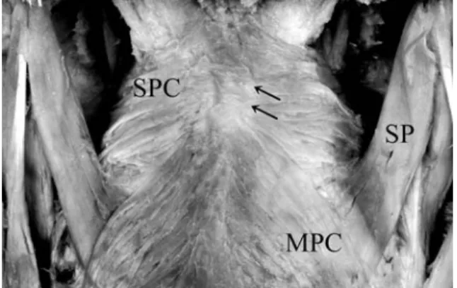
(7) 歯科学報. Vol.108,No.1(2008). 71. Fig. 13 Morphology of the insertion of the superior pharyngeal constrictor muscle as observed from the posterior direction (type B) . The posterior wall of the pharynx comprises right and left superior pharyngeal constrictor muscles. SPC = superior pharyngeal constrictor muscle, MPC = middle pharyngeal constrictor muscle, SP = stylopharyngeus muscle.. Fig. 12 Morphology of the insertion of the superior pharyngeal constrictor muscle as observed from the posterior direction (type A) . The posterior wall of the pharynx comprises membranous connective tissue. SPC = superior pharyngeal constrictor muscle, MPC = middle pharyngeal constrictor muscle, SP = stylopharyngeus muscle, arrow = membranous.. は茎突咽頭筋上縁を走行し,反対側の上咽頭収縮筋 と合して停止部を形成していた。 この停止部の形態は,結合組織性の膜状を呈して いたもの(A型) (Fig. 12) と左右の咽頭収縮筋が合流 したもの(B型) (Fig. 13) の2型に分けられた。37例 中,A型 は21例(56. 8%) ,B型 は16例(43. 2%) で あった。咽頭収縮筋を後方から観察すると中,下咽 頭収縮筋は斜走して停止していたのに対し,上咽頭 収縮筋は横走し停止していた。さらに,この上咽頭 収縮筋の背面下部は中咽頭収縮筋に覆われていた。 上咽頭収縮筋頬咽頭部の組織学的観察 肉眼観察で得られた3つの形態(上部から下部ま. Fig. 14 Histologic image of the origin of the superior pharyngeal constrictor muscle at the buccopharyngeal part(type A, Azan stain) . Muscle fibers are sporadically observed among rough connective tissue fibers.. で結合組織性の膜状のもの;A型,上部だけが膜状 のもの;B型,上部から下部まで膜が完全に欠如し ていたもの;C型) についてそれぞれ Azan 染色を 施し,組織学的観察を行った。. 成していた(Fig. 9) 。この舌咽頭部の 筋 は 起 始 直. その結果,A型は疎な結合組織線維の中に筋線維. 後,口蓋扁桃付近において上咽頭収縮筋顎咽頭部の. が散在していた(Fig. 14) 。B型は上部では疎な結合. 筋と交錯あるいは合流していた。両部の筋線維が交. 組織線維で,下部では横走する筋線維で構成されて. 錯し区別のつかなかったものを“交錯型” とし,両部. いた(Fig. 15) 。C型は結合組織線維が少なく,頬咽. の筋線維が交錯せず合流していたものを“合流型” と. 頭部の筋と頬筋の筋線維が互いに入り込んで構成さ. して両部の交錯状態を観察した。43例中,交錯型. れていた(Fig. 16) 。. (Fig. 10) は28例 (65. 1%) , 合流型 (Fig. 11) は15例(34. 9. 考 察. %) であった。 2)停止部の形態. 上咽頭収縮筋翼突咽頭部の筋は咽頭の最上部を形. 4部から起始した上咽頭収縮筋は起始直後,合流. 成していた。この翼突咽頭部の筋は蝶形骨翼突鈎か. し咽頭の上方側壁を形成していた。その後,この筋. ら起始すると報告されている5)∼8)12)。しかし今回の. ― 71 ―.
(8) 72. 津守, 他:嚥下機能に関連する上咽頭収縮筋の形態学的特徴. Fig.15 Histologic image of the origin of the superior pharyngeal constrictor muscle at the buccopharyngeal part(type B, Azan stain) . Rough connective tissue fibers are apparent in the superior area, and transverse muscle fibers are seen in the inferior area. 2 = superior pharyngeal constrictor muscle at the buccopharyngeal part, B = buccinator muscle.. Fig.16 Histologic image of the origin of the superior pharyngeal constrictor muscle at the buccopharyngeal part(type C, Azan stain) . Muscle fibers of the superior pharyngeal constrictor muscle at the buccopharyngeal part and buccinator muscle are not continuous and are separated. 2 = superior pharyngeal constrictor muscle at the buccopharyngeal part, B = buccinator muscle.. 観察結果から,この部の筋は蝶形骨翼突鈎から蝶形. 分かれており,出生後に縫線の形態が変化すると述. 骨翼状突起内側板後縁にかけて広い範囲から起始し. べている。この報告と今回の観察結果から出生後に. ていることが明らかとなった。. 咀嚼,嚥下,発音などの機能が発達し,上咽頭収縮. 上咽頭収縮筋頬咽頭部の筋の起始形態は,上部か. 筋頬咽頭部の筋と頬筋が協調的に作用した結果,そ. ら下部まで結合組織性の膜状を呈していたもの(A. れらの機能に適応する形態に翼突下顎縫線が変化す. 型) , 上部だけが膜状を呈していたもの(B型) ,上部. ると推測された。Casey12)によれば,上咽頭収縮筋. から下部まで膜が完全に欠如していたもの(C型) の. は頬筋,口輪筋と共に括約筋を形成しており,嚥下. 3型に分けられた。この部の筋は頬筋と同じ翼突下. の第一相において重要な働きを行っていると述べて. 顎縫線から起始するといわれている。これまで翼突. いる。今回の観察結果から,上咽頭収縮筋頬咽頭部. 下顎縫線については“the sphenoid tendon between. の筋と頬筋との間には膜の有無の違いはあったが,. the jaws”,腱のようなバンド,腱の線維,腱様構. 骨は存在せず,両筋が結合組織により接合してい. 4) 6) 13) 14). 造物な ど と 表 現 さ れ て い る. 。一 方,Gaugh-. た。これらのことから,形態的にみて嚥下時に上咽. ran によれば翼突下顎縫線は100%欠如し,従来記. 頭収縮筋と頬筋が協調的に作用すると推測された。. 載されている縫線は anatomical artifact であると述. さらに,咀嚼から嚥下運動に至る一連の動作がス. べている。今回の観察において腱は観察されなかっ. ムーズに行えるのは上咽頭収縮筋が頬筋,口輪筋と. た。また Gaughran は頬筋と上咽頭収縮筋は連続し. 共に一つの括約筋としての形態を形成してるからだ. ていて buccinatopharyngeus muscle を形成してお. と考えられた。. 15). り,今までの縫線の概念は誤っており,捨てなけれ. 上咽頭収縮筋顎咽頭部の筋の起始形態は,先端が. ばならないと述べている。この所見は,本研究の肉. 顎舌骨筋線上にみられたものと顎舌骨筋線から離れ. 眼解剖学的観察のC型に相当するものと考えられ. ていたものの2型に分けられた。この顎咽頭部の筋. た。しかし,組織学的観察においてC型は頬筋と上. の起始部は解剖学の教科書では顎舌骨筋線と記載さ. 咽頭収縮筋は連続せず,結合組織で分かれていた。. れている4)7)。しかし,今回,起始先端が顎舌骨筋線. Shimada と Gasser17)は胎児の翼突下顎縫線はすべ. から離れていたものの存在が明らかとなった。今. ての標本で頬筋と上咽頭収縮筋は幅広い筋膜により. 回,2つの起始形態が観察されたが,いずれの形態. ― 72 ―.
(9) 歯科学報. Vol.108,No.1(2008). も下顎骨から起始していた。このことから嚥下時に 下顎骨が動くと顎咽頭部の筋も作用すると推測され た。. 本論文の内容は,第11回日本摂食・嚥下リハビリテーショ ン学会学術大会(2005年9月,名古屋市) において学会奨励賞 を受賞した。. 文. 上咽頭収縮筋舌咽頭部の筋の起始部は,口蓋舌筋 や茎突舌筋とともに舌根部を形成していた。また, この舌咽頭部の筋は起始直後,顎咽頭部の筋と交錯 あるいは合流していた。今回,2つの型が観察され たが,解剖学的構造からみて嚥下運動において舌咽 頭部と顎咽頭部の筋がそれぞれ別々に作用するとは 考えづらく,両部の筋は協調しあって作用すると考 えられた。そして,この舌咽頭部の筋は食塊を舌根 部から咽頭へ送り込むのに重要な役割を果たしてい ると推測された。 上咽頭収縮筋の停止部の形態は,結合組織性の膜 状を呈していたものと左右の咽頭収縮筋が合流した も の の2型 に 分 け ら れ た。Shimada と Gasser18)の 報告と今回の観察を比べるとほぼ同様の結果となっ た。今回2つの型が観察されたが,いずれの型も軟 組織で形成されていた。このことから上咽頭収縮筋 の停止部は嚥下時に咽頭が自由に拡張や収縮できる のに適した形態であると推測された。また停止部に おいて上咽頭収縮筋で形成されていた部分は軟口蓋 とほぼ同じ高さであった。そして,この上咽頭収縮 筋は中,下咽頭収縮筋が斜走していたのに対し,横 走していた。これらのことから上咽頭収縮筋停止部 は嚥下運動の鼻咽腔閉鎖時に形成される Passavant 隆起に関与していると推測された。 今回の研究結果から上咽頭収縮筋は,咀嚼に重要 な役割を果たす頬筋と結合組織により接合し,下顎 骨,舌根部から起始していた。つまり口腔と咽頭が 頬筋や上咽頭収縮筋のような一連の筋によって囲ま れており,これらの筋は咀嚼,嚥下時に協調して, 機能すると考えられた。以上のことから,上咽頭収 縮筋の形態は,摂食の口腔期から咽頭期へスムーズ に行うのに適した形態であると示唆された。. 73. 献. 1)Donner, M. W., Bosma, J. F., Robertson, D. : Anatomy and physiology of the pharynx. Gastrosintest Radiol, 10: 196∼212,1985. 2)Bosma, J. F., Donner, M. W., Tanaka, E., Robertson, D. : Anatomy of the pharynx, pertinent to swallowing. Dysphagia, 1:23∼33,1986. 3)Snell, R. S. : Atlas of Clinical Anatomy. Boston : Little, Brown, 476∼477,1978 4)Williams, P. L. : Gray s Anatomy 38th ed. Philadelphia : W. B. Saunders, 1726∼1733,1995. 5)Agur, A. M. R., Lee, M. J. : Grant s Atlas of anatomy 10 th ed. Philadelphia : Lippincott Williams & Wilkins, 676 ∼684,1999. 6)Brand, R. W., Isselhard, D. E. : Anatomy of Orofacial Structures. Saint Louis : Mosby, 356∼363,2003. 7)Putz, R., Pabst, R. : Sobotta atlas of human anatomy Vol. 1 13th English ed. Philadelphia : Lippincott Williams & Wilkins, 136∼138,2001. 8)Hollinshead, W. H. : Anatomy of the pharynx and esophagus. In : English GM. (eds.) Otolaryngology. Philadelphia : Harper and Row, 1∼9,1985. 9)Bosma, J. F., Bartner, H. : Ligaments of the larynx and the adjacent pharynx and esophagus. Dysphagia, 8:23 ∼28,1993. 10)Mu, L., Sanders, I. : Muscle fiber-type distribution pattern in the human cricopharyngeus muscle. Dysphagia, 17:87∼96,2002. 11)Leaper, M., Zhang, M. : An anatomical protrusion exists on the posterior hypopharyngeal wall in some elderly cadavers. Dysphagia, 20:8∼14,2005. 12)Casey, D.M. : Palatopharyngeal anatomy and physiology. J Prosthet Dent 49:371∼378,1983. 13)Howland, J. P., Brodie, A. G. : Pressures exerted by the buccinator muscle. Angle Orthod, 36:1∼12,1966. 14)Sicher, H. : The anatomy of mandibular anesthesia. J Am Dent Assoc, 33:1541∼1557,1946. 15)Gaughran, G. R. L. : The pterygomandibular rapheanatomical artifact. Anat Rec, 184:410,1976. 16)Saigusa, H., Yamashita, K., Tanuma, K., Saigusa, M., Niimi, S. : Morphological studies for retrusive movement of the human adult tongue. Clin Anat, 17:93∼98,2004. 17)Shimada, K., Gasser, R.F. : Morphology of the pterygomandibular raphe in human fetuses and adults. Anat Rec, 224:117∼122,1989. 18)Shimada, K., Gasser, R.F. : Variations of the pharyngeal raphe. Clin Anat, 1:285∼294,1988.. ― 73 ―.
(10)
図


+2
関連したドキュメント
Then the change of variables, or area formula holds for f provided removing from counting into the multiplicity function the set where f is not approximately H¨ older continuous1.
It is suggested by our method that most of the quadratic algebras for all St¨ ackel equivalence classes of 3D second order quantum superintegrable systems on conformally flat
Keywords: continuous time random walk, Brownian motion, collision time, skew Young tableaux, tandem queue.. AMS 2000 Subject Classification: Primary:
In this work we give definitions of the notions of superior limit and inferior limit of a real distribution of n variables at a point of its domain and study some properties of
This paper develops a recursion formula for the conditional moments of the area under the absolute value of Brownian bridge given the local time at 0.. The method of power series
The main problem upon which most of the geometric topology is based is that of classifying and comparing the various supplementary structures that can be imposed on a
Then it follows immediately from a suitable version of “Hensel’s Lemma” [cf., e.g., the argument of [4], Lemma 2.1] that S may be obtained, as the notation suggests, as the m A
Our method of proof can also be used to recover the rational homotopy of L K(2) S 0 as well as the chromatic splitting conjecture at primes p > 3 [16]; we only need to use the
