Chemical study on structure and property
of C-terminal region of human prion protein
Chemical study on structure and property
of C-terminal region of human prion protein
using synthetic fragment peptides
SAKAGUCHI Yuko
Supervisor Professor TOYODA Hidenao
Doctoral Program in Pharmacy
Graduate School of Pharmacy
Ritsumeikan University
1 1
2 2.1 hPrP 6
2.2 hPrP180-192 8
2.2.1 Pull down assay 2.2.2 AFFINIX QNµ 2.3 hPrP180-192 16 2.3.1 2.3.2 Cu2+ 2.3.3 pH Cu2+ 2.4 hPrP180-192 V180I 22 2.4.1 Cu2+ 2.4.2 Cu2+ 2.5 AFFINIX QNµ 28 2.5.1 Tris-HCl 2.5.2 2.6 Thioflavin T 32 3 36 4 41 5 42 62 63
CD: circular dichroism hPrP: human prion protein
hPrP-CF: human prion protein C-terminal region fragment peptide MS: mass spectrometry
MT-MMP membrane type matrix metalloproteinase PBS: phosphate buffered salts
PrP: prion protein
PrPC: cellular prion protein PrPSc: scrapie-type prion protein QCM: quartz crystal microbalance ThT: thioflavin T
1 1
(transmissible spongiform encephalopathies: TSE)
(PrP) [1-4 100 1 [5] 1.5 100 [6] PrP [6-9] (PrPC ) (PrPSc) [1,2] PrPC PrPSc PrPC β-sheet 3 PrPSc 43 β-sheet [10,11] PrPC PrPSc Cu2+ [12] ( X) [13-15] PrPSc [2] [16] PrPSc PrPC
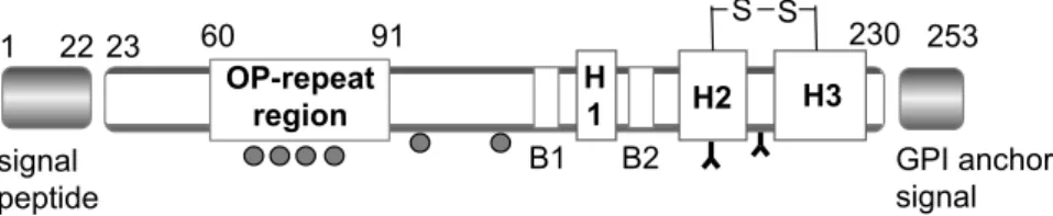
PrPSc PrPC PrPSc PrP (hPrPC) 253 27661 N 8 (PHGGGWGO) (OP-repeat) N- 2 β-sheet 1 -helix (H1) 2 -helix (H2 H3) (GPI)
(Fig. 1) [17, 18] OP-repeat His 4
His 2 Cu2+ [19] Cu2+ Cu2+ [20-37] PrPC PrP GPI [2,38,39] PrPC (NMR) (CD) C- PrPSc
3 [40-50] N-C- H2 H3 [40] PrP C- H2 (173-195) 16 -helix β-sheet [41] hPrP180-195 pH β-sheet H2 [42] hPrP180-193 hPrP178-193 [43] H2 pH H2 pH [44] PrPC PrPSc C- H2 pH Cu2+ 30 [51-53]
C- H2 hPrP180 Val Ile (V180I) [51] V180I
[54]
PrP [58] H2-H3 B2 - H2 [59] PrPSc PrPSc PrP (hPrP-CF) hPrP180-192 Cu2+ (MT-MMP) hPrP-CF [60-62] C- (Fig. 2) C- (CD) (HPLC) AFFINIX QNµ
Pull down assay Thioflavin T hPrP180-192
5
B1 : β-sheet region (hPrP 128-131), B2 : β-sheet region (hPrP 161-164), H1 : α-helix region (hPrP 144-154), H2 : α-helix region (hPrP 173-194), H3 : α-helix region (hPrP 200-228) S-S : disulfide bond hPrP179, 214 : N-type glycosylation hPrP181, 197 : Cu2+ binding sites hPrP61, 69, 77, 85, 96, 111 S S signal peptide OP-repeat region B1 B2 GPI anchor signal H 1 H2 H3 1 22 23 60 91 230 253
Fig. 1 Domain structure of the hPrP.
E N F T D K MA N GC WM LV L L F V A TW SD L GL C K K R PK P G G W N T G GS R N A G A A A A G D A V V G G L G G S Y M L G A M S R P I I H F G S Y E R Y Y R E N M H R Y P NQ V Y Y R P M D E YS N QNN F V H D C V N I T I KHQ T V T T T T K G T M M E R V V E Q M C I T Q Y E R E S Q AY Y R G S S M V L F S S P P V I L L I S F L I F L I V G D 22 60 91 160 230 1 253 GPI anchoring signal E V α- helix β− sheet (PNAS, Vol 97 (1), 145-150, 2000) His MT1-MMP MT3-MMP OP-repeat OP-repeat Cu2+ MT 1, 3-MMP Cu2+ C-Cu2+ pH β−sheet Signal peptide
2 2.1 hPrP C- 2 -helix (H2 H3) (Fig. 1) H2 [40-44] 2 His 177 His 187 His H2 24 hPrP169-192 177 His hPrP169-183 hPrP175-183 187 His hPrP180-192 180 Val Ile hPrP180-192 V180I 5 (hPrP-CF) F-moc HPLC HPLC (Fig. 3) (MS) (Table 1) 4
7 (a) hPrP169-192 (min.) (b) hPrP169-183 (d) hPrP180-192 (c) hPrP175-183 (e) hPrP180-192 V180I mAU (2 20 n m) mAU (2 20 n m) (min.) mAU (2 20 n m) (min.) mAU (2 20 n m) (min.) (min.) mAU (2 20 n m)
Fig. 3 Purity confirmation of each hPrP-CF.
Table 1 Amino acid sequence of each hPrP-CF and identification of each molecular weight.
hPrP-CF Sequence Detected ion m/z Experimental Mass Theoretical Mass hPrP169-192 YSNQNNFVHDCVNITIKQHTVTTT [ M + 4H ]4+ 2776.73 2776.32
hPrP169-183 YSNQNNFVHDCVNIT [ M + 2H ]2+ 1766.80 1766.77
hPrP180-192 V180I INITIKQHTVTTT [ M + 3H ]3+ 1468.41 1468.83
hPrP180-192 VNITIKQHTVTTT [ M + 3H ]3+ 1454.84 1454.81
2.2 hPrP180-192
hPrP180-192
[60-62] hPrP180-192
(hPrP-CF)
2.2.1 Pull down assay
hPrP180-192 (R-hPrP180-192) 1
hPrP-CF 25
Cu2+
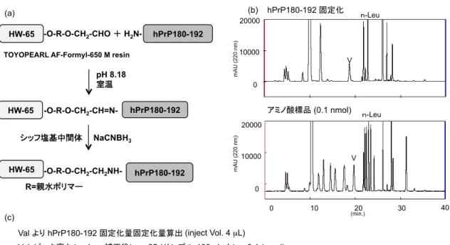
hPrP180-192 HW-65
(Fig. 4-a hPrP180-192
(Fig. 4-b n-Leu H (standard)
(Val)
9 hPrP180-192 HW-65 -O-R-O-CH2-CHO H2N- pH 8.18 -O-R-O-CH2-CH=N- NaCNBH3 -O-R-O-CH2-CH2NH- R=
TOYOPEARL AF-Formyl-650 M resin
HW-65
HW-65
hPrP180-192
hPrP180-192
Val hPrP180-192
Val ( n-Leu ) × 25 ( 100 µL ) × 0.1 (nmol) × 5 ( 5µL × ( V ) (inject Vol. 4 µL) (0.1 nmol) Val hPrP180-192 -Leu -Leu V 10000 20000 0 V 0 10 20 30 40 10000 20000 0 (min.) (a) (b) (c) mAU (2 20 n m) mAU (2 20 n m)
Fig. 4 Preparation of hPrP180-192 immobilized resin.
(a) Procedure of immobilization. (b) Chromatogram of amino acid analysis. (c) Calculating immobilized amount of hPrP180-192.
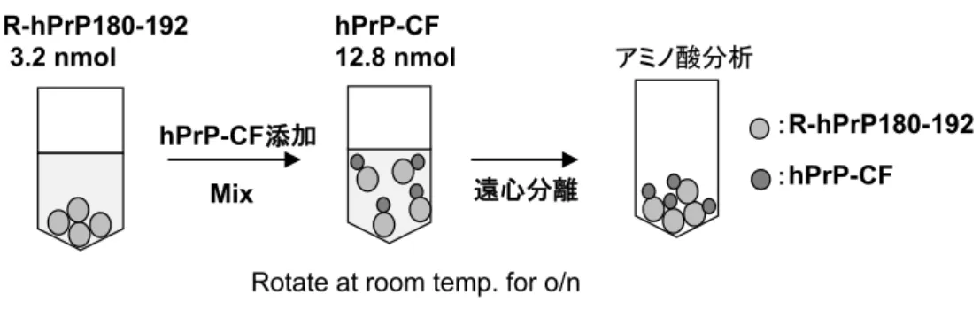
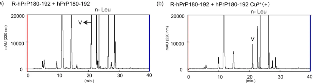
hPrP-CF R-hPrP180-192 (3.2 nmol) 4 hPrP-CF (12.8 nmol) 1 hPrP-CF (Fig. 5 Tris (R-Tris) R-hPrP180-192 hPrP180-192 Fig. 6 (V) Cu2+ Table. 2 R-Tris 2 + 4 ++ 2 - Cu2+ hPrP169-183 hPrP180-192 hPrP180-192 R-Tris 4 Cu2+ hPrP169-183 hPrP180-192 Cu2+ R-hPrP180-192 hPrP-CF R-hPrP180-192 3.2 nmol hPrP-CF 12.8 nmol hPrP-CF Mix
Rotate at room temp. for o/n
11 R-hPrP180-192 + hPrP180-192 V n- Leu 10000 20000 0 0 10 20 30 40 (min.) R-hPrP180-192 + hPrP180-192 Cu2+ + n- Leu V 10000 20000 0 0 10 20 30 40 (min.) (a) (b) mAU (2 20 n m) mAU (2 20 n m)
Fig. 6 The effects of Cu2+ on affinity between R-hPrP180-192 and hPrP180-192.
(a) In the absence of Cu2+ and (b) in the presence of Cu2+.
Table 2 Summary of pull down assay.
Pull down assay (nmol)
Cu2+ ( - ) Cu2+ ( + ) R-Tris R-hPrP180-192 R-Tris R-hPrP180-192 hPrP169-192 1.7 ± 0.2 1.9 ± 0 2.1 ± 0.1 3.0 ± 0.2 hPrP169-183 1.3 ± 0.2 2.7 ± 0.2 1.3 ± 0 0.8 ± 0.1 hPrP175-183 1.1 ± 0.2 N.D. 2.5 ± 0.1 1.1 ± 0.1 hPrP180-192 1.0 ± 0 4.8 ± 0.2 1.0 ± 0 0.3 ± 0.1
2.2.2 AFFINIX QNµ
Pull down assay 1
AFFINIX QNµ
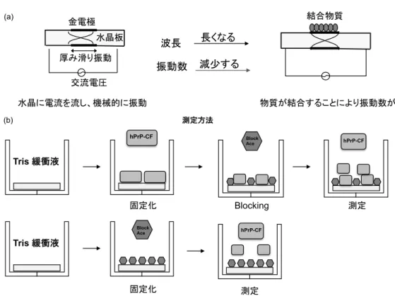
(AFFINIX QNµ (quartz crystal microbalance QCM)
(Fig. 7-a) AFFINIX QNµ
[63]
13 Blocking hPrP-CF Block Ace hPrP-CF Tris Tris Block Ace hPrP-CF (a) (b)
Fig. 7 Principle and measurement methods of AFFINIX QNµ. (a) Principle of AFFINIX QNµ. (b) Measurement methods.
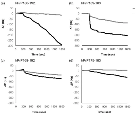
hPrP180-192 hPrP-CF Fig. 8 hPrP-CF H2 hPrP180-192 (Fig. 8-a H2 N- hPrP169-183 (Fig. 8-b H2 hPrP169-192 hPrP175-183 (Fig. 8-c and -d -300 -250 -200 -150 -100 -50 0 50 -300 -250 -200 -150 -100 -50 0 50 (d) hPrP169-192 (b) -300 -250 -200 -150 -100 -50 0 50 hPrP169-183 (c) (a) Δ F (H z) Time (sec) Time (sec) Δ F (H z) Δ F (H z) Time (sec) immobilized hPrP180-192 blocked -300 -250 -200 -150 -100 -50 0 50 hPrP175-183 Δ F (H z) Time (sec) hPrP180-192
Fig. 8 Intermolecular interaction of hPrP180-192 against hPrP-CF using an AFFINIX QNµ system. 5 kinds of hPrP-CF were injected into the hPrP180-192 immobilized QCM cuvette. (a) hPrP180-192, (b) hPrP169-183, (c) hPrP169-192 and (d) hPrP175-183 were injected in the absence of Cu2+
15
hPrP180-192 hPrP169-183
hPrP180-192 hPrP180-192
hPrP180-192
Pull down assay hPrP169-183 hPrP180-192
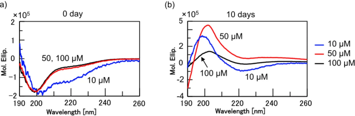
2.3 hPrP180-192 PrPC PrPSc β-sheet pH Cu2+ CD hPrP180-192 PrP 2.3.1 CD Cu2+ hPrP180-192 Tris-HCl (pH 7.5) hPrP180-192 CD (0 day) 50 µM 100 µM 197 nm
random coil (Fig. 9-a) 10 µM 208 nm
222 nm 191 nm 37 10 50 µM 100 µM 200 nm β-sheet 10 µM 192 nm (Fig. 9-b) 100 µM 200 nm β-sheet hPrP180-192
17 50 µM hPrP180-192 (a) (b) 0 day ×105 -4 -2 2 10 µM 50 µM 100 µM ×105 10 days 10 µM 50, 100 µM 50 µM 10 µM 100 µM
Fig. 9 CD spectra of hPrP180-192 at various concentrations at 0 and 10 days in the absence of Cu2+. Spectra were obtained for hPrP180-192 at 10, 50, and
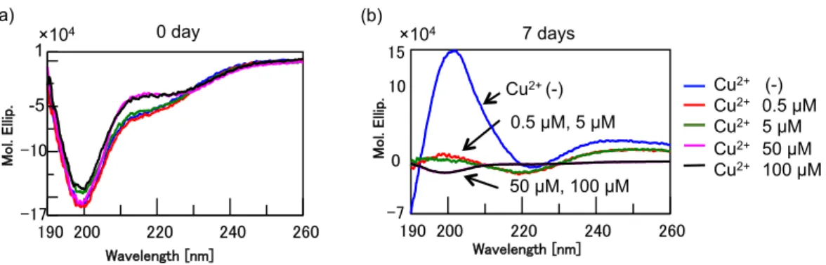
2.3.2 Cu2+ hPrP180-192 Cu2+ 50 µM hPrP180-192 Cu2+ 37 7 Cu2+ 197 nm random coil Cu2+ (Fig. 10-a) 7 CD Cu2+ 200 nm β-sheet hPrP180-192 50 µM 2 100 µM 197 nm β-sheet hPrP180-192 1/10 (Cu2+ 5 µM) 1/100 (Cu2+ 0.5 µM) 200 nm Cu2+ β-sheet Fig. 10-b hPrP180-192 187 His 1 Cu2+ 1 hPrP180-192 Cu2+ 2 Cu2+ (100 µM)
19 Cu2+ (-) Cu2+ 0.5 µM Cu2+ 5 µM Cu2+ 50 µM Cu2+ 100 µM ×104 Cu2+ (-) 0.5 µM, 5 µM 50 µM, 100 µM 1 -5 ×10⁴ (a) (b) 0 day 7 days
Fig. 10 The effects of Cu2+ concentration on CD spectra of hPrP180-192.
50 µM of hPrP180-192 was incubated with 0.5, 5, 50, and 100 µM of Cu2+ for
2.3.3 pH Cu2+ hPrP180-192 pH Cu2+ Cu2+ hPrP180-192 hPrP180-192 pH random coil 7 pH 7.0 pH 8.0 pH 9.0 200 nm β-sheet pH 7.0 200 nm β-sheet Cu2+ 7 pH 7.0 197 nm
random coil (Fig. 11-a) Cu2+
hPrP180-192 Cu2+ hPrP180-192 pH random coil 7 pH 9.0 200 nm β-sheet (Fig. 11-b) Cu2+ hPrP180-192 pH 6.0 Cu2+ pH 8.0 pH 7.0 Cu2+
random coil β-sheet Cu2+
21 7 days ×105 (a) ×105 pH 5 pH 6 pH 7 pH 8 pH 9 pH 5, 6, 7, 8, 9 pH 7 pH 8, 9 pH 5, 6 (b) ×105 0 day ×105 7 days pH 5, 6, 7, 8, 9 pH 9 pH 5, 6, 7, 8
14 days (Cu2+ 7days
×105
pH 8, 9 pH 5, 6, 7 0 day
Fig. 11 CD spectra of hPrP180-192 at increasing pH over time. Spectra were obtained in the (a) absence of Cu2+ and (right) following the addition of 100 µM
of Cu2+ after 7 days, obtained at 14 days. (b) Presence of Cu2+ at an initial time
2.4 hPrP180-192 V180I hPrP180-192 pH β-sheet PrP H2 2 180 Val Ile hPrP180-192 V180I pH 7.5 hPrP180-192 CD HPLC 2.4.1 Cu2+ Cu2+ hPrP180-192 hPrP180-192
V180I 197 nm random coil
HPLC 1 HPLC (Fig. 12 Cu2+ hPrP180-192 hPrP180-192 V180I 1 random coil HPLC (Fig. 13) Cu2+ hPrP180-192 hPrP180-192 V180I
23 0 day (a) (b) 1 day 0 day 1 day hPrP180-192 V180I 0 day 1 day (min.) mAU (2 20 n m) (min.) (min.) mAU (2 20 n m) (min.) hPrP180-192
Fig. 12 Time dependency of the structure and peak value changes of hPrP180-192 and hPrP 180-192 V180I in the absence of Cu2+.
Upper: CD spectra for 50 µM of (a) hPrP180-192 and (b) hPrP180-192 V180I incubated for 0 day and 1 day.
Lower: Detection of soluble (a) hPrP180-192 and (b) hPrP180-192 V180I by HPLC at 0 day and 1 day. Twenty µL of 50 µM of each peptide was injected for HPLC analysis.
4 ×10⁴ 4 ×10⁴ hPrP180-192 hPrP180-192 V180I (min.) mAU (2 20 n
m) 0 day 1 day 0 day 1 day
(min.) (min.) (min.)
(a) (b)
0 day 1 day
Fig. 13 Time dependency of the structure and peak value changes of hPrP180-192 and hPrP 180-192 V180I in the presence of Cu2+.
Upper: CD spectra for 50 µM of (a) hPrP180-192 and (b) hPrP180-192 V180I incubated for 0 day and 1 day.
Lower: Detection of soluble (a) hPrP180-192 and (b) hPrP180-192 V180I by HPLC at 0 day and 1 day. Twenty µL of 50 µM of each peptide was injected for HPLC analysis.
2.4.2 Cu2+ hPrP180-192 hPrP180-192 V180I Cu2+ 197 nm random coil 1 200 nm β-sheet Cu2+ CD β-sheet hPrP180-192 hPrP180-192 V180I Cu2+ hPrP180-192 0 hr random coil 2 hr 4 hr 6 hr 197 nm 26 hr 200 nm β-sheet 49 hr 200 nm 68 hr 49 hr Fig. 14-a hPrP180-192 68 hr Cu2+ Cu2+ 200 nm Cu2+ 2 197 nm random coil Cu2+ 8 197 nm Cu2+ 22 Cu2+ 8 (Fig. 14-b HPLC hPrP180-192 0 hr hPrP180-192
25 68 hr Cu2+ Cu2+ 60 % (Fig. 14-c and 16-b PrP180-192 V180I Cu2+ CD HPLC hPrP180-192 (Fig. 15-a Cu2+ hPrP180-192 (Fig. 15-b HPLC 15 % hPrP180-192 (Fig. 15-c and 16-b CD 200 nm (Fig. 16 CD 200 nm hPrP180-192 V180I Cu2+
68 (Cu2+ 0) 70 (Cu2+ 2) 76 (Cu2+ 8) 90 (Cu2+ 22) 68 (h) 8 (min.) 11 mAU (2 20 n m) 8 (min.) 11 0 h 4 h 26 h 68 h 76 h (Cu2+ 8h) 90 h (Cu2+ 22h) (min.) mAU (2 20 n m) (a) (c) (b)
Fig.14 Time dependency of the structure and peak value changes of hPrP180-192. (a) CD spectra for 50 µM of hPrP180-192 incubated for 0, 2, 4, 6, 26, 49, and 68 h without Cu2+. (b) CD spectra for hPrP180-192 following the addition of 100 µM of Cu2+ after 68 h, obtained at 70, 76, and 90 h. (c) Detection of soluble hPrP180-192 by HPLC at 0, 68, and 96 hours. Cu2+ was added at 68 h. Twenty µL of 50 µM hPrP180-192 was injected for HPLC analysis.
68 (Cu2+ 0) 70 (Cu2+ 2) 76 (Cu2+ 8) 90 (Cu2+ 22) 68 (h) 0 h 4 h 26 h mAU (2 20 n m) 68 h 76 h (Cu2+ 8h) 90 h (Cu2+ 22h)
(min.) (min.) (min.)
mAU (2 20 n m) (a) (c) (b)
Fig.15 Time dependency of the structure and peak value changes of hPrP180-192 V180I. (a) CD spectra for 50 µM of hPrP180-192 V180I incubated for 0, 2, 4, 6, 26, 49, and 68 h without Cu2+. (b) CD spectra for hPrP180-192 V180I following the addition of 100 µM of Cu2+ after 68 h, obtained at 70, 76, and 90 h. (c) Detection of soluble hPrP180-192 V180I by HPLC at 0, 68, and 96 hours. Cu2+ was added at 68 h. Twenty µL of 50 µM hPrP180-192 V180I was injected for HPLC analysis.
27 D ecre asi ng R at io (% )100 80 60 40 20 0 0 20 40 60 80 100 (h) + Cu2+ (a) (b) -15 -20 -5 0 5 0 20 40 60 80 100 (h) Mo l. El lip . (2 00 n m) + Cu2+ hPrP180-192 hPrP180-192 V180I hPrP180-192 hPrP180-192 V180I
Fig.16 Molar ellipticity at 200 nm and peak value changes of hPrP180-192 and hPrP180-192 V180I. (a) The molar ellipticity at 200 nm plotted for each CD spectrum. (b) The decreasing ratio of all peaks relative to their peak height at 0 h on HPLC were calculated.
2.5 AFFINIX QNµ hPrP180-192 hPrP180-192 V180I Cu2+ Cu2+ hPrP180-192 hPrP180-192 V180I AFFINIX QNµ Cu2+ 2.5.1 Tris-HCl hPrP180-192 Cu2+ hPrP180-192 (Fig. 17-a Cu2+ hPrP180-192 (Fig. 17-b hPrP180-192 V180I hPrP180-192 V180I Cu2+ (Fig. 17-c and -d hPrP180-192 V180I Cu2+ hPrP180-192 Cu2+
29 -300 -250 -200 -150 -100 -50 0 50 0 300 600 900 1200 1500 1800 -300 -250 -200 -150 -100 -50 0 50 0 300 600 900 1200 1500 1800 -300 -250 -200 -150 -100 -50 0 50 0 300 600 900 1200 1500 1800 -300 -250 -200 -150 -100 -50 0 50 0 300 600 900 1200 1500 1800 (d) hPrP180-192 V180I (b) hPrP180-192 + Cu2+ (c) (a) Δ F (H z) Time (sec) Time (sec) Δ F (H z) Δ F (H z) Time (sec) hPrP180-192 V180I + Cu2+ Δ F (H z) Time (sec) hPrP180-192 blocked immobilized hPrP180-192 blocked immobilized hPrP180-192 blocked immobilized hPrP180-192 V180I blocked immobilized hPrP180-192 V180I
Fig. 17 Intermolecular interaction of hPrP180-192 and hPrP180-192 V180I. Change in oscillation frequency of (a) hPrP180-192 and (b) hPrP180-192 V180I in the absence of Cu2+ and (c) hPrP180-192 and (d) hPrP180-192 V180I in the presence of Cu2+.
hPrP180-192 hPrP180-192 V180I hPrP180-192 V180I
hPrP180-192 hPrP180-192
(Fig. 18-a and -c)
Cu2+ (Fig. 18-b and -d) -300 -250 -200 -150 -100 -50 0 50 0 300 600 900 1200 1500 1800 -300 -250 -200 -150 -100 -50 0 50 0 300 600 900 1200 1500 1800 -300 -250 -200 -150 -100 -50 0 50 0 300 600 900 1200 1500 1800 -300 -250 -200 -150 -100 -50 0 50 0 300 600 900 1200 1500 1800 (a) hPrP180-192 V180I hPrP180-192 (c) Δ F (H z) Time (sec) Δ F (H z) Time (sec) blocked immobilized hPrP180-192 blocked immobilized hPrP180-192 V180I (b) hPrP180-192 V180I + Cu2+ Time (sec) Δ F (H z) blocked immobilized hPrP180-192 (d) hPrP180-192 + Cu2+ Δ F (H z) Time (sec) blocked immobilized hPrP180-192 V180I
Fig. 18 Intermolecular interaction between hPrP180-192 and hPrP180-192 V180I. Change in oscillation frequency after injection of each peptides. hPrP180-192 V180I injected in to the hPrP180-192-immobilized QCM cuvette (a) in the absence of Cu2+ and (b) in the presence of Cu2+. hPrP180-192 injected in to the hPrP180-192 V180I -immobilized QCM cuvette (c) in the absence of Cu2+ and (d) in the presence of Cu2+.
31 2.5.2
hPrP180-192 hPrP180-192 V180I Tris-HCl Tris-HCl
Phosphate buffered salts (PBS) (pH 7.4 hPrP180-192 Tris-HCl PBS (Fig. 19-a hPrP180-192 V180I Tris-HCl PBS (Fig. 19-b PBS Cu2+ PrPC PrPSc -600 -500 -400 -300 -200 -100 0 -600 -500 -400 -300 -200 -100 0 (b) hPrP180-192 V180I (a) Time (sec) Δ F (H z) Δ F (H z) Time (sec) hPrP180-192 blocked immobilized hPrP180-192 V180I blocked immobilized hPrP180-192
Fig. 19 Intermolecular interaction of hPrP180-192 and hPrP180-192 V180I in the PBS buffer. (a) hPrP180-192 and (b) hPrP180-192 V180I.
2.6 Thioflavin T CD HPLC hPrP180-192 hPrP180-192 V180I Thioflavin T ThT Tris-HCl pH 7.5 37 [64] ThT 490 nm [65] 10 (Fig. 20)
Random coil β-Sheet Aggregate
Soluble
Thioflavin T
Bind (Fluorescence)
Soluble Insoluble
33
Cu2+ hPrP180-192
Cu2+
(Fig. 21-a hPrP180-192 V180I 2
hPrP180-192 10 Cu2+ hPrP180-192 (Fig. 21-b hPrP180-192 PrP180-192 V180I Cu2+ hPrP180-192 hPrP180-192 V180I hPrP180-192 hPrP180-192 V180I β-sheet 3 Cu2+ hPrP180-192 Cu2+ 2 2 2 3 2 1 Cu2+ Cu2+ Cu2+ Fig. 21-c hPrP180-192 V180I PrP180-192 2 2 3 1 Cu2+ Cu2+ 1 Cu2+ (Fig. 21-d CD HPLC
hPrP180-192 V180I hPrP180-192 hPrP180-192 V180I Cu2+ hPrP180-192 0 100 200 300 400 500 600 700 0 1 2 3 4 5 6 7 8 9 10 0 20 40 60 80 100 120 0 1 2 3 4 5 6 7 8 9 10 0 100 200 300 400 500 600 700 0 1 2 3 4 5 6 7 8 9 10 0 20 40 60 80 100 120 0 1 2 3 4 5 6 7 8 9 10 F lu ore sce nce in te nsi ty F lu ore sce nce in te nsi ty F lu ore sce nce in te nsi ty F lu ore sce nce in te nsi ty Days Days Days Days (b) (a) hPrP180-192 hPrP180-192 V180I (d)
(c) hPrP180-192 Cu2+ (+) on 3day hPrP180-192 V180I Cu2+ (+) on 3day
Cu2+ (+) Cu2+ (-) Cu2+ (+) Cu2+ (-) + Cu2+ + Cu2+
Fig. 21 Aggregability of hPrP180-192 and hPrP180-192 V180I. Fluorescence intensity of (a) hPrP180-192 and (b) hPrP180-192 V180I in the absence or presence of Cu2+ and (c) hPrP180-192 and (d) hPrP180-192 V180I in the
35 PBS hPrP180-192 Tris-HCl Cu2+ (Fig. 22-a hPrP180-192 V180I (Fig. 22-b (Fig. 19) hPrP180-192 V180I hPrP180-192 0 20 40 60 80 100 120 0 1 2 3 4 5 6 7 8 9 10 0 500 1000 1500 2000 0 1 2 3 4 5 6 7 8 9 10 Cu2+ (+) Cu2+ (-) Cu2+ (+) Cu2+ (-) (b) (a) hPrP180-192 hPrP180-192 V180I
Fig. 22 Aggregability of hPrP180-192 in the PBS buffer. Fluorescence intensity of (a) hPrP180-192 and (b) hPrP180-192 V180I in the absence or presence of Cu2+.
3 PrP PrPC PrPC PrPSc PrPC pH ( ) [66] hPrP Cu2+ [19,20,67] PrPC PrPSc Cu2+ [68,69,70] OP-repeat hPrP Cu2+ β-sheet [68,69]
37 β-sheet [70] Cu2+ PrPC PrPSc PrP Cu2+ PrPC PrPSc β-sheet [1,2] 111 His 1 hPrP111-126 β-sheet His 1 Cu2+ β-sheet [71] Cu2+ PrP Cu2+ Mn2+ [20] OP-repeat C-C- H2 C-hPrP180-192 (Table 3 C- Cu2+ Cu2+ hPrP180-192 hPrP180-192 V180I Cu2+
Fig. 23 and Table 3 Cu2+
random coil Cu2+
-sheet Cu2+
random coil (Fig. 16-a)
PrP180-192 V180I -sheet Cu2+ random coil (Fig. 16-b) V180I Cu2+ C- (Fig. 24) Cu2+ OP-repeat PrPC random coil C--sheet Cu2+ Cu2+ random coil Cu2+
-sheet (Fig. 24-a)
V180I
Cu2+ (Fig. 24-b
C-
39 PrPSc Cu2+
PrPC PrPSc
Table 3 Characterization of hPrP-CF.
Name His (AFFINIX) (Pull down assay) (incubate 7
days
Cu2+ - Cu2+ + Cu2+ - Cu2+ +
169-192 2 β-sheet N.D
169-183 1 β-sheet β-sheet N.D
175-183 1 Random coil α-helix
180-192 1 Random coil β-sheet
hPrP180-192 hPrP180-192
random coil β-sheet (Aggregate)hPrP180-192 hPrP180-192 - Cu
2+ random coil Cu2+ (b) (a) Cu2+ hPrP180-192 V180I hPrP180-192 random coil hPrP180-192 - Cu 2+ random coil hPrP180-192 V180I random coil hPrP180-192 V180I β-sheet (Aggregate) Cu2+ Cu2+ hPrP180-192 V180I - Cu2+ random coil hPrP180-192 V180I random coil hPrP180-192 V180I β-sheet (Aggregate) hPrP180-192 V180I - Cu2+ random coil
Fig. 23 Structure changes and aggregate formation of hPrP180-192 and hPrP180-192 V180I. (a) hPrP180-192 V180I and (b) hPrP180-192.
OP-repeat region Middle region C-terminus region Cu2+ Enzymatic cleavage Endocytosis Cu2+ Cu2+ OP-repeat region Middle region C-terminal region Cu2+ Enzymatic cleavage Endocytosis Cu2+ Cu2+ PrPC V180I PrPC (b) (a) aggregate aggregate
:β-Sheet; :Random Coil; :β-Sheet / aggregate
Cu2+ × × × × × × × × ×× aggregate × × × ××
Fig. 24 Estimated role of C-terminus region for aggregation. (a) PrPC (b) V180I mutated PrPC
41 4 Fig. 23 1) hPrP180-192 hPrP180-192 V180I pH random coil β-sheet 2) β-sheet hPrP180-192 Cu2+ random coil 3) hPrP180-192 Cu2+
4) hPrP180-192 V180I β-sheet random coil
5) hPrP180-192 V180I hPrP180-192 Cu2+ 6) β-sheet 7) PrP hPrP-CF 8) PrP
5 5.1 5.1.1 Piperidine SIGMA O-(7-Azabenzo-triazol-1-yl)-N,N,N’,N’-tetra-methyluronium hezafluoro-phosphate (HATU) N,N Dimethylformamide (DMF) N-Diisopropy;lethylamine/N-Methylpyrrolidone N-Methylpyrrolidone Dichloromethane
Preloaded Resin F-moc-L-Thr (tBu) Preloaded Resin F-moc-L-Val
Applide Biosystems F-moc-L-Asn (Trt) F-moc-L-Asp (OtBu) F-moc-L-Cys (Trt)
F-moc-L-Gln (Trt) F-moc-L-His (Trt) F-moc-L-Ile F-moc-L-Lys (Boc) F-moc-L-Phe F-moc-L-Ser (tBu) F-moc-L-Thr (tBu) F-moc-L-Tyr (tBu) F-moc-L-Val
Aceonitrile ( )
43 Diethyl ether
Trifluoroacetic Acid (Peptide Synthesis Grade)
Thioanisole Acetic acid Crystalline Phenol 1,2-Ethandithiol 5.1.2 HPLC Aceonitrile ( ) HCl
Tris (hydroxymethyl) aminomethane (Tris)
5.1.3 Pull down assay
Tris (hydroxymethyl) aminomethane (Tris) HCl CuCl2 2H2O
TOYOPEARL AF-Formyl-650 M resin TOSOH BIOSCIENCE Sodium dihydrogen phosphate. Anhytrousb ( )
Amino acid standard (Type H) Methanol ( ) Acetonitrile ( ) CH3COONa HCl Acetic acid ( ) NaHCO3 Na2CO3 Dabsyl Chloride 5.1.4
Tris (hydroxymethyl) aminomethane (Tris) HCl CuCl2 2H2O
PBS (Phosphate Buffered Salts Tablet) TaKaRa
Block Ace Powder KAC (Japan)
5.1.5 CD
45
HCl CuCl2 2H2O
5.1.6 Thioflavin T
Tris (hydroxymethyl) aminomethane (Tris) HCl
CuCl2 2H2O
PBS (Phosphate Buffered Salts Tablet) TaKaRa
5.2 5.2.1 433 Applide Biosystems 5.2.2 HPLC HPLC ( ) Pump: 880-PU Mixer: HG-980-31 JASCO Integrater: C-R6A
Detector: SPD-6A SHIMADZU Column: CAPCELLPAK C18 (TYPE AQ 5 µm 10 mm I.D. x 250 mm) SHISEIDO
HPLC ( )
Column: CAPCELLPAK C18 (Type MGII 5 µm 4.6 mm i.d. x 150 mm) SHISEIDO Pump: LC-20AD SHIMADZU Oven: COLUMN HEATER U-620 TYPE30V Sugai Detector: MD-4017
47
HPLC ( )
Computer : FUJITSU FMV ESPRIMO FIJITSU Column: COSMOSIL Packed Column (4.6 mm I.D. x 250 mm)
(Type 5C118-MG- ) SHISEIDO Pump: PU-2089
Detector: UV-2075
Oven : CO-965 JASCO
5.2.3
JEOL JMS-700T JEOL QSTAR Elite Hybrid LC/MS/MS System Applide Biosystems
5.2.4
AFFINIX QNµ ULVAC
4.2.6 CD
5.2.7
FMP-825
FP-8300
49 5.3 5.3.1 hPrP-CF 5.3.1.1 ( ) (Applide Biosystems) 433A F-moc SynthAssist Software v3.1 5.3.1.2 (TFA ) 1.5 3 PTFE
< >
: TFA 9.5 mL MilliQ 0.5 mL
: hPrP169-192 hPrP169-183 hPrP175-183 hPrP180-192 hPrP180-192 V180I
5.3.1.3
50 % (0.1 % TFA) 3.0 mL/min.
SHISEIDO CAPCELLPAK C18 (TYPE AQ 5 µm 10 mm I.D.
x 250 mm) HPLC MilliQ (0.1 % TFA)
50 % (0.1 % TFA) 30
1.0 mL/min. 40 oC SHISEIDO
CAPCELLPAK C18 (Type MGII 5 µm 4.6 mm i.d. x 150 mm)
HPLC MilliQ (0.1 % TFA)
51 5.3.2 Pull down assay
5.3.2.1 hPrP180-192 (TOYOPEARL AF-Formyl-650 M) 3 mL 0.1 M buffer (pH 8.18) 10 mL 3 hPrP180-192 0.1 mM NaHCO3 0.2 mM 2000 rpm 5 NaCNBH3 90 mg 25 10 PrP180-192 MilliQ 1 M NaCl HPLC Val (Fig. 5) < > 0.1 M buffer (pH 8.18)
NaHCO3 4.2 g MilliQ 100 mL 0.5 M NaHCO3 MilliQ 40 mL 0.5 M NaHCO3 10 mL 0.1 M NaHCO3 (pH 8.42)
Na2CO3 5.3 g MilliQ 100 mL 0.5 M Na2CO3 MilliQ 40 mL 0.5 M Na2CO3 10 mL 0.1 M Na2CO3 (pH 11.21)
0.1 M NaHCO3 (pH 8.42) 0.1 M Na2CO3 (pH 11.21) pH 8.18
1 M NaCl
NaCl 58.44 g MilliQ 1 L
0.1 M Tris-HCl Buffer (pH 7.5)
Tris 12.1 g MilliQ HCl pH 7.5 MilliQ
1 L
5.3.2.2 Pull down assay hPrP180-192
hPrP180-192 (R-hPrP180-192) 15 mL Centrifuge
tube (NEST) hPrP180-192 (-NH2) TOYOPEARL
AF-Formyl-650 M resin (-CO)
NaCNBH3
R-hPrP180-192 20 µL (50 % suspension 3.2 nmol) 100 µM hPrP-CF 128 µL (12.8 nmol) 100 mM Tris-HCl Buffer (pH 7.5) 40 µL MilliQ 212 µL
400 µL Cu2 R-hPrP180-192 His 1
2 100 µM CuCl2 H2O 64 µL MilliQ
400 µL 24
100 mM Tris-HCL Buffer (pH 7.5) 3
53 hPrP180-192 6 HCL 100 µL 110 24 150 µL MilliQ 2 12.5 µM n-Leu 200 µL 40 µL pH 8 9 Dabsyl Chloride 2 mM 60 µL 70 10 HPLC HPLC hPrP-CF Tris R-Tris HPLC 4 µL 1.0 mL/min 40 CAPCELLPAK UG 120 (4.6 mm i.d. x 250 mm) UV 436 nm 10 mM NaH2PO4 buffer (4 % DMF) (pH 6.6) A CH3CN B A 73 % B 27 % 15 A 73 % B 27 % 24 A 40 % B 60 % 40 45 60 < > 100 mM Tris-HCl Buffer (pH 7.5)
100 µM Cu2+
CuCl2 H2O 0.34 mg MilliQ 1 mL 2 mM Cu2+
2 mM Cu2+ 50 µL MilliQ 950 µL 100µM Cu2+
12.5 µM n-Leu
n-Leu 1.33 mg MilliQ 1014 µL 10 mM n-Leu
10 1 mM n-Leu 8 125 µM n-Leu
10 12.5 µM n-Leu 2 mM Dabsyl Chloride
Dabsyl Chloride 6.5 mg 10 mL CH3CN 2 mM Dabsyl Chloride 1 mL
CH3CN 1 mL 25 mM buffer
NaHCO3 4.2 g MilliQ 100 mL 0.5 M NaHCO3 MilliQ 180 mL 0.5 M NaHCO3 10 mL 25 mM NaHCO3 (pH 8.42)
Na2CO3 5.3 g MilliQ 100 mL 0.5 M Na2CO3 MilliQ 180 mL 0.5 M Na2CO3 10 mL 25 mM Na2CO3 (pH 11.21)
25 mM NaHCO3 (pH 8.42) 25 mM Na2CO3 (pH 11.21) pH 9.0
55 5.3.3 AFFINIX QNµ 50 mM Tris-HCl (pH 7.5) hPrP180-192 1 mM PrP180-192 20 µL 5 % Block Ace 2 µL 500 µL 1 mM hPrP-CF 4 µL 5 25 1000 rpm 1 sec. Cu2+ 500 µL PrP180-192 20 nmol 20
nmol 40 nmol 2 CuCl2 H2O 160 µM
hPrP180-192 PrP180-192 V180I hPrP180-192 C-PrP180-192 V180I 1 mM hPrP180-192 V180I 20 µL Tris PBS PBS 50 mM Tris-HCl Buffer (pH 7.5) 500 µL PBS 500µL PBS Cu2+ < > 50 mM Tris-HCl Buffer (pH 7.5) Tris 121.4 g MilliQ HCl pH 7.5 1000 mL
1 M Tris-HCl Buffer (pH 7.5)
1 M Tris-HCl Buffer 500 µL MilliQ 9500 µL 50 mM Tris-HCl Buffer (pH 7.5)
2 mM Cu2+
CuCl2 H2O 0.34 mg MilliQ 1 mL
160 µM Cu2+, 50 mM Tris-HCl Buffer (pH 7.5)
1 M Tris-HCl Buffer 500 µL 2 mM Cu2+ 80 µL MilliQ 9420 µL
10 x PBS
PBS (Phosphate Buffered Salts) Tablets 1 MilliQ 10 mL 5 % Block Ace
57 5.3.4 CD 5.3.4.1 hPrP180-192 hPrP180-192 1 mM hPrP180-192 10 µL 50 µL 100 µL ( 10 µM 50 µM 100 µM) 100 mM Tris-HCl Buffer (pH 7.5) 100 µL MilliQ 1000 µL 37 10 hPrP180-192 Cu2+ 1 mM hPrP180-192 50 µL 100 mM Tris-HCl Buffer (pH 7.5) 100 µL Cu2+ 0.5 µM 5 µM 50 µM 100 µM CuCl2 2H2O MilliQ 1000 µL 37 7 hPrP180-192 pH Cu2+ 1 mM
hPrP180-192 50 µL 100 mM Tris-HCl Buffer (pH 5 6 7 8 9) 100 µL MilliQ
850 µL 1000 µL 37 7 2 mM CuCl2 2H2O 50 µL 37 7 Cu2+ 1 mM hPrP180-192 50 µL 100 mM Tris-HCl Buffer (pH 5 6 7 8 9) 100 µL 2 mM CuCl2 2H2O 50 µL MilliQ 800 µL 1000 µL 37 7
< > 100 mM Tris-HCl Buffer Tris 605.7 mg MilliQ HCl pH 5 6 7 8 9 50 mL 2 mM Cu2+ CuCl2 H2O 0.34 mg MilliQ 1 mL 5.3.4.2 hPrP180-192 V180I Cu2+ 1 mM hPrP-CF 50 µL 100 mM Tris-HCl Buffer (pH 7.5) 100 µL MilliQ 850µL 1000 µL Cu2+
1 mM hPrP-CF 50 µL 100 mM Tris-HCl Buffer (pH 7.5) 100 µL 2 mM CuCl2 2H2O 50 µL MilliQ 800 µL 1000 µL
37 1 CD
HPLC hPrP-CF
Cu2+ 1 mM hPrP-CF 75 µL 100 mM Tris-HCl Buffer (pH 7.5) 150 µL MilliQ 1275 µL 1500 µL
37
59 2H2O 37 CD HPLC hPrP-CF < > 100 mM Tris-HCl Buffer Tris 605.7 mg MilliQ HCl pH 7.5 50 mL 2 mM Cu2+ CuCl2 H2O 0.34 mg MilliQ 1 mL
5.3.5 Thioflavin T (ThT)
Cu2+ 1 mM hPrP-F 600 µL ( 50 µM) 1 mM ThT
1200 µL ( 100 µM) 100 mM Tris-HCl Buffer (pH 7.5) 1200 µL MilliQ
9000 µL 12000 µL 37 3 Cu2+ 3 Cu2+ 7000 µL Cu2+ 2 100 mM CuCl2 2H2O 7 µL Cu2+ 1 mM hPrP-CF 600 µL ( 50 µM) 1 mM ThT 1200 µL ( 100 µM) 100 mM Tris-HCl Buffer (pH 7.5) 1200 µL 2 mM CuCl2 2H2O 600 µL MilliQ 8400 µL 12000 µL 37 PBS 100 mM Tris-HCl Buffer (pH 7.5) 1200 µL 10 x PBS 1200 µL (FP-8300 JASCO) 440 nm 485 nm 37 1 < > 1 mM ThT ThT 1.59 mg MilliQ 1 mL 5 mM ThT 5 mM ThT 1 mL MilliQ 4 mL 1 mM ThT
61 10 x PBS
PBS (Phosphate Buffered Salts) Tablets 1 MilliQ 10 mL 100 mM Cu2+
CuCl2 H2O 17 mg MilliQ 1 mL
2 mM Cu2+
63
1. Prusiner SB: Novel proteinaceous infectious particles cause scrapie. Science, 1982,
216, 136-144.
2. Prusiner SB: Molecular biology of prion diseases. Science, 1991, 252, 1515-1522. 3. Bendheim PE, Brown HR, Rudelli RD, Scala LJ, Goller NL, Wen GY, Kascsak RJ,
Cashman NR, Bolton DC: Nearly ubiquitous tissue distribution of the scrapie agent precursor protein. Neurology, 1992, 42, 149-156.
4. Takada LT, Geschwind MD: Prion diseases. Semin Neurol, 2013, 33, 348-356. 5. Nozaki I, Hamaguchi T, Sanjo N, Noguchi-Shinohara M, Sakai K, Nakamura Y,
Sato T, Kitamoto T, Mizusawa H, Moriwaka F, Shiga Y, Kuroiwa Y, Nishizawa M, Kuzuhara S, Inuzuka T, Takeda M, Kuroda S, Abe K, Murai H, Murayama S, Tateishi J, Takumi I, Shirabe S, Harada M, Sadakane A, Yamada M: Prospective 10-year surveillance of human prion diseases in Japan. Brain, 2010, 133, 3043-3057.
6. Nakamaura Y, Ae R, Takumi I, Sanjo N, Kitamoto T, Yamada M, Mizusawa H: Descriptive epidemiology of prion disease in Japan: 1999-2012. J Epidemiol, 2015,
25, 8-14.
7. Masters CL, Richardson EP Jr: Subacute spongiform encephalopathy (Creutzfeldt-Jakob disease). The nature and progression of spongiform change. Brain, 1978, 101, 333-344.
8. Mikol J: Neuropathology of prion diseases. Biomed Pharmacother, 1999, 53, 19-26. 9. Geschwind MD: Prion Diseases. Continuum (Minneap Minn), 2015, 21, 1612-1638.
10. : . , 2005, 215, 877.
11. K M Pan, M Baldwin, J Nguyen, M Gasset, A Serban, D Groth, I Mehlhorn, Z Huang, R J Fletterick, F E Cohen: Conversion of alpha-helices into beta-sheets features in the formation of the scrapie prion proteins. Proc Natl Acad Sci U S A, 1993, 90, 10962-10966.
12. Wong BS, Vénien-Bryan C, Williamson RA, Burton DR, Gambetti P, Sy MS, Brown DR, Jones IM. Copper refolding of prion protein. Biochem Biophys Res Commun, 2000, 276, 1217-1224.
13. Wen-QuanZou, Pierluigi Gambetti: Modeling of human prions and prion diseases in vitro and in vivo. Drug Discovery Today. Disease Models, 2004, 1, 157-164.
14. Telling GC, Scott M, Mastrianni J, Gabizon R, Torchia M, Cohen FE, DeArmond SJ, Prusiner SB: Prion propagation in mice expressing human and chimeric PrP transgenes implicates the interaction of cellular PrP with another protein. Cell, 1995,
83, 79-90.
15. Kaneko K, Zulianello L, Scott M, Cooper CM, Wallace AC, James TL, Cohen FE, Prusiner SB: Evidence for protein X binding to a discontinuous epitope on the cellular prion protein during scrapie prion propagation. Proc Natl Acad Sci U S A, 1997, 94, 10069-10074.
65
pathogenic mechanism in Alzheimer's disease and scrapie? Cell, 1993, 73, 1055-1058.
17. ; ( ). ,
1998, 43, 65-74.
18. Zahn R, Liu A, Lührs T, Riek R, von Schroetter C, López García F, Billeter M, Calzolai L, Wider G, Wüthrich K: NMR solution structure of the human prion protein. Proc Natl Acad Sci U S A, 2000, 97, 145-150.
19. Jackson GS, Murray I, Hosszu LL, Gibbs N, Waltho JP, Clarke AR, Collinge J: Location and properties of metal-binding sites on the human prion protein. Proc Natl Acad Sci U S A, 2001, 98, 8531-8535.
20. Brown DR, Qin K, Herms JW, Madlung A, Manson J, Strome R, Fraser PE, Kruck T, von Bohlen A, Schulz-Schaeffer W, Giese A, Westaway D, Kretzschmar H: The cellular prion protein binds copper in vivo. Nature, 1997, 390, 684-687.
21. Pauly PC, Harris DA: Copper stimulates endocytosis of the prion protein. J Biol Chem, 1998, 273, 33107-33110.
22. Ruiz FH, Silva E, Inestrosa NC. The N-terminal tandem repeat region of human prion protein reduces copper: role of tryptophan residues. Biochem Biophys Res Commun, 2000, 269, 491-495.
23. Mouillet-Richard S, Ermonval M, Chebassier C, Laplanche JL, Lehmann S, Launay JM, Kellermann O: Signal transduction through prion protein. Science, 2000, 289, 1925-1928.
24. Wulf MA, Senatore A, Aguzzi A: The biological function of the cellular prion protein: an update. BMC Biol, 2017, 15, 34.
25. Castle AR, Gill AC: Physiological Functions of the Cellular Prion Protein. Front Mol Biosci, 2017, 4, 19.
26. Brown DR, Schulz-Schaeffer WJ, Schmidt B, Kretzschmar HA: Prion protein-deficient cells show altered response to oxidative stress due to decreased SOD-1 activity. Exp Neurol, 1997, 146, 104-112.
27. Wong BS, Liu T, Paisley D, Li R, Pan T, Chen SG, Perry G, Petersen RB, Smith MA, Melton DW, Gambetti P, Brown DR, Sy MS: Induction of HO-1 and NOS in doppel-expressing mice devoid of PrP: implications for doppel function. Mol Cell Neurosci, 2001, 17, 768-775.
28. Sakaguchi S, Katamine S, Nishida N, Moriuchi R, Shigematsu K, Sugimoto T, Nakatani A, Kataoka Y, Houtani T, Shirabe S, Okada H, Hasegawa S, Miyamoto T, Noda T: Loss of cerebellar Purkinje cells in aged mice homozygous for a disrupted PrP gene. Nature, 1996, 380, 528-531.
29. Nishida N, Tremblay P, Sugimoto T, Shigematsu K, Shirabe S, Petromilli C, Erpel SP, Nakaoke R, Atarashi R, Houtani T, Torchia M, Sakaguchi S, DeArmond SJ, Prusiner SB, Katamine S: A mouse prion protein transgene rescues mice deficient for the prion protein gene from purkinje cell degeneration and demyelination. Lab Invest, 1999, 79, 689-697.
67
Karunaratne A, Pasternak SH, Chishti MA, Liang Y, Mastrangelo P, Wang K, Smit AF, Katamine S, Carlson GA, Cohen FE, Prusiner SB, Melton DW, Tremblay P, Hood LE, Westaway D: Ataxia in prion protein (PrP)-deficient mice is associated with upregulation of the novel PrP-like protein doppel. J Mol Biol, 1999, 292, 797-817.
31. Li A, Sakaguchi S, Atarashi R, Roy BC, Nakaoke R, Arima K, Okimura N, Kopacek J, Shigematsu K: Identification of a novel gene encoding a PrP-like protein expressed as chimeric transcripts fused to PrP exon 1/2 in ataxic mouse line with a disrupted PrP gene. Cell Mol Neurobiol, 2000, 20, 553-567.
32. Kuwahara C, Takeuchi AM, Nishimura T, Haraguchi K, Kubosaki A, Matsumoto Y, Saeki K, Matsumoto Y, Yokoyama T, Itohara S, Onodera T: Prions prevent neuronal cell-line death. Nature, 1999, 400, 225-226.
33. Bounhar Y, Zhang Y, Goodyer CG, LeBlanc A: Prion protein protects human neurons against Bax-mediated apoptosis. J Biol Chem, 2001, 276, 39145-39149. 34. Nishida N, Katamine S, Shigematsu K, Nakatani A, Sakamoto N, Hasegawa S,
Nakaoke R, Atarashi R, Kataoka Y, Miyamoto T: Prion protein is necessary for latent learning and long-term memory retention. Cell Mol Neurobiol, 1997, 17, 537-545.
35. Collinge J, Whittington MA, Sidle KC, Smith CJ, Palmer MS, Clarke AR, Jefferys JG: Prion protein is necessary for normal synaptic function. Nature, 1994, 370, 295-297.
36. Tobler I, Gaus SE, Deboer T, Achermann P, Fischer M, Rülicke T, Moser M, Oesch B, McBride PA, Manson JC: Altered circadian activity rhythms and sleep in mice devoid of prion protein. Nature, 1996, 380, 639-642.
37. Cashman NR, Loertscher R, Nalbantoglu J, Shaw I, Kascsak RJ, Bolton DC, Bendheim PE: Cellular isoform of the scrapie agent protein participates in lymphocyte activation. Cell, 1990, 61, 185-192.
38. Basler K, Oesch B, Scott M, Westaway D, Wälchli M, Groth DF, McKinley MP, Prusiner SB, Weissmann C: Scrapie and cellular PrP isoforms are encoded by the same chromosomal gene. Cell, 1986, 46, 417-428.
39. Stahl N, Baldwin MA, Burlingame AL, Prusiner SB: Identification of glycoinositol phospholipid linked and truncated forms of the scrapie prion protein. Biochemistry, 1990, 29, 8879-8884.
40. Colacino S, Tiana G, Broglia RA, Colombo G: The determinants of stability in the human prion protein: insights into folding and misfolding from the analysis of the change in the stabilization energy distribution in different conditions. Proteins, 2006,
62, 698-707.
41. Tizzano B, Palladino P, De Capua A, Marasco D, Rossi F, Benedetti E, Pedone C, Ragone R, Ruvo M: The human prion protein alpha2 helix: a thermodynamic study of its conformational preferences. Proteins, 2005, 59, 72-79.
42. Ronga L, Palladino P, Saviano G, Tancredi T, Benedetti E, Ragone R, Rossi F: Structural characterization of a neurotoxic threonine-rich peptide corresponding to
69
the human prion protein alpha 2-helical 180-195 segment, and comparison with full-length alpha 2-helix-derived peptides. J Pept Sci, 2008, 14, 1096-1102.
43. Thompson A, White AR, McLean C, Masters CL, Cappai R, Barrow CJ: Amyloidogenicity and neurotoxicity of peptides corresponding to the helical regions of PrP(C). J Neurosci Res, 2000, 62, 293-301.
44. Yamaguchi KI, Kuwata K: Formation and properties of amyloid fibrils of prion protein. Biophys Rev, 2018, 10, 517-525.
45. Singh J, Udgaonkar JB: Molecular Mechanism of the Misfolding and Oligomerization of the Prion Protein: Current Understanding and Its Implications. Biochemistry, 2015, 54, 4431-4442.
46. Dima RI, Thirumalai D: Exploring the propensities of helices in PrP(C) to form beta sheet using NMR structures and sequence alignments. Biophys J, 2002, 83, 1268-1280.
47. Dima RI, Thirumalai D: Probing the instabilities in the dynamics of helical fragments from mouse PrPC. Proc Natl Acad Sci U S A, 2004, 101, 15335-15340. 48. Lu X, Wintrode PL, Surewicz WK: Beta-sheet core of human prion protein amyloid
fibrils as determined by hydrogen/deuterium exchange. Proc Natl Acad Sci U S A, 2007, 104, 1510-1515.
49. : , 2007, 26, 151
50. Honda R, Kuwata K: Evidence for a central role of PrP helix 2 in the nucleation of amyloid fibrils. FASEB J, 2018, 32, 3641-3652.
51.
. , , 2010
52. Mead S: Prion disease genetics. Eur J Hum Genet. 2006, 14, 273-281.
53. Mastrianni JA: The genetics of prion diseases. Genet Med, 2010, 12, 187-195. 54. Higuma M, Sanjo N, Satoh K, Shiga Y, Sakai K, Nozaki I, Hamaguchi T,
Nakamura Y, Kitamoto T, Shirabe S, Murayama S, Yamada M, Tateishi J, Mizusawa H: Relationships between clinicopathological features and cerebrospinal fluid biomarkers in Japanese patients with genetic prion diseases. PLoS One, 2013,
8, e60003.
55. Qina T, Sanjo N, Hizume M, Higuma M, Tomita M, Atarashi R, Satoh K, Nozaki I, Hamaguchi T, Nakamura Y, Kobayashi A, Kitamoto T, Murayama S, Murai H, Yamada M, Mizusawa H: Clinical features of genetic Creutzfeldt-Jakob disease with V180I mutation in the prion protein gene. BMJ Open, 2014, 4, e004968.
56. Akagi A, Iwasaki Y, Mimuro M, Kitamoto T, Yamada M, Yoshida M: Pathological progression of genetic Creutzfeldt-Jakob disease with a PrP V180I mutation. Prion, 2018, 12, 54-62.
57. Iwasaki Y, Kato H, Ando T, Akagi A, Mimuro M, Miyahara H, Kitamoto T, Yoshida M: Autopsy case of V180I genetic Creutzfeldt-Jakob disease presenting with early disease pathology. Neuropathology, 2018, 38, 638-645.
71
Inhibits the Aggregation of Human Prion Protein and Decreases Its Cytotoxicity. Sci Rep, 2018, 8, 12603.
59. Meli M, Gasset M, Colombo G: Dynamic diagnosis of familial prion diseases supports the β2-α2 loop as a universal interference target. PLoS One, 2011, 6, e19093.
60. Kojima A, Mabuchi Y, Konishi M, Okihara R, Nagano M, Akizawa T: Metal-binding ability of hu-man prion protein fragment peptides analyzed by column switch HPLC. Chem Pharm Bull (Tokyo), 2011, 59, 965-971.
61. Kojima A, Konishi M, Akizawa T: Prion fragment peptides are digested with membrane type matrix metalloproteinases and acquire enzyme resistance through Cu ⁺-binding. Biomolecules, 2014, 4, 510-526.
62. Kojima A, Sakaguchi Y, Toyoda H, Taniguchi M, Konishi M, Akizawa T: C-terminal Region of the hPrP Can Be the Core for the Structural Conversion and Aggregation. Peptide Science, 2015, 52, 122-125.
63. Takahashi T, Tanaka T, Tsushima Y, Muragaki K, Uehara K, Takeuchi S, Maeda H, Yamagata Y, Nakayama M, Yoshimi A, Abe K: Ionic interaction of positive amino acid residues of fungal hydrophobin RolA with acidic amino acid residues of cutinase CutL1. Mol Microbiol, 2015, 96, 14-27.
64. Honda RP, Kuwata K: The native state of prion protein (PrP) directly inhibits formation of PrP-amyloid fibrils in vitro. Sci Rep, 2017, 7, 562.
fibrils in vitro using the fluorescent dye, thioflavin T1. Anal Biochem, 1989, 177, 244-249.
66. Wetzel R, Shivaprasad S, Williams AD: Plasticity of amyloid fibrils. Biochemistry, 2007, 46, 1-10.
67. Brown DR: Brain proteins that mind metals: a neurodegenerative perspective. Dalton Trans, 2009, 21, 4069-4076.
68. Hornshaw MP, McDermott JR, Candy JM, Lakey JH: Copper binding to the N-terminal tandem repeat region of mammalian and avian prion protein: structural studies using synthetic peptides. Biochem Biophys Res Commun, 1995, 214, 993-999.
69. Pushie MJ, Rauk A, Jirik FR, Vogel HJ: Can copper binding to the prion protein generate a misfolded form of the protein? Biometals, 2009, 22, 159-175.
70. Kawahara M, Koyama H, Nagata T, Sadakane Y: Zinc, copper, and carnosine attenuate neurotoxicity of prion fragment PrP106-126. Metallomics, 2011, 3, 726-734.
71. Inayathullah M, Satheeshkumar KS, Malkovskiy AV, Carre AL, Sivanesan S, Hardesty JO, Rajadas J: Solvent microenvironments and copper binding alters the conformation and toxicity of a prion fragment. PLoS One, 2013, 8, e85160.
73
SAKAGUCHI Yuko, NAKAMURA Rina, KONISHI Motomi, TANIGUCHI Masanari, TOYODA Hidenao and AKIZAWA Toshifumi: The effects of Cu2+ on conformational changes of hPrP180-192 derived from the C-terminal region of prion protein. Biomedical Research on Trace Elements, 2019, 30, 1-8
SAKAGUCHI Yuko, NAKAMURA Rina, KONISHI Motomi, HATAKAWA Yusuke, TOYODA Hidenao and AKIZAWA Toshifumi: Effects of Cu2+ on conformational change and aggregation of hPrP180-192 with a V180I mutation of the prion protein. Biochemical and Biophysical Research Communications, 2019,
514,798-802
1. KOJIMA Aya, SAKAGUCHI Yuko, TOYODA Hidenao, TANIGUCHI Masanari, KONISHI Motomi and AKIZAWA Toshifumi: C-terminal Region of the hPrP Can Be the Core for the Structural Conversion and Aggregation. Peptide Science, 2015,