緒 言
非出血性解離性椎骨動脈瘤(non-hemorrhagic vertebral artery dissecting aneurysm;nhVADA)の治療は血圧の コントロールを主体とした保存的治療が第一選択とされ るが,神経症状または画像上の変化を認めた場合は外科 的もしくは血管内治療が考慮される8).血管内治療によ る方法として,虚血耐性がある場合は internal trapping が選択されるが,耐性がないために親血管を温存しなけれ ばならない場合はステントが非常に有効なデバイスとな る.実際に,ステント支援下にコイルを留置したり13,20), ステントそのものによる flow diverter 効果4,13,14,16,21)を利 用したりすることにより,従来では治療が困難であった 動脈瘤も治療可能になってきた. 今回,我々は nhVADA に対してステント単独治療を 行い 10 年間にわたって経過観察することができた 2 症 例につき報告し,同疾患に対するステント単独治療の有 用性について考察する.
症例呈示
1.対象および方法 2001 年 1 月から 2002 年 12 月までの 2 年間に経験しステント単独で治療した非出血性椎骨動脈解離性動脈瘤の 2 例
-ステント単独治療の長期成績-
井坂文章1) 樋口一志2)Long-term results of non-hemorrhagic vertebral artery dissecting aneurysms
treated by stent monotherapy
Fumiaki ISAKA1) Kazushi HIGUCHI2)
1) Department of Neurosurgery,Ako City Hospital 2) Department of Neurosurgery,Shiga University of Medical Science
●Abstract●
Objective: To report the long-term results of non-hemorrhagic vertebral artery dissecting aneurysms
(nhVADAs) treated by stenting alone.
Methods: Between January 2001 and December 2002, single or dual stents were placed in 2 of 5 patients
with nhVADA who gave their informed consent. The patients were followed by 3-dimensional computed tomography (3D-CT), cerebral angiography, and intravascular ultrasonography.
Results: No ischemic or hemorrhagic complications were observed. At the 10-year follow-up, total
or subtotal occlusion of the aneurysmal sac was achieved, and ectopic calcification was found in the thrombosed aneurysmal sac.
Discussion: In these cases, the use of balloon-mounted coronary stents led to straightening of the parent
artery as well as alteration of the in-flow, which can contribute to flow stagnation and subsequent thrombosis of the aneurysms.
Conclusion: The use of stenting alone may afford a favorable long-term outcome for nhVADAs and could
be a durable therapeutic choice.
●Key Words●
dissecting aneurysm, flow diverter (FD), stent monotherapy, vertebral artery (VA)
1)赤穂市民病院 脳神経外科 2)滋賀医科大学 脳神経外科
<連絡先:井坂文章 〒678-0232 兵庫県赤穂市中広 1090 E-mail: [email protected]>
た nhVADA 5 例のうち,ステント単独治療を行うこと に同意を得た 2 例に対し冠動脈ステントを 1 本ないし 2 本挿入し,経時的に瘤の血栓化状態を 3D-CT と血管撮 影を用いて確認し,7 年目に血管内エコーを用いて内膜 の状態を観察した.これらの症例では術前の balloon test occlusion(BTO)で虚血耐性を確認しており必要時 には母血管閉塞が可能であった.なお,本治療を行うに 当たり院内の倫理委員会の許可を得,患者および家族に 十分なインフォームドコンセントを行った後,治療を行 った.同意の得られた 2 例については発症 1ヵ月後に治 療を行い,同意の得られなかった 3 例については経過観 察とした. 2.症例 1 患者:53 歳,男性. 主訴:突然の後頚部痛. 現病歴:仕事中に突然の後頚部痛を自覚し近医を受診し た.MRI にて右 VADA が発見され入院となった.入院 時,神経学的に異常を認めず,頭部 MRI にて頭蓋内出 血や脳梗塞を認めなかった.脳血管撮影にて右椎骨動脈 (vertebral artery;VA) に 大 き さ 14 mm × 27 mm の VADA を認めた(Fig. 1A).右後下小脳動脈(posterior inferior cerebellar artery;PICA)は明らかでなく,その 支配領域は右前下小脳動脈(anterior inferior cerebellar artery;AICA)によって支配されていた.BTO にて 20 分間の虚血耐性を確認した.治療 1 週間前より Aspirin
81mg および Ticlopidine 200 mg を投与した.
治療 1 回目:全身麻酔後,7Fr パトリーブ(テルモ・ク リニカルサプライ,岐阜)を右 V2 segment に留置した. 全身ヘパリン化を行った後(activated clotting time; ACT=270 秒以上),RapidTransit 150cm 2 marker(Cordis Endovascular, Johnson & Johnson, Miami, FL, USA)お よび Agility14 soft(Cordis Endovascular, Johnson & Johnson)にて解離腔を慎重に越えて microcatheter を basilar top に置いた.次いで guidewire を STABILIZER14- 300cm(Cordis Endovascular, Johnson & Johnson)に交 換し,microcatheter を抜去した.血管内エコー上,瘤 は VA union から 7 mm 近位より約 25 mm にわたって 存在した.血管径は瘤近位部 4.8 mm × 4.9 mm,遠位部 4.4 mm × 4.6 mm であった.解離腔を確実にカバーする た め に Bx Velocity 33 mm 長(Cordis Endovascular, Johnson & Johnson)を選択し,nominal 圧 10atm(拡張 径 4.5 mm)よりも低圧の 6 atm(拡張径 4.1 mm)で拡 張した(Fig. 1B).確認の血管撮影を行ったところ依然 として造影剤の解離腔への流入を認めたが血管内エコー 上,ステントの両端部分は正常血管壁に密着していたの で手技を終了した(Fig. 1C). 1 回目治療後の経過:抗血小板薬は継続投与し,定期的 に血管撮影を行った(Fig. 2).3ヵ月目には瘤の半分が 血栓化し,その状態は 6ヵ月目まで持続したが,1 年後 の血管撮影で再開通が認められたため,再治療を行った. 治療 2 回目:全身麻酔後,右 V4 segment に 6Fr ENVOY 100cm(Cordis Endovascular, Johnson & Johnson)を留 置.STABILIZER14 300 cm を P2 に進めて血管内エコ ーを施行した.Entry 部を確認し,この部分を十分にカ バーできるように BX velocity 23 mm 長を nominal 圧 10atm(拡張径 4 mm)で1分間拡張した.確認のため の血管撮影を行うと依然として瘤の造影を認めたので, さらに 13 atm(拡張径 4.20 mm)で 3 分間拡張したとこ ろ瘤に造影剤が停滞した(Fig. 2E).また,ステントに より血管が直線化しているのが確認できた.なお,2 回 の治療に関連した合併症は認めなかった. 2 回目治療後の経過:1 年後の 3D-CT で偽腔の血栓化 が不十分であったため(Fig. 3A),抗血小板薬を中止し た.3 年目の 3D-CT では瘤はほとんど閉塞していた(Fig. 3B).7 年目に施行した 3D-CT では 3 年目とほぼ同様 の所見であった(Fig. 3C)が,血管撮影では一部,ステ ントストラットから解離腔への造影剤の leak を認めた (saw-like image:Fig. 3E).血管内エコーではその部分 を除いてステント内腔はしっかりとした新生内膜に覆わ れていた(Fig. 3F).これらの所見は 10 年目の 3D-CT でも確認された(Fig. 3D).10 年間の経過観察中に神経 学的異常を認めなかった. 3.症例 2 患者:43 歳,男性. 主訴:めまい,意識障害. 現病歴:突然のめまい,嘔吐,それに引き続く意識障害 のため近医搬送された.症状より右 Wallenberg 症候群 と診断され,MRI,脳血管撮影にて大きさ 7.0 mm × 15mm の右 VADA(Fig. 4A)を認めたため加療目的で 転院となった. 入院時には右顔面と左体幹の温痛覚脱失(感覚解離), 嗄声および右 Horner 症候群を認めたがいずれも改善傾 向にあった.頭部 MRI にて右延髄外側に脳梗塞を認め た.脳血管撮影上,PICA は明瞭でなく同血管の支配領
域は同側の AICA が灌流していた.BTO にて 20 分間の 虚血耐性を確認した.すでに Aspirin 81 mg の投与を受 けていたので,さらに Ticlopidine 200 mg を治療 1 週間 前に追加した. 治療:全身麻酔後,7Fr パトリーブを右 V2 segment に 留置した.全身ヘパリン化を行った後(ACT=270 秒以 上),RapidTransit 150 cm 2 marker および Agility14 soft にて解離腔を慎重に越えて,microcatheter を basilar top に置いた.次いで guidewire を STABILIZER14-300 cm に交換し microcatheter を抜去した.血管内エコー上,
A B C
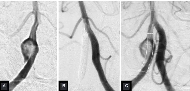
Fig. 1 Case 1
A: Initial diagnostic angiogram of the right vertebral artery (anteroposterior view) shows a dissecting aneurysm. B:Left vertebral angiogram during vessel reconstruction using a coronary stent.
C:Angiogram obtained immediately after stent deployment (dotted frame).
A B C D E
Fig. 2 Case 1
Serial vertebral angiography findings (A) 1 day, (B) 3 months, and (C) 6 months after reconstruction show progressive thrombosis; however, at 1 year (D) the aneurysmal sac is recanalized. Arrows indicate in-flow into the aneurysmal sac. E: Angiogram immediately after deployment of a stent in the originally placed stent. Straightening of the parent artery
with the stent facilitates flow stagnation in the aneurysm. The normalization of flow direction is shown by estimates of flow direction at each stage (indicated by arrows).
A B C
A B C D
Fig. 3 Case 1
Serial MIP (maximum intensity projection) images of 3D-CT (A) 1 day, (B) 3 years, (C) 7 years, and (D) 10 years after stent deployment in the originally placed stent demonstrate progressive thrombosis (arrows).
E: “Saw-like image”(arrows) on a right vertebral angiogram 7 years after reconstruction indicates slight protrusion of contrast medium through the stent struts.
F: Intravascular ultrasound shows neointima formation around the stent struts (arrows).
E F
Fig. 4 Case 2
A: Anteroposterior view of a right vertebral angiogram shows a dissecting aneurysm presenting as Wallenberg's syndrome.
B: Fluoroscopic image during reconstruction with a coronary stent (between the arrows) shows stasis of contrast medium within the aneurysm.
C: Angiogram a few minutes after stent deployment shows the straightened parent vessel (white arrow) and persistent stasis of the contrast medium at the venous phase.
entry は VA union より 10 mm 近位から約 10 mm にわ たって存在した.血管径は瘤近位部 4.2 mm × 4.3 mm, 遠位部 3.9 mm × 3.7 mm であった.解離部を十分カバ ー し な が ら BX velocity 18 mm 長 を nominal 圧 10 atm (拡張径 4.0 mm)よりも低圧の 6 atm(拡張径 3.7 mm) で拡張した.血管撮影で,依然として瘤の造影を認めた ため nominal 圧 10 atm で後拡張を行った.その結果, 瘤内に造影剤が停滞した(Fig. 4B, C).また,ステン トにより血管が直線化しているのが確認できた(Fig. 4C).治療に関連した合併症は認めなかった. 治療後の経過:抗血小板薬は 3ヵ月で中止した.6ヵ月 後の血管撮影では瘤はほとんど血栓化しており,一部 saw-like image を呈していた(Fig. 5A).治療 7 年目に 施行した血管撮影では造影剤の解離腔への leak(saw-like image)は減少しており(Fig. 5B),血管内エコーで同 部を確認することができた(Fig. 5B).10 年目の 3D-CT では動脈瘤は完全に血栓化しており,その一部は石灰化 していた(Fig. 5C).10 年間の経過観察中に新たな神経 学的異常を認めなかった.
考 察
脳動脈瘤に対するステント治療は 1990 年代初頭から 模索されてきた18).ステントには,1)コイル逸脱防止 効果,2)flow diverter 効果,3)瘤頚部の内皮化の足場 となる framework 効果,の 3 つの効果があると考えら れ18),これらの効果はさまざまな実験モデルによって 示されてきた2,11).1997 年,Higashida らは fusiform タ イプの脳底動脈瘤症例に初めてステントとコイルを用い て治療した5).以来,動脈瘤(特に巨大動脈瘤や解離性 動脈瘤など)に対する治療はステントを用いた血行再建 的血管内治療に関心が移り,ステントを用いたコイル塞 栓術が多数報告されるようになった13,20).その中に,コ イルを用いなくてもステント単独で動脈瘤が閉塞できる 症例が散見されるようになり,その頻度は VADA で高 かった13,16,21).Park らは 27 例(29 動脈瘤)の解離性椎骨 脳 底 動 脈 瘤(vertebrobasilar dissecting aneurysm; VBDA)に対してステント単独治療を行い,治療に関連 した合併症はなく,過去に報告された VBDA に対する 同治療の review においても合併症は後頭葉の梗塞のみ (4.7%)で,VBDA に対するステント単独治療は安全な 治療法である,と述べている16). 我々はステントの持つこのような特性と過去の知見に 注目し,nhVADA に対してステント単独治療を行った. nhVADA を選択した理由は,VADA のステント単独治 療がすでに有効性と安全性を両立しうる modality と考 えられていたこと,破裂症例では短時間に止血効果を得 る必要があり,ゆっくり瘤内血栓を誘発させることを目 的としているステント単独治療は不適切と判断したこ と,当時,当疾患の治療法として母血管閉塞が確立して おり,対側が正常である椎骨動脈病変ならステント留置 A B C enhanced plain Fig. 5 Case 2
A: Right vertebral angiogram 6 months after reconstruction demonstrates“saw-like image”(arrows).
B: Protrusion of contrast medium 7 years after the reconstruction is reduced compared with (A). (Inset) Intravascular ultrasound (IVUS) reveals protrusion of contrast medium through the stent struts (arrowhead).
C: MIP image of the 3D-CT 10 years after the stenting shows complete obliteration of the aneurysm. Part of the thrombosed aneurysmal sac is calcified (arrows).
によりトラブルが発生した場合でも救済措置として母血 管を閉塞させることが可能と考えたからである. ステントを留置すると動脈瘤への in-flow が変化する. その結果,瘤内の血流は stagnation し血栓化が誘発され る.症例 1 の場合,1 本のステントでは完全な血栓化を 誘発することができなかったが,“stent-in-stent”の形 で 2 本目のステントを留置することで血栓化が得られ た.これはステントストラットがより細かくなった (porosity が低下した)ことも血栓形成が促された理由 の一つであるが1,10),母血管の直線化により瘤への in flow が変化し,瘤内の stagnation が助長された結果,よ り血栓化が誘発されやすくなったものと推察される.実 際,症例 2 ではステントは 1 本しか挿入されていないが, 直後よりステントによる血管の直線化を認め,瘤内 stagnation が起こった.これはステントメッシュの細か さのみが血栓化を誘発する因子ではないことを示唆して いる.Meng らはステントが誘発する flow diverter 効果 は血管の彎曲度が高くなればなるほど減少すると報告し ており,ステントによる瘤の血栓化には母血管の直線化 が重要であることを指摘した15).Zenteno らも同様のこ とを述べている21). 母血管の直線化は瘤破裂や穿通枝損傷の危険性を孕ん でいるが,少なくとも動脈瘤に対してバルーン拡張型ス テント(バルーン拡張時に血管が一時的にも直線化した り,血管そのものが移動したりする)を用いた過去の報 告において,同合併症を指摘した報告はなく,自己拡張 型ステントであればさらに安全が期待できる.母血管の 直線化が得られず,依然として有効な flow stagnation を 認めない症例においては後述するようにステントをさら に追加するかコイル塞栓術を追加した方がよいと思われ る.なお,解離性動脈瘤の場合は intimal flap を realigning させることにより,瘤内血栓を促す可能性も指摘されて いる1)ので,本症例ではこの因子も関与しているかも しれない. ステントの持つこのような特性をさらに進化させたも のが Flow Diverter(FD)であり,最近, 種々の報告が なされている4,14).FD は冠動脈ステントや脳動脈瘤コ イ ル 塞 栓 術 支 援 の ス テ ン ト よ り porosity を 下 げ た
cylindrical mesh device で4,7),コイルによる瘤内塞栓で
は治療が困難な巨大動脈瘤や wide-neck な動脈瘤を,母 血管を温存しながら血行再建することができる. FD についてその有用性を述べている報告もあるが4,14), 合併症の高さからごく限られた症例に用いるべきである と主張している報告もある12).中でも FD 留置後に遅発 性に発生する動脈瘤破裂は当初予期しなかった合併症 で,その原因として Cebral らは FD 留置後に瘤内の圧 が上昇し,破裂が生じると考察しており3),Kulscar ら は瘤内血栓の自己融解が原因であろうと推察している9). このようにステント留置後に生じる血栓化の過程とその 終末像については不明な点が多く,同治療の経年経過報 告が待たれる.我々は症例 2 の 10 年目の評価において 血栓化した動脈瘤の一部が石灰化しているのを観察する ことができた.ステント留置後,血栓化に成功した動脈 瘤すべてに同様の所見が認められるかどうかは不明であ るが,動脈硬化性病変の終末像が石灰病変である19)こ とと類似しておりステント留置後に血栓化した動脈瘤の 治癒過程を推察する上で興味深い.今後,FD の導入に より冠動脈用のステントが動脈瘤の治療に使用されるこ とは少なくなるが,血管の直線化が瘤の血栓化を引き起 こす重要な因子のひとつであるなら冠動脈ステントの “硬い”という欠点は,長所となりうる.FD は柔軟で あるためそれ単独で in flow を変化させることが難しい 場合も想定され,先述した FD 留置後の破裂事例も勘案 すると,より早期に血栓化を促すべくコイルを留置した り,double stent によってメッシュをさらに細かくした りする等の工夫をした方がよい場合もあると考えられ る. また,FD を含むステント単独治療の問題点としてス テント内血栓がある.ステント内血栓はステント留置に 伴う重大な合併症の一つであるため,術前から抗血小板 薬の 2 剤投与が推奨される21).先の Park らの review によれば過去に 3 例のステント内血栓症例があり,3 例 とも抗血小板薬が投与されていないか,もしくは服薬で きていなかった症例であった16).本症例では術前から Aspirin 81 mg と Ticlopidine 200 mg の 2 剤が投与され ており虚血性合併症はなかった.一方,抗血小板薬の効 果により瘤内の血栓化が遅れるとも言われている21).症 例 1 ではステント留置後,抗血小板薬を 1 年間服用させ ていた.最初の半年は順調に血栓化が進んだが最終的に は再開通してしまった.この事実はステントメッシュに よる flow stagnation 効果のみで瘤の血栓化を安定させる ことが難しいことを示唆している.ステント内血栓を回 避しつつ瘤の血栓化を促進するという正反対の現象を同 時に可能にすることは極めて難しく , 抗血小板薬の 2 剤
投与に関しては異論はないまでも,同薬を中止するもし くは減量する時期に関しては症例毎にきめ細かい調整を 行わざるを得ない. ステント内血栓を防止するために抗血小板薬が必須で ある以上,破裂 VBDA 症例に対してステント単独治療 を応用することは慎重であるべきであるとの意見がある が1),過去の報告を見ると渉猟し得た範囲ではステント 単独治療後に再破裂を起こした例は Kaku らの報告以外 なく6),実際には破裂 VADA に対するステント単独治 療の成績は良好であるため16,21),治療後の再破裂を懸念 して抗血小板薬を早く中止する必要性はないと考えられ る.ただし再出血した場合は止血困難な状態が容易に予 想されるので,瘤内にコイルを入れることにより早期血 栓化を促すのも一つの方法である17).コイルの追加は 先述した FD 治療後の予期せぬ瘤破裂を回避する方法と しても提案されている9). ステント単独治療の成績については,治療後 2 年間経 過を追った報告はあるが21),10 年にわたる長期経過を 追った報告はない.症例数は 2 例と少ないが,本結果は 長期予後の観点からも nhVADA に対するステント単独 治療の有用性を示したものであり,同様の治療コンセプ トである FD も同等以上の効果が期待される.
結 語
nhVADA に対するステント単独治療は適応を十分吟 味すれば有効な治療法であり,その効果は長期的に見て も十分安定していると思われた. 本論文に関して,開示すべき利益相反状態は存在しない. 文 献1) Ansari SA, Thompson BG, Gemmete JJ, et al: Endovascular treatment of distal cervical and intracranial dissections with the neuroform stent. 62:636-646, 2008. 2) Bai H, Masuda J, Sawa Y, et al: Neointima formation after
vascular stent implantation.Spatial and chronological distribution of smooth muscle cell proliferation and phenotypic modulation. 14:1846-1853, 1994.
3) Cebral JR, Mut F, Raschi M, et al: Aneurysm rupture following treatment with flow-diverting stents: computational hemodynamincs analysis of treatment. 32:27-33, 2011.
4) Fiorella D, Kelly ME, Albuquerque FC, et al: Curative reconstruction of a giant midbasilar trunk aneurysm with the pipeline embolization device. 64:212-217,
2009.
5) Higashida RT, Smith W, Gress D, et al: Intravascular stent and endovascular coil placement for a ruptured fusiform aneurysm of the basilar artery. Case report and review of the literature. 87:944-949, 2007.
6) Kaku Y, Yoshimura S, Yamakawa H, et al: Failure of stent-assisted endovascular treatment for ruptured dissecting aneurysms of the basilar artery. 45:22-26, 2003.
7) Kallmes DF, Ding YH, Dai D, et al: A new endoluminal, flow-disrupting device for treatment of saccular aneurysms.
38:2346-2352, 2007.
8) 加藤祥一,米田浩,石原秀行,他:非出血発症の解離性動脈 瘤における臨床症状,画像の長期観察による検討.脳卒中 の外科 33:193-199, 2005.
9) Kulcsar Z, Houdart E, Bonafe A, et al: Intra-aneurysmal thrombosis as a possible cause of delayed aneurysm rupture after flow-diversion treatment. 32:20-25, 2011.
10) Lieber BB, Stancampiano AP, Wakhloo AK: Alteration of hemodynamics in aneurysm models by stenting: influence of stent porosity. 25:460-469, 1997. 11) Lieber BB, Gounis MJ: The physics of endoluminal stenting
in the treatment of cerebrovascular aneurysms. 24:33-42, 2002.
12) Lubicz B, Collignon L, Raphaeli G, et al: Flow-diverter stent for the endovascular treatment of intracranial aneurysms: a prospective study in 29 patients with 34 aneurysms.
41:2247-2253, 2010.
13) Lylyk P, Cohen JE, Ceratto R, et al: Endovascular reconstruction of intracranial arteries by stent placement and combined techniques. 97:1306-1313, 2002. 14) Lylyk P, Miranda C, Ceratto R, et al: Curative endovascular
reconstruction of cerebral aneurysms with the pipeline embolization device: the Buenos Aires experience.
64:632-643, 2009.
15) Meng H, Wang Z, Kim M, et al: Saccular aneurysms on straight and curved vessels are subject to different hemodynamics: implications of intravascular stenting.
27:1861-1865, 2006.
16)Park SI, Kim BM, Kim DI, et al: Clinical and angiographic follow-up of stent-only therapy for acute intracranial vertebrobasilar dissecting aneurysms. 30:1351-1356, 2009.
17) Sadato A, Maeda S, Hayakawa M, et al: Endovascular treatment of vertebral artery dissection using stents and coils: its pitfall and technical considerations.
53:243-249, 2010.
18) Szikora I, Nelson PK, Berentei Z, et al: The potential of flow modification in the treatment of intracranial aneurysms.
14:77-80, 2008.
19) Virmani R, Kolodgie FD, Burke AP, et al: Lessons from sudden coronary death: a comprehensive morphological classification scheme for atherosclerotic lesions.
20:1262-1275, 2000.
20) 山崎信吾,橋本邦雄,重田恵吾,他:ステントで治療した椎 骨動脈と脳底動脈の解離性動脈瘤.脳外誌 11:484-491, 2002. 21) Zenteno MA, Santos-Franco JA, Freitas-Modenesi JM, et
of aneurysms in the posterior circulation in a prospective
JNET 7:111-118, 2013 要 旨
【目的】非出血性椎骨動脈解離性動脈瘤(non-hemorrhagic vertebral artery dissecting aneurysm;nhVADA) に対してステント単独治療を行い,10 年間経過を追跡しえた 2 例を経験したので報告する.【症例】2001 年から 2002年の 2 年間に経験した nhVADA 5 例のうち,ステント単独治療を行うことに同意を得た 2 例に対しステン トを 1 本ないし 2 本挿入し,瘤の血栓化状態と内膜形成をそれぞれ 3 次元 CT angiography(3D-CTA),脳血管 撮影および血管内エコーを用いて観察した.【結果】観察期間中,塞栓性,出血性合併症を認めなかった.治療 10 年目の評価において,動脈瘤はほとんどもしくは完全に血栓化しており,その一部は石灰化していた.【考察】瘤 の血栓化はステント留置により母血管が直線化した結果,in-flow が変化し,造影剤の停滞が得られた場合に起こ ると考えられた.【結論】nhVADA に対するステント単独治療の自験例は長期成績も良好であり,有用な方法に なり得ると思われた.