Endocrine Journal Advance Publication
GLUCOSE acts as a primary stimulator of insulin secretion in pancreatic β-cells [1]. Glucose has been thought to exert its action solely through its metabo- lism [2, 3]. Thus, glucose enters the cells via glucose transporters and is then metabolized through the gly- colytic pathway and in mitochondria. The resultant increase in the ATP/ADP ratio leads to closure of the ATP-sensitive potassium (KATP) channel and thereby depolarizes the plasma membrane [4, 5]. This would activate the voltage-dependent Ca2+ channel and pro- mote entry of Ca2+ into β-cells. Exocytosis of insu- lin granules is then triggered by the elevation of sub- plasma membrane Ca2+ concentration. Additionally, exocytosis of insulin granules is amplified by another mechanism, which is not dependent on the KATP chan- nel [3, 6].
Sucralose, an activator of the glucose-sensing receptor, increases ATP by calcium-dependent and -independent mechanisms
Longfei Li1), Yoshiaki Ohtsu2), Yuko Nakagawa1), Katsuyoshi Masuda3) and Itaru Kojima1)
1) Institute for Molecular and Cellular Regulation, Gunma University, Maebashi 371-8512, Japan
2) Department of Pediatrics, Graduate School of Medicine, Gunma University, Maebashi 371-8512, Japan
3) Suntory Global Innovation Center, Kyoto 619-0284, Japan
Abstract. Sucralose is an artificial sweetener and activates the glucose-sensing receptor expressed in pancreatic β-cells.
Although sucralose does not enter β-cells nor acts as a substrate for glucokinase, it induces a marked elevation of intracellular ATP ([ATP]c). The present study was conducted to identify the signaling pathway responsible for the elevation of [ATP]c induced by sucralose. Previous studies have shown that sucralose elevates cyclic AMP (cAMP), activates phospholipase C (PLC) and stimulates Ca2+ entry by a Na+-dependent mechanism in MIN6 cells. The addition of forskolin induced a marked elevation of cAMP, whereas it did not affect [ATP]c. Carbachol, an activator of PLC, did not increase [ATP]c. In addition, activation of protein kinase C by dioctanoylglycerol did not affect [ATP]c. In contrast, nifedipine, an inhibitor of the voltage-dependent Ca2+ channel, significantly reduced [ATP]c response to sucralose. Removal of extracellular Na+ nearly completely blocked sucralose-induced elevation of [ATP]c. Stimulation of Na+ entry by adding a Na+ ionophore monensin elevated [ATP]c. The monensin-induced elevation of [ATP]c was only partially inhibited by nifedipine and loading of BAPTA, both of which completely abolished elevation of [Ca2+]c. These results suggest that Na+ entry is critical for the sucralose-induced elevation of [ATP]c. Both calcium-dependent and -independent mechanisms are involved in the action of sucralose.
Key words: Sweet taste receptor, Glucose metabolism, β-cell, Calcium, ATP
We have recently postulated the involvement of another signaling pathway activated by glucose.
Thus, glucose acts as a ligand and binds to and acti- vates the cell-surface glucose-sensing receptor [7, 8].
This receptor is comprised of the T1R3 subunit of the sweet taste receptor [9] expressed in taste cells of the tongue. An intriguing feature of the glucose-sens- ing receptor is that activation of the receptor facili- tates glucose metabolism in β-cells and increases ATP [10]. In fact, when an artificial sweetener sucralose is administered to β-cells to activate the glucose-sensing receptor, intracellular ATP concentration ([ATP]c) is markedly elevated [10]. Sucralose is an analogue of sucrose and does not enter β-cells, nor it is metabolized through the glycolytic pathway [11]. It is reasonable to expect that sucralose does not affect [ATP]c in β-cells.
Nevertheless, when changes in [ATP]c were monitored in MIN6 cells, sucralose induced a marked elevation of [ATP]c and is, in fact, much more potent in increas- ing [ATP]c compared to glucose. Furthermore, sucra-
Submitted Apr. 26, 2016; Accepted May 11, 2016 as EJ16-0217 Released online in J-STAGE as advance publication May 31, 2016 Correspondence to: Itaru Kojima, M.D., IMCR, Gunma University, Maebashi 371-8512, Japan. E-mail: [email protected]
©The Japan Endocrine Society
Original
Endocrine Journal Advance Publication
NaOH (pH 7.4). To remove extracellular sodium, sodium was replaced with NMDG.
Measurement of cytoplasmic free calcium and cyclic AMP concentrations
Cytoplasmic Ca2+ concentration ([Ca2+]c) was mea- sured in a single MIN6 cell using a Ca2+ indicator Fura-2/AM. Cells were loaded with Fura-2 by incubat- ing in HBSS containing 2 µM Fura-2/AM for 20 min at room temperature. For measurement of cytoplasmic cyclic AMP concentration ([cAMP]c), MIN6 cells were transfected with plasmid encoding Epac1-camps [15], kindly provided by Dr. Martin Lohse of Würzburg University (Würzburg, Germany). Cells were placed on a 35 mm glass bottom culture dish and the medium was changed to HBSS. Cells were visualized with a 40 Uapo/340 objective lens (Olympus, Tokyo, Japan). To detect the fluorescence image, the AQUACOSMOS/
ASHURA, 3CCD based fluorescence energy transfer imaging system (Hamamatsu Photonics, Hamamatsu, Japan) was used. Images (40 ms exposure) were captured with a C7780-22 ORCA3CCD camera (Hamamatsu photonics) at 10 sec intervals [16]. These values (F) were normalized to each initial value (F0), and the relative fluorescence change was referred to as F/F0. In each experiment, recordings were obtained in more than ten cells.
Measurement of cytoplasmic ATP
MIN6 cells expressing luciferase [10] were seeded into a culture plate-96 and incubated for 2 to 3 days before the measurement of [ATP]c. Cell were then incubated for 20 min in HBSS containing 50 µM D-luciferin potassium salt in a 37°C incubator. To measure basal [ATP]c (L0), we counted the emitted photons with EnSpire (Perkin Elmer, Waltham, MA, U.S.A) for 20 min. The cells were then incubated in the presence and absence of stimulatory agents. We counted the emitted photon for 1 h. These values (L) were normalized to each value (L0) so that the rela- tive luminescent changes were referred to as L/L0 [10].
Measurement was done in quadruplicate and means of the four determinations are presented. Statistical sig- nificance was assessed by using the Student t-test.
Results
When MIN6 cells were stimulated by 10 mM sucra- lose in the presence of 5.5 mM glucose, a rapid eleva- lose increases [ATP]c even in the absence of ambient
glucose [10]. This implies that activation of the glu- cose-sensing receptor by sucralose greatly facilitates glucose metabolism in β-cells and increases ATP pro- duction. Collectively, sucralose provides a good tool to assess the mechanism by which the glucose-sensing receptor modulates glucose metabolism.
The present study was conducted to determine the mechanism of action of sucralose on [ATP]c in β-cells.
Our previous study showed that sucralose activates phospholipase C (PLC), and thereby induces Ca2+
release from an intracellular pool and activates protein kinase C [12]. Sucralose also stimulates Ca2+ influx by a mechanism totally dependent on extracellular Na+ [12]. Presumably, sucralose stimulates Na+ entry, causes depolarization of the plasma membrane and augments Ca2+ entry. In addition, sucralose increases cyclic AMP (cAMP) by activating Gs [12, 13]. Using glucose-responsive MIN6 cells [14], we tried to iden- tify the signaling pathway(s) responsible for the eleva- tion of [ATP]c induced by sucralose.
Materials and Methods
Materials
Lactisole, monensin, 1,2-Bis(2-aminophenoxy) ethane-N, N, N’, N 7-tetraacetic acid tetrakis-ace- toxymethylester (BAPTA/AM), 1,2-dioctanoyl-sn- glycerol (diC8) and sucralose were obtained from Sigma-Aldrich (St. Louise, MO, U.S.A). Nifedipine, D-luciferin potassium salt, and N-methyl-D-glucamine (NMDG) were from Wako Pure Chemicals (Osaka, Japan). Forskolin was obtained from EMD Millipore (Billerica, MA, U.S.A). Fura-2/AM was obtained from Dojindo (Kumamoto, Japan).
Cell culture
MIN6 cells [14] were grown in Dulbecco’s modifies Eagle’s medium (DMEM) containing a high concentra- tion of glucose (Invitrogen, Waltham, MA, U.S.A) and 10% fetal bovine serum (Sigma-Aldrich): cells were cultured in a humidified incubator with 95% air and 5% CO2 at 37°C.
Solution
Hanks’ balanced salt solution (HBSS) contained 138 mM NaCl, 5.4 mM KCl, 1.3 mM CaCl2, 0.44 mM KH2PO4, 0.5 mM MgCl2, 0.38 mM MgSO4, 0.34 mM Na2HPO4, 5.5 mM D-glucose and 10 mM HEPES/
Endocrine Journal Advance Publication Endocrine Journal Advance Publication
tion of [ATP]c was observed (Fig. 1A). Elevation of [ATP]c was detected at 20 sec and peaked at around 2 min. [ATP]c levels remained elevated for as long as 30 min. The effect of sucralose was completely blocked by lactisole, an inhibitor of T1R3 [17, 18] (Fig. 1B and 1C), indicating that the effect of sucralose was medi- ated by the glucose-sensing receptor [18].
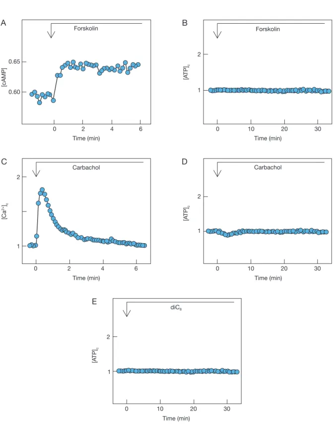
Previous studies have shown that sucralose increases [cAMP]c and activates PLC, which causes Ca2+ release from an intracellular trigger pool and activation of protein kinase C [12, 13]. Sucralose also stimulates Ca2+ entry by a mechanism dependent on Na+ entry [12, 13]. We first assessed the role of cAMP in the action of sucralose. To this end, we added for- skolin, an activator of adenylyl cyclase, and increased [cAMP]c in MIN6 cells. As shown in Fig. 2A, 10 µM forskolin rapidly increased [cAMP]c in MIN6 cells.
However, the addition of forskolin did not affect [ATP]c levels (Fig. 2B). We then assessed the role of PLC activation in sucralose-induced elevation of [ATP]c. We activated PLC by adding carbachol, an agonist of the muscarinic receptor. As shown in Fig. 2C, addition of 10 µM carbachol evoked a rapid transient elevation of [Ca2+]c, which may be induced by carbachol-mediated activation of PLC. When changes in [ATP]c were measured, there was a small decrease in [ATP]c (Fig. 2D). The decrease in [ATP]c was only transient and elevation of [ATP]c was not observed subsequently. We then activated protein kinase C by adding a synthetic diacylglycerol diC8. As shown in Fig. 2E, application of diC8, which activates protein kinase C in β-cells [19], did not affect [ATP]c.
We then assessed the role of Ca2+ entry in the action of sucralose. To this end, we measured the effect of nifedipine, an inhibitor of the L-type voltage-depen- dent calcium channel, in sucralose-induced elevation of [ATP]c. In the presence of nifedipine, sucralose induced a small and transient elevation of [Ca2+]c (Fig.
3A). [Ca2+]c levels returned to the basal level within 1 min. In this condition, sucralose induced a small and sustained elevation of [ATP]c (Fig. 3B). Nifedipine markedly inhibited [ATP]c response (Fig. 3C). When BAPTA was loaded to clamp the [Ca2+]c levels, sucra- lose was not able to induce any changes in [Ca2+]c (Fig.
4A). In this condition, sucralose induced a small but sustained elevation of [ATP]c (Fig. 4B). BAPTA sig- nificantly inhibited [ATP]c response induced by sucra- lose (Fig. 4C). When extracellular Na+ was removed, sucralose induced a small and transient elevation of
B A
C
Time (min) [ATP]c
0 10 20 30
1
Sucralose
2
Time (min) [ATP]c
0 10 20 30
1
Sucralose 2
Lactisole
* Sucralose (mM) 10 10
AUC
10 20 30
Lactisole (mM) 0 5
Fig. 1 Effect of sucralose on [ATP]c
A. Luciferase-expressing cells were stimulated by 10 mM sucralose and changes in [ATP]c were monitored. Results are the mean of four determinations and representative of four experiments. B. Cells were incubated for 10 min with 5 mM lactisole and were then stimulated by 10 mM sucralose in the presence of lactisole. Results are the representative of four experiments. C. Experiments are carried out as shown in A and B and area under the curve (AUC) from 0 to 20 min was calculated. Values are the mean ± S.E. for four experiments. * P < 0.01 vs control.
Endocrine Journal Advance Publication
B A
C
2 4 6
0 0.60
Forskolin
0.65
Time (min) 10
0 20 30
1
Forskolin
2
Time (min)
2 4
0 6
1
Carbachol 2
Time (min) [ATP]c
20 30
10 0
1
Carbachol
2
Time (min)
20 30
10 0
1
diC8
2
D
E
Time (min)
[cAMP] [ATP]c
[Ca2+]c [ATP]c
Fig. 2 Effect of forskolin, carbachol and diC8
A. Cells expressing Epac1-camps were stimulated by 10 µM forskolin. The results are representative of three experiments.
B. Luciferase-expressing cells were stimulated by 10 µM forskolin. The results are representative of three experiments.
C. Fura-2-loaded cells were stimulated by 10 µM carbachol and changes in [Ca2+]c were monitored. The results are the representative of three experiments. D. Luciferase-expressing cells were stimulated by 10 µM carbachol and changes in [ATP]c were monitored. The results are the representative of four experiments. E. Luciferase-expressing cells were stimulated by 10 µM diC8 and changes in [ATP]c were monitored. The results are the representative of three experiments.
Endocrine Journal Advance Publication Endocrine Journal Advance Publication
B A
C
Time (min)
4 6
2 0
1
Sucralose 2
Nifedipine
Time (min)
20 30
10 0
1
Sucralose
2
Nifedipine
*
10 0
AUC
20 40 [Ca2+]c[ATP]c
Nifedipine (µM)
Fig. 3 Effect of nidedipine on sucralose-induced chnages in [Ca2+]c and [ATP]c
A. Fura-2-loaded cells were stimulated by 10 mM sucralose in the presence ( ) and absence ( ) of 10 µM nifedipine. Nifedipine was added 10 min prior to the stimulation by sucralose. B. Luciferase-expressing cells were stimulated by 10 mM sucralose in the presence ( ) and absence ( ) of 10 µM nifedipine, which was added 10 min prior to the stimulation. C. Changes in [ATP]c were measured as shown in B and AUC from 0 to 20 min was calculated. Values are the mean ± S.E. for four experiments. * P < 0.01 vs without nifedipine.
B A
C
Time (min)
2 4 6
0 1
Sucralose 2
BAPTA
Time (min) [ATP]c
10 20 30
0 1
Sucralose
2
BAPTA
*
BAPTA (–) (+)
AUC
20 40 [Ca2+]c
Fig. 4 Effect of BAPTA-loading on sucralose-induced changes in [Ca2+]c and [ATP]c
A. Fura-2-loaded cells were stimulated 10 mM sucralose in the presence ( ) and absence ( ) of BAPTA and changes in [Ca2+]c were monitored. B. Luciferase- expressing cells were stimulated by 10 mM sucralose in the presence ( ) and absence ( ) of BAPTA and changes in [ATP]c were monitored. Results are the representative of three experiments. C. Changes in [ATP]c were measured as shown in B and AUC from 0 to 20 min was calculated. Values are the mean ± S.E. for three experiments. * P < 0.05 vs without BAPTA.
Endocrine Journal Advance Publication
[Ca2+]c (Fig. 5A). Indeed, sucralose-mediated eleva- tion of [ATP]c was nearly completely blocked by the removal of extracellular Na+ (Fig. 5B and 5C).
We then assessed the role of Ca2+ entry in elevation of [ATP]c in MIN6 cells. We added 40 mM potassium chloride, which depolarizes the plasma membrane and augments Ca2+ entry via the voltage-dependent calcium channel. As shown in Fig. 6A, 40 mM potassium chlo- ride induced biphasic changes in [ATP]c. Immediately after the addition of potassium chloride, a rapid eleva- tion of [ATP]c was observed. After the peak of [ATP]c, [ATP]c decreased rapidly to a level far below the basal level. [ATP]c remained decreased for 30 min. A simi- lar pattern of changes in [ATP]c was observed when 20 mM potassium chloride was administered, although the magnitude of the responses was smaller (Fig. 6B). We then blocked Ca2+ entry induced by potassium chloride by adding a calcium channel blocker nifedipine. As shown in Fig. 6C, nifedipine completely blocked ele- vation of [Ca2+]c induced by 40 mM potassium chlo- ride. When 40 mM potassium chloride was adminis- tered in the presence of nifedipine, [ATP]c increased rapidly and after the peak of [ATP]c, [ATP]c levels then decreased gradually. The decrease in [ATP]c was blunted and was never below the basal level. Thus, a large decrease in [ATP]c was considerably inhibited by nifedipine (Fig. 6D). We then blocked the eleva- tion of [Ca2+]c by applying a Ca2+ chelator BAPTA.
As shown in Fig. 6E, 40 mM potassium chloride did not affect [Ca2+]c in cells loaded with BAPTA. In this condition, 40 mM potassium chloride induced a mono- phasic elevation of [ATP]c (Fig. 6F). Thus, potassium chloride increased [ATP]c, which was followed by a gradual decrease to the basal level. The peak value was lower than that in the absence of BAPTA.
Finally, we studied the role of Na+ entry in the ele- vation of [ATP]c. To this end, we administered a Na+ ionophore monensin [20]. Monensin induces Na+ entry and thereby depolarizes the plasma membrane. Then the voltage-dependent calcium channel is activated and Ca2+ entry is augmented. As shown in Fig. 7A, monen- sin induced a sustained elevation of [ATP]c in MIN6 cells. To determine the role of Ca2+ entry in monensin- evoked elevation of [ATP]c, we administered nifedipine.
As shown in Fig. 7B, nifedipine completely blocked the elevation of [Ca2+]c induced by monensin. Nifedipine, however, did not completely abolish elevation of [ATP]c evoked by monensin. As shown in Fig. 7C, monensin induced a gradual but sustained elevation of [ATP]c in
B A
C
Time (min)
2 4 6
0 1
Sucralose 2
Time (min) [ATP]c
10 20 30
0 1
Sucralose
2
138 (–)
AUC
20 40
Na+ free [Ca2+]c
Na+ free
Na+ (mM)
Fig. 5 Effect of removal of extracellular Na+ on sucralose- induced changes in [Ca2+]c and [ATP]c
A. Fura-2-loaded cells were incubated in HBSS ( ) or Na+-free HBSS ( ) for 30 min and then stimulated by 10 mM sucralose. Changes in [Ca2+]c were monitored.
B. Luciferase-expressing cells were incubated in HBSS ( ) or Na+-free HBSS ( ) for 30 min and then stimulated by 10 mM sucralose. C. Changes in [ATP]c were measured as shown in B and AUC from 0 to 20 min was calculated. Values are the mean ± S.E. for four experiments. * P < 0.01 vs with Na+.
Endocrine Journal Advance Publication Endocrine Journal Advance Publication
B A
C
Time (min) [ATP]c
10 20 30
0 1 2
Time (min) [ATP]c
10 20 30
0 1 2
Time (min)
2 4
0 6
1 2
Nifedipine
Time (min) [ATP]c
10 20 30
0 1 2
Nifedipine
Time (min) 2
0 4 6
1 2
BAPTA
Time (min) [ATP]c
10 20 30
0 1 2
BAPTA
D
E F
40 mM KCl 20 mM KCl
40 mM KCl
[Ca2+]c
40 mM KCl
[Ca2+]c
40 mM KCl 40 mM KCl
Fig. 6 Effect of potassium chloride on [Ca2+]c and [ATP]c
A, B. Luciferase-expressing cells were stimulated by 40 mM (A) or 20 mM (B) potassium chloride and changes in [ATP]c were monitored. C. Fura-2-loaded cells were stimulated by 40 mM potassium chloride in the presence ( ) and absence ( ) of 10 µM nifedipine added 10 min prior to the stimulation. Results are representative of three experiments. D. Luciferase- expressing cell were stimulated by 40 mM potassium chloride in the presence ( ) and absence ( ) of 10 µM nifedipine added 10 min prior to the stimulation. Results are the representative of three experiments. E. Fura-2-loaded cells were stimulated by 40 mM potassium chloride in the presence ( ) and absence ( ) of BAPTA. Results are the representative of four experiments.
F. Luciferase-expressing cells were stimulated by 40 mM potassium chloride in the presence ( ) and absence ( ) of BAPTA.
Results are the representative of four experiments.
Endocrine Journal Advance Publication
B A
C D
E F
Time (min) [ATP]c
10 20 30
0 1
Monensin
2
Time (min)
2 4 6
0 1
Monensin 2
Nifedipine
Time (min) [ATP]c
10 20 30
0 1
Monensin
2
Nifedipine
Time (min)
2 4 6
0 1
Monensin 2
BAPTA
AUC
20 40
(–) (–)
(–) (–) (–) (–)
* *
10 10 10 10
Nifedipine BAPTA
(+)
(+) Time (min)
[ATP]c
10 20 30
0 1
Monensin
2
BAPTA
[Ca2+]c[Ca2+]c
Monensin (µM)
Fig. 7 Effect of monensin on [Ca2+]c and [ATP]c
A. Luciferase-expressing cells were stimulated by 10 µM monensin and changes in [ATP]c were monitored. Results are the representative of three experiments. B. Fura-2-loaded cells were stimulated by 10 µM monensin in the presence ( ) and absence ( ) of 10 µM nifedipine added 10 min prior to the stimulation. Results are the representative of four experiments.
C. Luciferase-expressing cells were stimulated by 10 µM monensin in the presence ( ) and absence ( ) of 10 µM nifedipine added 10 min prior to the stimulation. Results are the representative of four experiments. D. Fura-2-loaded cells were stimulated by 10 µM monensin in the presence ( ) and absence ( ) of BAPTA. Results are the mean ± S.E. for three experiments. E. Luciferase-expressing cells were stimulated by 10 µM monensin in the presence ( ) and absence ( ) of BAPTA. Results are the representative of four experiments. F. Quantitative analysis of the effects of nefedipine and BAPTA on [ATP]c. Experiments were carried out as in C and E. AUC was calculated. * P < 0.05 vs monensin alone.
Endocrine Journal Advance Publication Endocrine Journal Advance Publication
the presence of nifedipine. To confirm that monensin was able to increase [ATP]c in the absence of elevation of [Ca2+]c, we chelated [Ca2+]c by adding BAPTA. As shown in Fig. 7D, BAPTA-loading completely abol- ished elevation of [Ca2+]c induced by monensin. In this condition, monensin was still able to induce sustained elevation of [ATP]c (Fig. 7E). Quantitative analyses of the results are shown in Fig. 7F.
Discussion
The present study was conducted to identify the sig- naling pathway responsible for the elevation of [ATP]c induced by sucralose. Sucralose is a potent agonist for the glucose-sensing receptor and induces elevation of [ATP]c in the presence and even absence of glucose [10]. With regard to the signaling pathways, sucralose increases [cAMP]c presumably by activating Gs [12, 13]. Interestingly, sucralose also activates PLC through activation of Gq leading to mobilization of Ca2+ from endoplasmic reticulum and activation of protein kinase C [12, 13]. Sucralose also stimulates Ca2+ entry, which is dependent on extracellular Na+ [13]. We assessed the role of these signaling pathways using pharmaco- logical agents modifying these pathways. First, results obtained by using forskolin negate the involvement of cAMP in sucralose action on [ATP]c (Fig. 2B). To assess the role of PLC activation, we applied a musca- rinic agonist carbochol. Although carbachol increased [Ca2+]c to a level comparable to that induced by sucra- lose, carbachol failed to increase [ATP]c. Additionally, activation of protein kinase C by diC8 failed to increase [ATP]c. Collectively, it is unlikely that sucralose ele- vates [ATP]c by activating PLC.
As shown in Fig. 3B, inhibition of Ca2+ entry through the voltage-dependent calcium channel by nifedipine significantly reduced the [ATP]c response to sucralose. The result suggests that stimulation of Ca2+
entry is involved in the sucralose action on [ATP]c. Ca2+ entry rather than Ca2+ release from endoplas- mic reticulum is important for facilitation of glucose metabolism by sucralose. Importance of Ca2+ entry was supported by the fact that removal of extracellular sodium, which abolishes Ca2+ entry by sucralose [12, 13], nearly completely abolished elevation of [ATP]c by sucralose. We have to point out that stimulation of Ca2+ entry is not the sole mechanism by which sucra- lose increases [ATP]c. As shown in Fig. 4B and 4C, loading of BAPTA, which completely abolishes ele-
vation of [Ca2+] induced by sucralose, did not com- pletely inhibit elevation of [ATP]c. There must be a Ca2+-independent mechanism to elevate [ATP]c. This mechanism is dependent on Na+ entry since removal of extracellular Na+ completely blocked sucralose action on [ATP]c (Fig. 5C).
When depolarizing concentration of potassium was applied, there was a peak of [ATP]c which was fol- lowed by a profound decrease in [ATP]c (Fig. 6A).
This decrease in [ATP]c may be due to consumption of ATP by the plasma membrane Ca2+ pump [21]. In accordance with this notion, the profound decrease in [ATP]c was blocked by either nifedipine or loading of BAPTA (Fig. 6D and 6F). Collectively, stimulation of Ca2+ influx causes facilitation of glucose metabolism and increases [ATP]c. This is probably because Ca2+
entry leads to elevation of Ca2+ concentration in mito- chondria, which activates Ca2+-dependent dehydro- genases in mitochondria [22, 23]. It should be men- tioned that high concentration of potassium increased [ATP]c even in the presence of nifedipine or BAPTA.
As shown in Fig. 6C and 6E, elevation of [Ca2+]c was completely blocked by either nifedipine or BAPTA- loading. These results indicate that there may be a calcium-independent action of high concentration of potassium. A possible candidate signal is depolariza- tion of the plasma membrane induced by a high con- centration of potassium chloride.
To further assess the role of depolarization and Ca2+
entry, we administered a sodium ionophore monensin.
As shown in Fig. 7A, monensin induced a sustained increase in [ATP]c. The monensin-induced elevation of [ATP]c was only partially inhibited by nifedipine and loading of BAPTA, both of which blocked eleva- tion of [Ca2+]c induced by monensin. Hence, stimula- tion of Na+ increased [ATP]c by both Ca2+-dependent and -independent mechanisms. It seems likely that depolarization of the plasma membrane by monen- sin may cause elevation of [ATP]c to some extent. At present, the mechanism by which depolarization of the plasma membrane increases [ATP]c is not certain.
In this regard, it is known that depolarization itself affects cellular functions by a mechanism independent of the changes in Ca2+ fluxes [24-26]. Furthermore, enzymes regulated by membrane potential are also described [26]. Accordingly, it is not impossible that depolarization of the plasma membrane controls glu- cose metabolism. Further studies are clearly needed to identify the mechanism by which depolarization
Endocrine Journal Advance Publication
affects glucose metabolism.
We have reported that activators of the glucose- sensing receptor induce different patterns of changes in intracellular signals depending upon the types of agonists [13]. Accordingly, signals evoked by sucra- lose is slightly different from those evoked by glucose [13, 27]. In this regard, the present results obtained by sucralose cannot be directly applied to the action of glucose. However, some of the present results are quite important for consideration of the action of glucose. For example, it is well known that glucose induces oscillatory changes in the membrane potential [4]. As shown in the present study, depolarization of the plasma membrane facilitates glucose metabolism by both calcium-dependent and -independent mecha- nisms. Therefore, oscillatory changes in the membrane
potential may be quite important for promotion of the production of ATP in β-cells. In any case, the mecha- nism of action of glucose in pancreatic β-cells should be revised considerably.
Disclosure
Note of the authors have any potential conflicts of interest associated with this research.
Acknowledgements
The authors are grateful to Mayumi Odagiri for her secretarial assistance during preparation of the manu- script.
References
1. Rasmussen H, Zawalich KC, Ganesan S, Calle R, Zawalich WS (1990) Physiology and pathophysiology of insulin secretion. Diabetes Care 13: 655-666.
2. Matschinsky FM (1990) Glucokinase as glucose sen- sor and metabolic signal generator in pancreatic β-cells and hepatocytes. Diabetes 39: 647-652.
3. Henquin JC (2009) Regulation of insulin secretion:
a matter of phase control and amplitude modulation.
Diabetologia 52: 739-751.
4. Dean PM, Matthews EK (1968) Electrical activity in pancreatic islet cells. Nature 219: 389-390.
5. Cook DL, Hales CN (1984) Intracellular ATP directly blocks K+-channels in pancreatic B-cells. Nature 311:
271-273.
6. Komatsu M, Takei M, Ishii H, Sato Y (2013) Glucose- stimulated insulin secretion: A newer perspective. J Diabetes Investig 4: 511-516.
7. Kojima I, Nakagawa Y, Ohtsu Y, Hamano K, Medina J, et al. (2015) Return of the glucoreceptor: Glucose acti- vates the glucose-sensing receptor T1R3 and facilitates metabolism in pancreatic β-cells. J Diabetes Investig 6:
256-263.
8. Kojima I, Nakagawa Y, Hamano K, Medina J, Li L, et al. (2015) Glucose-sensing receptor T1R3: A new signaling receptor activated by glucose in pancreatic β-cells. Biol Pharm Bull 38: 674-679.
9. Nelson G, Hoon MA, Chandrasekar J, Zhang Y, Ryba NJP, et al. (2001) Mammalian sweet taste receptors.
Cell 106: 381-390.
10. Nakagawa Y, Ohtsu Y, Nagasawa M, Shibata H, Kojima I (2014) Glucose promotes its own metabolism by acting on the cell-surface glucose-sensing receptor.
Endocr J 61: 119-131.
11. Knight I (1994) The development and applications of sucralose, a new high-intensity sweetener. Can J Physiol Pharmacol 72: 435-439.
12. Nakagawa Y, Nagasawa M, Yamada S, Hara A, Mogami H, et al. (2009) Sweet taste receptor expressed in pan- creatic β-cells activates the calcium and cyclic AMP signaling system and stimulates insulin secretion. PLoS One 4: e5106.
13. Nakagawa Y, Nagasawa M, Mogami H, Lohse MJ, Ninomiya Y, et al. (2013) Multimodal function of the sweet taste receptor expressed in pancreatic β-cells:
generation of diverse patterns of intracellular signals by sweet agonists. Endocr J 60: 1191-1206.
14. Miyazaki J, Araki K, Yamato E, Ikegami H, Asano T, et al. (1990) Establishment of a pancreatic β-cell line that retains glucose-inducible insulin secretion.
Endocrinology 127: 126-132.
15. Nikolaev VO, Bünemann M, Hein L, Hannawacker A, Lohse MJ (2004) Novel single chain cAMP sensor for receptor-induced signal propagation. J Biol Chem 279:
37215-37218.
16. Pin JP, Galvez T, Prézeau L (2003) Evolution, struc- ture and activation mechanism of family 3/C G-protein- coupled receptors. Pharmacol Ther 98: 325-354.
17. Jiang P, Cui M, Zhao B, Liu Z, Snyder LA, et al.
Lactisole interacts with the transmembrane domains of human T1R3 to inhibit sweet taste. J Biol Chem 280:
15238-15246.
18. Hamano K, Nakagawa Y, Ohtsu Y, Li LF, Medina J, Tanaka Y, et al. (2015) Lactisole inhibits the glucose- sensing receptor T1R3 expressed in mouse pancreatic β-cells. J Endocrinol 226: 57-66.
19. Suzuki Y, Zhang H, Saito N, Kojima I, Urano T, et
Endocrine Journal Advance Publication Endocrine Journal Advance Publication
al. (2006) Glucagon-like peptide 1 activates protein kinase C through Ca2+-dependent activation of phos- pholipase C in insulin-secreting cells. J Biol Chem 281: 28499-28507.
20. Cussler EL, Evans DF, Matesich MA (1971) Theoretical and experimental basis for a specific counter transport system in membranes. Science 172: 377-379.
21. Li J, Shuai HY, Gylfe E, Tengholm A (2013) Oscillations of sub-membrane ATP in glucose-stim- ulated beta cells depend on negative feed back from Ca(2+). Diabetologia 56: 1577-1586.
22. McCormack JG, Halestrap AP, Denton RM (1990) Role of calcium ions in regulation of mammalian intra- mitochondrial metabolism. Physiol Rev 70: 391-425.
23. McCormack JG, Longo EA, Corkey BE (1990) Glucose-induced activation of pyruvate dehydrogenase
in isolated rat pancreatic islets. Biochem J 267: 527-530.
24. Schutz JE, Klumpp S, Benz R, Schürhoff-Goeters WJ, Schmid A (1992) Regulation of adenylyl cyclase from paramecium by an intrinsic potassium conductance.
Science 255: 600-603.
25. Zhang C, Zhou Z (2002) Ca(2+)-independent but volt- age-dependent secretion in mammalian dorsal root gan- glion neurons. Nat Neurosci 5: 425-430.
26. Murata Y, Iwasaki H, Sasaki M, Inaba K, Okamura Y (2005) Phosphoinositide phosphatase activity coupled to an intrinsic voltage sensor. Nature 435: 1239-1243.
27. Nakagawa Y, Nagasawa M, Medina J, Kojima I (2015) Glucose evokes rapid Ca2+ and cyclic AMP signals by activating the cell-surface glucose-sensing receptor in pancreatic β-cells. PLoS One 10: e0144053.
![Fig. 1 Effect of sucralose on [ATP] c](https://thumb-ap.123doks.com/thumbv2/123deta/6240817.1092439/3.892.458.762.146.902/fig-effect-sucralose-atp-c.webp)
![Fig. 4 Effect of BAPTA-loading on sucralose-induced changes in [Ca 2+ ] c and [ATP] c](https://thumb-ap.123doks.com/thumbv2/123deta/6240817.1092439/5.892.462.767.143.887/fig-effect-bapta-loading-sucralose-induced-changes-atp.webp)
![Fig. 5 Effect of removal of extracellular Na + on sucralose- sucralose-induced changes in [Ca 2+ ] c and [ATP] c](https://thumb-ap.123doks.com/thumbv2/123deta/6240817.1092439/6.892.457.763.141.908/fig-effect-removal-extracellular-sucralose-sucralose-induced-changes.webp)
![Fig. 6 Effect of potassium chloride on [Ca 2+ ] c and [ATP] c](https://thumb-ap.123doks.com/thumbv2/123deta/6240817.1092439/7.892.457.769.145.939/fig-effect-potassium-chloride-ca-c-atp-c.webp)
![Fig. 7 Effect of monensin on [Ca 2+ ] c and [ATP] c](https://thumb-ap.123doks.com/thumbv2/123deta/6240817.1092439/8.892.96.758.126.931/fig-effect-monensin-ca-c-atp-c.webp)
