INTRODUCTION
Reduction of oxygen by normal cellular metabo-lism leads to the production of reactive oxygen
species (ROS) that include superoxide anion (!O2-),
hydrogen peroxide (H2O2), and hydroxyl radical
(OH!) (1). These species are now believed to par-ticipate in a variety of cellular signaling mechanisms that transmit transcriptional/translational regulation, cell growth, differentiation, and apoptosis (2, 3). In this regard, extracellular signal-regulated kinases (ERK) and other members of the mitogen-activated protein kinase (MAPK) family are up-regulated as a result of ROS stimulation (4, 5). In addition, several
ORIGINAL
Hydrogen peroxide inhibits insulin-induced ATP-sensitive
potassium channel activation independent of insulin
signaling pathway in cultured vascular smooth muscle
cells
Sonoko Yasui
a, Kazuaki Mawatari
a,b, Ran Morizumi
a, Hiroko Furukawa
a,c,
Takaaki Shimohata
a,b, Nagakatsu Harada
a, Akira Takahashi
a,b, and Yutaka Nakaya
a aDepartment of Nutrition and Metabolism, andb
Department of Preventive Environment and Nutrition, Institute of Health Biosciences, the University of Tokushima Graduate School, Tokushima, Japan ; andc
Department of Health Science, University of Kochi, Kochi, Japan.
Abstract : Both reactive oxygen species (ROS) and insulin resistance have been reported to play essential pathophysiological roles in cardiovascular diseases, such as hyperten-sion and atherosclerosis. However, the mechanistic link between ROS and insulin resis-tance in the vasculature remains unclear. Recently we have shown that insulin causes membrane hyperpolarization via ATP-sensitive potassium (KATP) channel activation,
which is mediated by phosphatidylinositol 3-kinase (PI3-K) in cultured vascular smooth muscle cells (VSMCs). KATPchannel in the vasculature is critical in the regulation of
vas-cular tonus. Here we examined the effects of ROS induced by hydrogen peroxide (H2O2)
on insulin-induced KATPchannel activities in cultured VSMCs, A10 cells. H2O2(10μM)
in-creased significantly intercellular ROS in A10 cells. By using a cell-attached patch clamp experiment, 10μM H2O2suppressed significantly insulin-induced KATPchannel activation
without inhibition of insulin receptor signal transduction component including IRS and Akt in A10 cells. Furthermore 10μM H2O2suppressed significantly pinacidil-induced KATP
channel activation in A10 cells. These data suggest that H2O2might inhibit directly KATP
channel independent of insulin signaling pathway. This study may contribute to our understanding of mechanisms of insulin resistance-associated cardiovascular disease. J. Med. Invest. 59 : 36-44, February, 2012
Keywords : insulin, KATPchannel, ROS, oxidative stress, VSMCs
Sonoko Yasui and Kazuaki Mawatari contributed equally to this work.
Received for publication August 24, 2011 ; accepted September 8, 2011.
Address correspondence and reprint requests to Yutaka Nakaya, MD, Ph.D., Department of Nutrition and Metabolism, Institute of Health Biosciences, the University of Tokushima Graduate School, Kuramoto - cho - 3 - 18 - 15, Tokushima 770 - 8503, Japan
protein tyrosine kinases (PTK) are reported to be activated by ROS in various cell lines (6, 7). Accu-mulating evidence indicates that ROS play impor-tant roles in cardiovascular diseases such as hy-pertension, atherosclerosis, and restenosis after an-gioplasty (8, 9). Besides cardiovascular disease, oxi-dative stress, exerted by the intracellular accumula-tion of ROS, has been implicated in Type 2 diabetes as well as in beta cell failure and insulin resistance (10, 11). There is now much emerging evidences suggesting that several factors that cause cardiovas-cular diseases and insulin resistance have a common pathway in the excessive formation of ROS (12, 13). This seems to be risen from inflammation, glucotox-icity, lipotoxicity and some endocrine mediators.
Insulin exerts important biological roles on vas-culature as well as conventional insulin-responsible tissue such as skeletal muscle and adipose tissue (14-17). Insulin enhances glucose uptake in skeletal muscle cells and adipocytes, and induces vasodila-tion through nitric oxide producvasodila-tion from vascular endothelial cells (14-17). These effects of insulin mediate insulin receptor signaling pathway-catalyzed tyrosine phosphorylation of insulin receptor sub-strate (IRS) and the subsequent activation of phos-phatidylinositol 3 kinase (PI3-K) and downstream molecules such as protein kinase B (Akt) (14-17). Recently we had reported that insulin activates ATP-sensitive potassium (KATP) channel and causes
mem-brane hyperpolarization through insulin receptor signaling pathway including PI3-K in cultured vas-cular smooth muscle cells (VSMCs) (18). KATP
chan-nel in vascular smooth muscle likely plays an im-portant role in vaso-dilation and control of blood pressure (19, 20). Activation of KATP channels
in-duces membrane hyperpolarization, lowered intra-cellular calcium concentrations, and vaso-relaxation (19, 20). In addition to vaso-dilative effect, the open-ing of KATPchannels has a protective function on the
cardiovascular systems such as ischemic precon-ditioning (21). Thus insulin has important roles in vascular relaxation by activation of PI3-K/Akt sig-naling pathways. However, the relationship between oxidative stress and the effects of insulin on the vasculature remains unclear.
The KATPchannel consists pore-forming
hetero-octamer containing four inwardly rectifying K+
chan-nel (Kir) subunit and four sulfonylurea receptor (SUR) subunits (22). The KATPchannels have
dif-ferent isotypes among expressed tissue, such as VSMCs (Kir6.1/SUR2B), pancreaticβ-cells (Kir6.2/ SUR1), and skeletal muscle cells (Kir6.2/SUR2A)
(23). Pinacidil is a well known KATPchannel opener
which binds SUR subunits and induces relaxation of endothelium-denuded aorta through activating KATPchannel in smooth muscle cells (24). Erdös et
al. reported that insulin-resistant rats with oxidative
stresses were observed weaker pinacidil-induced vaso-dilation than control subjects (25). Further-more Yang et al. recently reported that H2O2
sup-pressed pinacidil-induced activity of smooth muscle isotype KATPchannel, Kir6.1/SUR2B, which was
ex-pressed in human embryonic kidney (HEK) cells by genetically modification technique, because H2O2
induced S -glutathionyl modulation of KATPchannel
(26). However the effect of H2O2on the activity of
native KATP channel expressed in VSMCs remains
unknown. In this study, we demonstrated the effect of extracellular administration of H2O2on
pinacidil-induced KATPchannel activities in cultured VSMCs
by using patch-clamp experiments. Furthermore to investigate the effect of H2O2on insulin’s action in
vasculature, we also observed insulin-induced KATP
channel activities and signaling pathway in cultured VSMCs.
MATERIALS AND METHODS
Cell culture
A10 cells, which are embryonic rat thoracic aortic smooth muscle cells from DB1X rat, were obtained from American Type Culture Collection (ATCC, Manassas, VA, USA). A10 cells is an useful model of VSMCs for the observation about KATPchannel
activity, because A10 cells showed close similar ac-tivities about pinacidil- and insulin-induced KATP
channel with primary isolated VMSCs from Wistar rat thoracic aorta (18). Cells were cultured at 37!! in a humidified atmosphere with 95% air and 5% CO2
in Dulbecco’s modified Eagle medium (DMEM, Sigma, St. Louis, MO, USA) containing 0.5 mg/mL gentamicin (Sigma) and 10% fetal bovine serum (Invitrogen, Carlsbad, CA, USA). Cells were serum-starved for 1 hour before experiments.
Determination of intercellular reactive oxygen spe-cies
Changes in intercellular ROS were indicated us-ing the fluorescence dye, carboxy-H2DCFDA
[6-carboxy-2’,7’-dichlorodihydrofluorescein diace-tate, di (acetoxymethyl ester)] (Molecular Probes, Eugene, OR, USA). When the intercellular ROS was increased, the dye was deacetylated and oxidated
into the fluorescent product (DCF) and fluores-cence intensities increased (27). A10 cells were subcultured on glass cover - slips. After loading with carboxy - H2DCFDA at 37#"for 30 minutes,
cells were placed in a chamber filled with HEPES buffered solution (HBS) containing (mM) NaCl 145, KCl 5, MgCl21, CaCl21, D-glucose 5.5, HEPES
10, pH 7.2. Fluorescence intensities at an excita-tion wavelength of 490 nm were obtained by mi-croscopy (IX71, Olympus, Tokyo, Japan) and ana-lyzed with Meta Fluor software (Molecular Devices, Downingtown, PA, USA). All experiments were per-formed at 37#". Fluorescence intensities were con-tinuously recorded every 30 seconds. Fluorescence intensities of an identical area adjacent to cells were subtracted as background. To evaluate changes in fluorescence intensity, we calculated their ratio be-fore and after H2O2treatment.
Cell preparation and Western blotting analysis
After incubation in HBS for 1 hour, cells were pretreated with or without H2O2for 5 minutes and
then treated with 100 nM insulin for 5 minutes. Af-ter treatment with insulin, the cells were rinsed with cold phosphate buffered saline (PBS) and solubi-lized in cold cell lysis buffer containing (mM) Tris-HCl 20, pH 8.0, NaCl 140, MgCl21, CaCl21,
dithio-threitol 1, sodium vanadate 0.5, sodium pyrophos-phate 20, phenylmethanesulfonyl fluoride 0.02, 10% glycerol (v/v), 1% Nonidet P40 (v/v).
The lysate samples were normalized for protein concentration by using Bicinchoninate (BCA) method (PIERCE, Rockford, IL, USA) and subjected to immunoblotting. The normalized lysates were electrophoresed on a sodium dodecyl sulfate (SDS)-polyacrylamide gel and transferred to nitrocellu-lose membranes (Schleicher & Schnell Bioscience, Dassel, Germany), which were incubated with spe-cific primary antibodies for 12 hours at 4#". Follow-ing incubation with horseradish-peroxydase conju-gated secondary antibodies, immunoreactive bands were visualized using an enhanced chemi-lumines-cence substrate. The membranes were stripped by incubating with stripping buffer containing (mM) Tris-HCl 62.5, and 2-mercaptoethanol 100, 2% SDS, pH 6.8, at 50#"for 30 minutes for a second round immunoblotting.
Patch-clamp experiments
Single channel activities were measured using cell-attached patch-clamp recordings as described by Hamill et al (28). Cells on glass cover - slips
were placed in a chamber in a solution containing (mM) KCl 140, MgCl21, CaCl20.1, D-glucose 5.5,
HEPES 10, pH 7.2. The buffered solution in patch pipettes contained (mM) KCl 140, D-glucose 5.5, HEPES 10, pH 7.2. Patch pipettes were made from soft-glass capillaries (DRUMMOND SCIENTIFIC, Broomall, PA, USA) using an electrode puller (PP-830, Narishige, Tokyo, Japan). The resistance of pi-pettes filled with buffered solution was 7-10 MΩ. All drugs were added into the dishes. The inside of the pipette was voltage-clamped at +50 mV. Experi-ments were performed at 37#".
Currents were recorded with a patch clamp ampli-fier (L/M-EPC7, List-Medical, Darmstadt, Germany) and converted into digital files using DigiData 1200 (Axon Instruments, Foster, CA, USA). The noises of current signals were removed using a low-pass filter of 1 kHz. pClamp version 6 software (Axon Instruments) was used for recording data. BIO-PATCH Ver. 3.42 software (BIO - LOGIC, Claix, France) was used to analyze recorded data. The channel activities were expressed as NPo. NPo was determined from current amplitude histograms and calculated as follows, !"$! # #!! ! %## ! ""#
where Po is the open probability ; T, the duration of the measurement ; tj, the time spent at the current level corresponding to j = 1, 2 ... N channels in the open state ; N, the number of the channels active in the patch. The NPo values were determined from recording data for longer than 120 seconds.
Reagents
Pinacidil and glibenclamide were purchased from SIGMA. Insulin and H2O2was from WAKO
chemi-cals (Tokyo, Japan). Anti-phosphorylated (Tyr612) and total insulin receptor substrate (IRS) anti-bodies were from Santa Cruz Biotechnology (Santa Cruz, CA, USA). Antibodies of anti-phosphorylated Akt (Ser478), anti-total Akt, anti-phosphorylated ERK (Thr202/Tyr204), and anti-total ERK were from Cell Signaling (Beverly, MA, USA).
Statistical analysis
Statistical analysis of differences was estimated using ANOVA plus Bonferroni multiple comparison tests. Student’s t- tests for paired data were used when appropriate. A value of P!0.05 was consid-ered statistically significant.
RESULTS
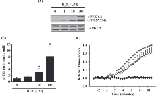
H2O2increases ERK phosphorylation and ROS
gen-eration in A10 cells
It is well known that stimulation of ROS enhanced the phosphorylation of ERK (4, 5). To demonstrate the effects of extracellular administration with H2O2
on ERK phosphorylation, we treated A10 cells with various concentration (1-100μM) of H2O2for 5
min-utes by using Western blot analysis (Fig. 1A). H2O2
treatment increased Thr202/Tyr204 phosphoryla-tion of ERK from the concentraphosphoryla-tion in 10 μM. By using densitometric analysis for Western blotting, the effect of H2O2on ERK phosphorylation was
sig-nificant increased in both 10 and 100μM (Fig. 1B). Next, to check effect of extracellular administra-tion with H2O2on ROS generation in A10 cells, we
observed the change of intercellular ROS by using a ROS-reactive fluorescent dye, carboxy-H2DCFDA
(Fig. 1C). Treatment with 10μM H2O2increased
intercellular ROS after 2.5 minutes and intercellular ROS was kept increasing for 10 minutes (Fig. 1C). In addition, we observed obvious morphological changes of A10 cells with 100 μM H2O2 treatment
under a microscopy, but did not in the cells with un-der 10μM (data not shown). These data suggested that 10 μM H2O2had sufficient effect on elevation
of both intercellular ROS and ERK phosphorylation without morphological changes in cultured vascular smooth muscle A10 cells.
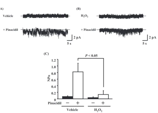
H2O2suppresses pinacidil-induced KATPchannel
ac-tivity in A10 cells.
Recent report showed that H2O2 suppressed
pinacidil-induced activity of smooth muscle KATP
channels, Kir6.1/SUR2B isotype, which were ex-pressed in HEK cells by genetically modification technique (26). To consider direct effect of H2O2
on the activities of native KATPchannel expressed
in VSMCs, we measured the effect of H2O2 for
pinacidil-induced KATP activities in cultured A10
cells using cell-attached patch clamp experiments (Fig. 2). The cells were pretreated with vehicle or 10μM H2O2for 5 minutes and then stimulated with
100μM pinacidil for 5 minutes. Pinacidil increases channel activation without H2O2treatment (Fig. 2A).
Treatment with 3μM glibenclamide, a KATPchannel
blocker, significantly inhibited pinacidil-induced channel activation (data not shown). Treatment with H2O2 suppressed significantly pinacidil-induced
channel activity in A10 cells (Figs. 2B and 2C). These results suggested that H2O2suppress KATP
channel activity in cultured VSMCs.
Fig. 1 Measurements of reactive oxygen species (ROS). (A) Representative data of ROS - induced phosphorylated extracellular regulated kinase (ERK) by extracellular administration with H2O2in A10 cells. The cells were treated with the indicated concentra-tions of H2O2for 5 minutes. Whole cell lysates were subjected to immunoblot analysis with specific antibodies to phosphorylated form (upper panels) and total (lower panels) ERK protein. (B) Densitometric analysis for phosphorylated forms of ERK. (n = 5). (*)P! 0.05 versus vehicle treatment. (C) Intercellular ROS measurements in A10 cells. Intercellular ROS measurements were measured using the ROS reactive fluorescence dye, carboxy - H2DCFDA [6 - carboxy - 2’,7’-dichlorodihydrofluorescein diacetate, di (acetoxymethyl ester)]. The data indicate relative fluorescence intensity for the following conditions. (#) Vehicle treatment. (") 10 μM H2O2. Values are shown as means!S.E.. All curves are representative of triplicate independent experiments and each point is the mean of tripli-cate values. Independent experiment typically included 6 cells. (*) P!0.01 versus vehicle treatment.
Insulin activates KATPchannels in A10 cells.
We previously have reported that insulin causes membrane hyperpolarization via KATPchannels
acti-vation in cultured VSMCs (18). To observe the effect of H2O2for insulin-induced potassium ion transport
on the membrane of A10 cells, we directly meas-ured single channel activities using cell-attached
patch-clamp experiments (Fig. 3). Ion channels were activated 5 minutes after insulin stimulation without H2O2treatment (Fig. 3A). Insulin-activated
currents showed inward rectification ; the conduc-tance of the positive pipette voltage was 28.6 pS, whereas that of the negative pipette voltage was 21.8 pS. The conductance of insulin-induced cur-rents in A10 cells was similar to that of vascular
Fig. 2 Effect of H2O2for pinacidil - induced KATPchannel activities in A10 cells. Single channel currents were measured by the cell -attached patch- clamp experiments. The pipette solution was voltage - clamped at + 50 mV. The cells were pretreated with (A) vehicle, or (B)10μM H2O2for 5 min and then stimulated with 100μM pinacidil for 5 min. (C) KATPchannel activities are shown as NPo. Values are shown as means!S.E. (n = 5).
Fig. 3 Effect of H2O2for insulin - induced KATPchannel activities in A10 cells. Single channel currents were measured by the cell -attached patch- clamp analysis as Fig. 2. The cells were pretreated with (A) vehicle, or (B) 10μM H2O2for 5 min and then stimu-lated with 100 nM insulin for 5 min. (C) KATPchannel activities are shown as NPo. Values are shown as means!S.E. (n = 12 - 19).
smooth muscle type KATPchannel (18). These data
indicate that insulin activates KATPchannel in A10
cells. Treatment of 10μM H2O2significantly
sup-pressed insulin-induced channel activities (Figs. 3B and 3C).
H2O2has no effects for insulin-induced IRS-1 and
Akt phospholyration in A10 cells
We previously have reported that insulin activates KATPchannels via insulin receptor signaling pathway,
which contains insulin receptor substrate (IRS) and Akt, in cultured VSMCs (18). To determine the ef-fect of H2O2treatment on insulin receptor signaling
pathway in A10 cells, we examined the phosphoryla-tion state of IRS-1 and that downstream molecule Akt using Western blotting analysis (Fig. 4). The cells were pretreated with vehicle or 1-10μM H2O2
for 5 minutes and then stimulated with 100 nM insu-lin for 5 minutes. Treatment with H2O2had no effect
for insulin-stimulated IRS phosphorylation (Figs. 4A and 4B). Similarly with IRS, insulin-stimulated Akt phosphorylation was not changed by treatment with H2O2(Figs. 4A and 4C). From these results, we
sug-gested that H2O2 inhibited insulin-induced KATP
channel activation independent of insulin receptor signaling pathway including IRS and Akt.
DISCUSSION
Here we found that 10μM H2O2increases
inter-cellular ROS and inhibits pinacidil-induced KATP
channel activities in cultured VSMCs. This result was similar with the report using smooth muscle
Fig. 4 Effect of H2O2treatment for insulin signaling pathway in A10 cells. The cells were pretreated with the indicated concentration of H2O2for 5 minutes and then stimulated with 100 nM insulin for 5 minutes. Whole cell lysates were subjected to immunoblot analy-sis with specific antibodies to phosphorylated forms (upper panels) and total (lower panels) insulin receptor substrate (IRS) and protein kinase B (Akt) protein. (B - C) Densitometric analysis for phosphorylated forms of IRS and Akt respectively (n = 5). Values are shown as means!S.E..
KATPchannel expressed HEK cells (26).
Further-more we found that H2O2 suppressed insul
in-duced KATPchannel activities independent of insulin
signaling pathway, including IRS and Akt, in cul-tured VSMCs. To our knowledge, this is the first report that H2O2inhibits both pinacidil- and
insulin-induced KATPchannel activities in VSMCs. The in
vitro studies have reported that a KATP channel
blocker glibenclamide increases muscle tone and causes depolarization in VSMCs (29, 30). Further-more, the in vivo studies also have showed that glibenclamide significantly increases vascular resis-tance and decreased arterial diameter (31). In the study for the mouse disrupted the gene encoding Kir6.1, vascular smooth muscle type KATP channel
is critical in the regulation of vascular tonus, espe-cially in the coronary arteries (32). Our data may provide further insight into the relationship among KATPchannel and oxidative stress on cardiovascular
diseases.
There are several reports that insulin activates KATPchannels in neuronal cells (33, 34), pancreatic
β-cells (35), and skeletal muscle cells (36). KATP
channels are sensitive to intracellular ATP levels : their activity is suppressed by increases in ATP and activated by increases in ADP and other nucleoside diphosphates (37). In previous study in VSMCs, we reported that insulin-activated KATPchannel
activa-tion was not mediated the changes of intracellular ATP levels (18). Furthermore we and others have found that insulin-induced KATPchannel activities are
mediated by PI3-K in VSMCs and other cells (18, 34-36). PI3-K is main downstream molecule in in-sulin receptor signaling pathway (14-17). Some re-ports showed ROS-generating agents, such as an-giotensin II, could suppress insulin-receptor signal-ing pathways (38). We have also previously reported that diamide, a thiol-oxidizing agent, inhibited criti-cal insulin signal transduction component including IRS and Akt (39). In addition, diamide suppressed insulin-induced KATPchannel activities, but did not
change pinacidil-induced activities in A10 cells (39). However here we showed 10μM H2O2suppressed
insulin-induced KATP channel activities without
in-hibition of insulin-receptor signaling (Figs. 3 and 4). Yang et al. recently reported that H2O2 inhibited
the activities of VSMC type KATPchannels (Kir6.1/
SUR2B) expressed in HEK cells, because H2O2
in-duced conformational changes of the channels as closed state via S -glutathionylation of Kir6.1 Cys176,
a cysteine residue in the core domain (40). From these data, some agents of oxidative stress may
induce oxidation to different target molecules for KATPchannel activities in VSMCs.
In conclusion, we have demonstrated that H2O2
suppresses insulin-induced KATPchannel activities in
vascular smooth muscle A10 cells. Oxidative stress is increased in the diabetic state ; hyperglycemia leads to production of H2O2within the cells (41).
Our findings suggest that insulin has maybe impor-tant roles of the regulation of vascular tonus, which mediates KATPchannel activation, but ROS induces
impairment these effects on the vasculature. Our data should provide further insight into the effect of ROS on insulin’s action in the vasculature.
CONFLICT OF INTEREST
The authors declared no conflict of interest.
ACKNOWLEDGEMENTS
This study was supported in part by the Grant-in-Aid for the 21st Century COE Program, Human Nu-tritional Science on Stress Control, to Tokushima University from the Ministry of Education, Science, Sports, and Culture and Technology, Japan.
REFERENCES
1. Li Y, Trush MA : Diphenyleneiodonium, an NAD(P)H oxidase inhibitor, also potently inhib-its mitochondrial reactive oxygen species pro-duction. Biochem Biophys Res Commun 253 : 295-299, 1998
2. Palmer HJ, Paulson KE : Reactive oxygen spe-cies and antioxidants in signal transduction and gene expression. Nutr Rev 55 : 353-361, 1997 3. Finkel T : Oxygen radicals and signaling. Curr
Opin Cell Biol 10 : 248-253, 1998
4. Fialkow L, Chan CK, Rotin D, Grinstein S, Downey GP : Activation of the mitogen-acti-vated protein kinase signaling pathway in neu-trophils. Role of oxidants. J Biol Chem 269 : 31234-42, 1994
5. Sundaresan M, Yu ZX, Ferrans VJ, Irani K, Finkel T : Requirement for generation of H2O2
for platelet-derived growth factor signal trans-duction. Science 270 : 296-299 1995
6. Abe J, Berk BC : Reactive oxygen species as me-diators of signal transduction in cardiovascular
disease. Trends Cardiovasc Med 8 : 59-64, 1998 7. Kamata H, Hirata H : Redox regulation of
cel-lular signaling. Cell Signal 11 : 1-14, 1999 8. Westhuyzen J : The oxidation hypothesis of
atherosclerosis : an update. Ann Clin Lab Sci 27 : 1-10, 1997
9. Nunes GL, Robinson K, Kalynych A, King SB, Sgoutas DS, Berk BC : Vitamins C and E in-hibit O2-production in the pig coronary artery :
Circulation 96 : 3593-3601, 1997
10. Robertson RP : Chronic oxidative stress as a central mechanism for glucose toxicity in pan-creatic islet beta cells in diabetes. J Biol Chem 279 : 42351-42354, 2004
11. Brownlee, M : The pathobiology of diabetic complications : a unifying mechanism. Diabetes 54 : 1615-1625, 2005
12. Evans JL, Goldfine ID, Maddux BA, Grodsky GM : Oxidative stress and stress-activated sig-naling pathways : a unifying hypothesis of type 2 diabetes. Endocr Rev 23 : 599-622, 2002 13. Evans JL, Maddux BA, Goldfine ID : The
mo-lecular basis for oxidative stress-induced in-sulin resistance. Antioxid Redox Signal 7 : 1040-1052, 2005
14. Isenovic ER, Meng Y, Jamali N, Milivojevic N, Sowers JR : Ang II attenuates IGF-1-stimulated Na+, K+-ATPase activity via PI3-K/Akt pathway
in vascular smooth muscle cells. Int J Mol Med 12 : 1-12, 2003
15. Li D, Sweeney G, Wang Q, Klip A : Participa-tion of PI3-K and atypical PKC in Na+, K+-pump
stimulation by IGF-1 in VSMC. Am J Physiol Heart Circ Physiol 276 : 2109-2116, 1999 16. Zeng G, Quon MJ : Insulin-stimulated
produc-tion of nitric oxide is inhibited by wortmannin. Direct measurement in vascular endothelial cells. J Clin Invest 15 : 894-898, 1996
17. Zeng G, Nystrom FH, Ravichandran LV, Cong LN, Kirby M, Mostowski H, Quon MJ : Roles for insulin receptor, PI3-kinase and Akt in in-sulin-signaling pathways related to production of nitric oxide in human vascular endothelial cells. Circulation 101 : 1539-1545, 2000 18. Yasui S, Mawatari K, Kawano T, Morizumi R,
Hamamoto A, Furukawa H, Koyama K, Nakamura A, Hattori A, Nakano M, Harada N, Hosaka T, Takahashi A, Oshita S, Nakaya Y : Insulin activates ATP-sensitive potassium chan-nels via phosphatidylinositol 3-kinase in cul-tured vascular smooth muscle cells. J Vasc Res 45 : 233-243, 2008
19. Brayden JE : Functional roles of KATPchannels
in vascular smooth muscle. Clin Exp Pharma-col Physiol 29 : 312-316, 2002
20. Quayle JM, Standen NB : KATPchannels in
vas-cular smooth muscle. Cardiovasc Res 28 : 797-804, 1994
21. Cole WC, McPherson CD, Sontag D : ATP-regulated K+channels protect the myocardium
against ischemia/reperfusion damage. Circ Res 69 : 571-581, 1991
22. Yamada M, Isomoto S, Matsumoto S, Kondo C, Shindo T, Horio Y, Kurachi Y : Sulfonylurea receptor 2B and Kir6.1 form sulfonylurea sen-sitive but ATP-insensen-sitive K channel. J Physiol 499 : 715-720, 1997
23. Teramoto N : Physiological roles of ATP-sen-sitive K+ channels in smooth muscle. J Physiol 572 : 617-624, 2006
24. Moreau C, Prost AL, Dérand R, Vivaudou M : SUR, ABC proteins targeted by KATPchannel
openers. J Mol Cell Cardiol 38 : 951-963, 2005 25. Erdös B, Simandle SA, Snipes JA, Miller AW, Busija DW : Potassium channel dysfunction in cerebral arteries of insulin-resistant rats is me-diated by reactive oxygen species. Stroke 35 : 964-969, 2004
26. Yang Y, Shi W, Cui N, Wu Z, Jiang C : Oxida-tive stress inhibits vascular KATPchannels by
S-glutathionylation. J Biol Chem 285 : 38641-38648, 2010
27. Vrbacky M, Drahota Z, Mracek T, Vojtiskova A, Jesina P, Stopka P, Houstek J : Respiratory chain components involved in the glycerophos-phate dehydrogenase-dependent ROS produc-tion by brown adipose tissue mitochondria. Biochim Biophys Acta 1767 : 989-997, 2007 28. Hamill OP, Marty A, Neher E, Sakmann B,
Sigworth FJ : Improved patch-clamp techniques for high-resolution current recording from cells and cell-free membrane patches. Pflugers Arch 391 : 85-100, 1981
29. Nelson MT, Huang Y, Brayden JE, Hescheler J, Standen NB : Arterial dilations in response to calcitonin generelated peptide involve activation of K+channels. Nature 344 : 770-773, 1990
30. Nakashima M. Vanhoutte PM : Isoproterenol causes hyperpolarization through opening of ATP-sensitive potassium channels in vascular smooth muscle of the canine saphenous vein. J Pharmacol Exp Ther 272 : 379-384, 1995 31. Quayle JM, Nelson MT, Standen NB :
channels in smooth muscle. Physiol Rev 77 : 1165-1232. 1997
32. Miki T, Suzuki M, Shibasaki T, Uemura H, Sato T, Yamaguchi K, Koseki H, Iwanaga T, Nakaya H, Seino S : Mouse model of Prinzmetal angina by disruption of the inward rectifier Kir6.1. Nat Med 8 : 466-472, 2002
33. Mirshamsi S, Laidlaw HA, Ning K, Anderson E, Burgess LA, Gray A, Sutherland C, Ashford ML : Leptin and insulin stimulation of signaling pathways in arcuate nucleus neurones : PI3K dependent actin reorganization and KATP
chan-nel activation. BMC Neurosci 5 : 54, 2004 34. Spanswick D, Smith MA, Mirshamsi S, Routh
VH, Ashford ML : Insulin activates ATP-sen-sitive K+channels in hypothalamic neurons of
lean, but not obese rats. Nat Neurosci 3 : 757-758, 2000
35. Khan FA, Goforth PB, Zhang M, Satin LS : In-sulin activates ATP-sensitive K+channels in
pancreatic beta-cells through a phosphatidyli-nositol 3-kinase-dependent pathway. Diabetes 50 : 2192-2198, 2001
36. Tricarico D, Mallamaci R, Barbieri M, Conte Camerino D : Modulation of ATP-sensitive K+
channel by insulin in rat skeletal muscle fibers. Biochem Biophys Res Commun 232 : 536-539,
1997
37. Moreau C, Prost AL, Derand R, Vivaudou M : SUR, ABC proteins targeted by KATPchannel
openers. J Mol Cell Cardiol 38 : 951-963, 2005 38. Frank GD, Eguchi S, Yamakawa T, Tanaka S, Inagami T, Motley ED : Involvement of reactive oxygen species in the activation of tyrosine kinase and extracellular signal-regulated kinase by angiotensin II. Endocrinology 141 : 3120-3126, 2000
39. Mawatari K, Yasui S, Morizumi R, Hamamoto A, Furukawa H, Koyama K, Hattori A, Yoshioka E, Yoshida M, Nakano M, Teshigawara K, Harada N, Hosaka T, Takahashi A, Nakaya Y : Reactive oxygen species induced by diamide in-hibit insulin-induced ATP-sensitive potassium channel activation in cultured vascular smooth muscle cells. Asia Pac J Clin Nutr 17 : S162-S166, 2008
40. Yang Y, Shi W, Chen X, Cui N, Konduru AS, Shi Y, Trower TC, Zhang S, Jiang C : Molecu-lar basis and structural insight of vascuMolecu-lar KATP
channel gating by S-glutathionylation. J Biol Chem 286 : 9298-9307, 2011
41. Paolisso G, Giuglianoc D : Oxidative stress and insulin action : Is there a relationship? Diabe-tologia 39 : 357-363, 1996