Butyric Acid in Saliva of Chronic Periodontitis Patients Induces Reactivation of EBV
Ryo Koike
Nihon University Graduate School of Dentistry, Major in Oral and Maxillofacial Surgery
(Directors: Prof. Morio Tonogi and Prof. Kenichi Imai)
Contents
Page
Abstract ….. 3
Introduction ….. 5
Materials and Methods ….. 8
Results ….. 13
Discussion ….. 16
Acknowledgements ….. 20
References ….. 21
Figures ….. 25
This doctoral thesis was prepared using the original article “Butyric Acid in Saliva of Chronic
Periodontitis Patients Induces Transcription of the EBV Lytic Switch Activator BZLF1: A
Pilot Study” (Ryo Koike, Keiko Nodomi, Norihisa Watanabe, Yorimasa Ogata, Osamu
Takeichi, Masami Takei, Tadayoshi Kaneko, Morio Tonogi, Ai Kotani and Kenichi Imai. In
Vivo, 2020 in press) with new unpublished data (Figure 2E and Table 1).
3
Abstract
Background/Aim: Human chronic periodontitis (CP) is a major health concern
worldwide. Epstein–Barr virus (EBV) was recently reported to be associated with the progression of CP. Previous report has demonstrated that butyric acid (BA) produced by periodontopathic bacteria can inhibit histone deacetylase, thus increasing the EBV lytic switch activator BZLF1 expression. Because higher levels of periodontopathic bacteria were detected in the saliva of CP patients, it was considered whether the saliva of CP patients can reactivate EBV. Materials and Methods: Saliva was collected from seven CP patients and five periodontally healthy individuals. BA was quantified using HPLC.
Real-time PCR, luciferase assay, and western blotting were performed to determine the
mRNA expression level and transcriptional activity of BZLF1 and the phosphorylation
of histone H3, respectively. Results: BZLF1 mRNA expression and transcriptional
activity were significantly higher in EBV-infected Daudi cells treated with the saliva of
CP patients than with that of the healthy controls. Interestingly, although significantly
higher levels of BA, propionic acid, and acetic acid were detected in the saliva of CP
patients, only BA correlated significantly with the amount of BZLF1 transcripts (r =
0.88; p < 0.02). In addition, the saliva of CP patients induced acetylation of histone H3
in Daudi cells. Conclusion: These findings suggest that BA in saliva may play a role in
4
the initiation of EBV reactivation and contribute to the clinical progression of EBV- related diseases, including periodontitis.
Keywords: Epstein–Barr virus; Periodontitis; butyric acid; BZLF1; reactivation
5
Introduction
Chronic periodontitis (CP) is a complex chronic inflammatory microbial disease that is prevalent in humans worldwide (1, 2). Severe CP can result in the loosening of teeth, occasional pain, periodontal bone loss, and eventual tooth loss (1, 2). Mounting evidence has indicated that CP is a risk factor for aspiration pneumonia, chronic obstructive pulmonary disease, diabetes, and pre-term birth (1, 2). Although no single etiological agent has been identified, a number of putative bacteria, such as
Porphyromonas gingivalis and Fusobacterium nucleatum, are considered to be
associated with CP and, thus, are used as diagnostic markers (2, 3). Recently, members of the herpes virus family, such as Epstein–Barr virus (EBV), have been suggested to be involved in the aetiology of CP because bacterial activity alone does not adequately explain the clinical characteristics of CP (4-8).
Similar to other herpes viruses, EBV establishes a persistent infection in the human host, and its life cycle has both lytic and latent phases (9, 10). The EBV-encoded immediate-early BZLF1 gene encodes ZEBRA, a sequence-specific DNA-binding protein that is a member of
the bZIP family of leucine-zipper transcriptional activators (9, 10). Since ZEBRA can
transactivate early and late genes of EBV, and thereby induce the lytic cycle, this viral
transcriptional activator is a master regulator of the transition from latency to the lytic
6
replication cycle (9, 10). EBV is frequently reactivated in immunocompromised hosts and can induce infectious mononucleosis, as well as several malignancies, such as Burkitt lymphoma and nasopharyngeal carcinoma (9-12).
Many studies have demonstrated that the amount of EBV DNA detected in the periodontal pockets and gingival tissues of CP patients is correlated with disease severity (4- 8). Accordingly, Kato et al. previously reported that EBV DNA was more frequently detected in deep, rather than shallow, periodontal pockets among Japanese patients with CP and healthy controls (13, 14). In addition, a large number of EBV-encoded small RNA-positive B-cells were observed in the gingival tissues of CP patients (13). Although EBV is epidemiologically involved in the aetiology of CP, the process by which latent EBV is reactivated in the oral cavity remains unclear.
EBV is usually transmitted through saliva and replicates in the salivary glands, oral
mucosal membrane, nasopharyngeal epithelium, and B cells (6, 7, 11, 12, 15, 16). In
addition, the saliva of CP patients contains EBV-infected B cells, higher levels of EBV
DNA, and greater concentrations of periodontopathic bacteria (6, 7, 15-17), suggesting
a relationship between microbial interactions and the aetiology of CP. Imai et al. have
also reported that although short-chain fatty acids (SCFAs) are secreted extracellularly
by P. gingivalis and F. nucleatum, only butyric acid (BA) can induce reactivation of
7
EBV (18). These observations suggest that EBV reactivation may be caused by BA in the saliva of CP patients. However, no studies have yet to evaluate the amount of BA in the saliva of CP patients and determine whether the saliva can reactivate EBV.
Therefore, the aim of the present study was to assess the levels of BA in the saliva
of CP patients, which could efficiently induce the expression of the EBV lytic switch
activator BZLF1, and to determine for the first time whether there is a possible
pathophysiological link with EBV reactivation. In addition, this article discusses how
this relationship may pertain to the aetiology of CP.
8
Materials and Methods
Study approval and participants
The study protocol was approved by the Institutional Internal Review and Ethics Board of the Nihon University School of Dentistry (Tokyo, Japan; approval no. EP17D006) and conducted in accordance with the tenets of the Declaration of Helsinki . The study cohort included seven CP patients (mean age, 53.1 ± 13.7 years) and five periodontal healthy individuals (mean age, 32.6 ± 6.1 years). Written informed consent was obtained from each study participant after all procedures had been fully explained.
Reagents
BA was purchased from Wako Pure Chemical Industries, Ltd. (Osaka, Japan).
Antibodies (Abs) against ZEBRA and -actin were purchased from Santa Cruz
Biotechnology, Inc., Dallas, TX, USA), whereas those against acetylated histone 3 were obtained from Thermo Fisher Scientific (Waltham, MA, USA) and those against non- acetylated histone 3 were purchased from Abcam (Cambridge, UK).
Saliva collection
All of the study participants received dental care at Nihon University School of
Dentistry. Periodontal status was assessed based on the probing pocket depth (PPD),
clinical attachment level (CAL), and bleeding on probing. The PPD and CAL were
measured using a PCP11 probe (Hu-Friedy Mfg. Co., LLC, Chicago, IL, USA). CP was
9
defined as the presence of at least two sites with a PPD of ≥5 mm and attachment loss of
>6 mm.
The healthy controls had no clinical signs of gingivitis, attachment loss, or detectable bone loss on radiographic examinations, and PPD was ≤3 mm. All patients were systemically healthy with no history of periodontal treatment or any type of antibiotic therapy for at least 3 months prior to participation in this study. About 10 ml of saliva were collected from each participant. After centrifugation to remove cells and debris, the supernatant of the collected saliva was sterilised by passing through a 0.22- μm pore filter membrane and then either immediately analysed or stored at –80°C for
future use.
Cell culture
Daudi cells, which are well-characterised EBV-positive human Burkitt lymphoma- derived cells, and B95-8-221 Luc cells (18, 19), which were stably transfected with the BZLF1 promoter, were maintained at 37°C in Roswell Park Memorial Institute 1640
medium (Sigma-Aldrich Corporation, St. Louis, MO, USA) containing 10% heat-
inactivated fetal bovine serum (Sigma-Aldrich Corporation), penicillin (100 U/ml), and
streptomycin (100 µg/ml). For the stimulation experiments, cells (1.0 × 10
6cells /1.0-ml
well) were treated with saliva or BA.
10
Quantification of SCFAs
SCFAs [BA, propionic acid (PA), acetic acid (AA), isoBA, and isovaleric acid] were quantified using ion exclusion high-performance liquid chromatography (HPLC), as described previously (18, 20). Briefly, each saliva sample was mixed with 12%
perchloric acid, filtered through a cellulose acetate membrane filter (Cosmonice Filter W, pore size: 0.45 µm; Nacalai Tesque, Inc., Kyoto, Japan), and then injected into a SIL-10 auto injector (Shimadzu Corporation, Kyoto, Japan). SCFAs were separated using a serial organic acid column and a guard column with isocratic elution of p- toluene sulfonic acid aqueous solution and detected using an electronic conductivity detector.
Preparation of mRNA and real-time polymerase chain reaction (PCR)
The experimental procedures for RNA purification and real-time PCR were performed
as previously described (18, 21). Briefly, Daudi cells were washed once with ice-cold
phosphate-buffered saline (PBS) and homogenised using a QIAshredder (QIAGEN,
Alameda, CA, USA), while total RNA was purified using an RNeasy Mini Kit
(QIAGEN). For cDNA synthesis, total RNA (1 μg) was reverse transcribed using an
RNA PCR kit (PrimeScript; Takara Bio, Shiga, Japan). The resulting cDNA mixture
was subjected to real-time PCR analysis using SYBR Premix Ex Taq solution (Takara
11
Bio) containing 5 μM sense and antisense primers. The primer sequences used for the amplification of each gene were as follows: BZLF1 forward (5-TTC CAC AGC CTG CAC CAG TG-3) and reverse (5- GGC AGC AGC CAC CTC ACG GT -3); and glyceraldehyde-3-phosphate dehydrogenase (GAPDH), forward (5-ACC AGC CCC AGC AAG AGC ACA AG-3) and reverse (5-TTC AAG GGG TCT ACA TGG CAA CTG-3). PCR assays were performed using a TP-800 Thermal Cycler Dice Real-Time System (Takara Bio) and analysed using the software provided by the device
manufacturer. The thermal cycling conditions were 40 cycles at 95°C for 5 sec, 60°C for 30 sec, and 72°C for 1 min. All real-time PCR experiments were performed in triplicates, and the specificity of each product was verified via a melting curve analysis.
The calculated gene expression levels were normalised to GAPDH mRNA levels.
Luciferase assay
Luciferase assay was then performed using a Dual-Luciferase Reporter Assay System
(Promega), according to the manufacturer’s instructions. The experimental procedure
for the luciferase assay has been previously reported (20, 21). B95-8-221 Luc. cells
were harvested and the extracts were subjected to luciferase assay using the Dual-
Luciferase Assay System
TM(Promega). All the experiments were carried out in
12
triplicates and the data were presented as the fold increase in luciferase activities (means
± S.D.) relative to the control of three independent transfections.
Immunoblot assay
The experimental procedures for immunoprecipitation and immunoblotting were performed according to previously published protocols (22, 23). Briefly, cells were harvested with lysis buffer (25 mM HEPES-NaOH [pH 7.9], 150 mM NaCl, 1.5 mM MgCl
2, 0.2 mM EDTA, 0.3 % NP-40, 1 mM dithiothreitol, 0.5 mM
phenylmethylsulfonyl fluoride), the proteins were separated by SDS-PAGE and
transferred to a polyvinylidene fluoride membrane (EMD Millipore Corporation, Billerica, MA, USA). The protein content was measured by a detergent-compatible protein assay kit (Bio-Rad, Hercules, Hercules, CA, USA). All membranes were treated
with ECL prime detection reagent (Thermo Fisher Scientific) prior to examination. All bands were visualised using a ChemiDoc XRS System (Bio-Rad).
Statistical analysis
Comparison of two groups was performed using the two-tailed Student’s t-test. The
correlation coefficient (r) was calculated where applicable. A probability (p) value of <0.05
was considered statistically significant.
13
Results
Saliva of CP patients contains relatively high levels of SCFAs
Previous studies have reported that the periodontal pockets and dental plaques of CP patients contain high concentrations (mM levels) of SCFAs (24-26). However, the amounts of SCFAs have not been investigated in the saliva of Japanese CP patients. Therefore, this study measured the concentrations of SCFAs in the saliva of seven CP patients and five healthy controls by HPLC. As presented in Figure 1, the saliva of CP patients contained significantly higher levels (p< 0.01) of BA, PA, and AA. On the other hand, the amounts of isoBA and isovaleric acid in the saliva were very low. The concentrations of BA, PA, and AA in the saliva of CP patients were 0.31–1.37, 0.49–1.35, and 2.12–5.81 mM, respectively. On average, the saliva of CP patients contained about 33.3-, 3.3-, and 2.4-fold higher levels of BA, PA, and AA than that of the healthy controls. The concentration of SCFAs in the saliva of each individuals were shown Table1.
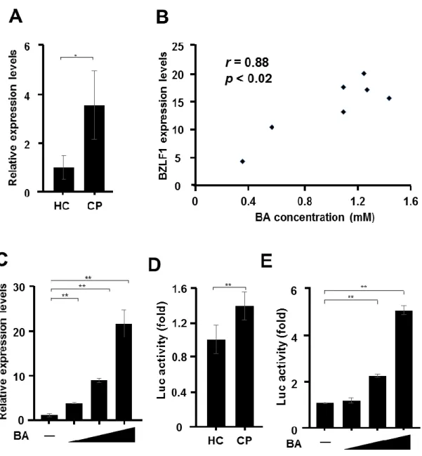
Saliva of CP patients induces expression of BZLF1
Since high concentrations of SCFAs were found in the saliva of CP patients, whether the
saliva can reactivate EBV was investigated. Real time-PCR was conducted to evaluate the
effect of the saliva of seven CP patients and five healthy controls at a 1:2 dilution on BZLF1
mRNA expression in Daudi cells. As presented in Figure 2A, mRNA levels of the EBV lytic
14
gene BZLF1 were significantly higher in cells treated with the saliva of CP patients than of that of the healthy controls. Interestingly, there was a significant correlation between BA concentrations and BZLF1 transcript levels (r=0.88; p<0.02; Figure 2B). The concentration of BA in the saliva of CP patients (0.31–1.37 mM) induced BZLF1 expression in a concentration-dependent manner (Figure 2C). In contrast, no such effect was observed with PA and AA (data not shown). Next, the effect of saliva on gene expression of the BZLF1 promoter was examined using the luciferase assay. As demonstrated in the results presented in Figure 2D, the saliva of CP patients transactivated the BZLF1 promoter in B95-8-221 Luc cells. In addition, although no such effect was observed with PA and AA (data not shown), the concentration of BA in the saliva of CP patients transactivated the BZLF1 promoter in B95-8-221 Luc cells. (Figure 2E).
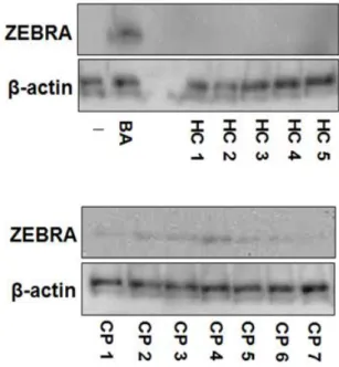
Saliva of CP patients induces expression of ZEBRA
The expression of the lytic switch transactivator ZEBRA by exposure to the saliva of CP patients was examined. As presented in Figure 3, the saliva of the healthy controls had no effect on the expression of ZEBRA in Daudi cells. However, the addition of the saliva of CP patients increased the expression of ZEBRA.
Hyperacetylation of histones by the saliva of CP patients
15
BA is known to inhibit the enzymatic activity of histone deacetylase (HDAC) by
competing with the HDAC substrate for the enzyme’s active site pocket, which contains the catalytic center (27), thus stimulating transcription of various genes, including BZLF1 (9, 10, 28). Imai et al. have previously demonstrated that the culture supernatant from periodontopathic bacteria, which contains high concentrations of BA, can inhibit HDACs, thereby increasing the level of histone acetylation and the transcriptional activity of the BZLF1 gene (18). Next, the effects of saliva and BA on histone
acetylation were examined by western blotting with Abs specific for acetylated histone
H3. As presented in Figure 4, although there was no effect by the saliva of the healthy
controls, both the saliva of CP patients and BA induced acetylation of histone H3
(Figure 4A, B). In contrast, no such effect was observed with the other tested SCFAs
(data not shown).
16
Discussion
Reactivation of latent EBV is associated with progeny virus production and several human diseases (9-12). Therefore, elucidation of the mechanisms that promote or disrupt EBV latency in infected individuals is required to understand the pathobiology of EBV infection and to develop preventive measures and novel therapies. However, the trigger that is responsible for the switch from latency to the lytic cycle in individuals latently infected with EBV remains unclear. This study examined the biological actions of the saliva of CP patients and healthy controls on the reactivation of EBV infection.
It has been reported that more than sufficient concentrations of BA are present in
the dental plaques (range= 4.7–13.8 mM) (25, 26) and periodontal pockets (mean, 2.6 ±
0.4 mM) of patients with periodontal disease (24), whereas the BA concentration is
below the detection limits in healthy sites, suggesting that BA may play a role in the
initiation of EBV reactivation and contribute to the clinical progression of EBV-related
diseases. The results of the present study revealed significantly higher levels of BA in
the saliva of CP patients, which could efficiently induce BZLF1 transcription. These
observations suggest that the BA content in the saliva of CP patients might be involved
in the progression of EBV-related diseases as well as periodontitis.
17
Since BZLF1 expression is a key step in the reactivation from latency in EBV- infected cells (9, 10), this study focused on the expression of this transcriptional activator. Although, neither BZLF1 mRNA nor ZEBRA protein was detectable during latency, high levels of anti-BZLF1 Abs in blood are associated with an increased EBV serum load in EBV-infected patients (29). Figure 1 shows that high concentrations (mM levels) of BA, AA, and PA are present in the saliva of Japanese CP patients (Figure 1).
Interestingly, there was a significant correlation between BA concentrations and the levels of BZLF1 transcripts (r=0.88; p<0.02; Figure 2B). In fact, BA was the only acid in the saliva of CP patients that induced BZLF1 expression (Figure 2C). These results support the findings of a previous in vitro study, which reported that among several SCFAs in the culture supernatant of periodontopathic bacteria, only butyrate reactivated EBV, whereas non-butyrate-producing bacteria did not (18).
Recent studies have revealed a new mechanism that regulates the maintenance and reversal of EBV latency, which involves nucleosome configurations and histone modifications. In the latent state, the BZLF1 gene promoter is bound by histone proteins into a chromatin structure that serves to repress the transcription of BZLF1 (9, 10).
Hyperacetylation of core histone proteins adjacent to the BZLF1 promoter was correlated
with transcriptional activation of BZLF1, whereas hypoacetylation mediated by HDAC was
18
correlated with its repression, which is considered responsible for the maintenance of latency (9, 10). Since BA is one of the most potent inhibitors of HDACs and previous report indicated that BA in the culture supernatant of periodontopathic bacteria promotes histone acetylation and the transcriptional activity of the BZLF1 gene (18), in this study whether the saliva of CP patients induced histone acetylation was examined. Figure 4 shows that the saliva of CP patients induced Lys acetylation of histone H3 in EBV-infected cells. Imai et al. have reported that no such activity occurred with P. gingivalis or bacterial components, such as lipopolysaccharide and fimbriae (16). Although it is necessary to assess other factors contained in saliva, such as cytokines and enzymes, these findings suggested that H3 histone acetylation and BZLF1 expression are ascribable to BA contained in the saliva of CP patients.
The saliva of patients with periodontitis contains EBV-infected B cells, and bleeding of the gums is often observed in these patients (7, 15-17). In addition, it was recently reported that EBV infects the oral epithelial cells of patients with periodontitis in addition to the epithelial cells of the upper aerodigestive tract (30). The extent of gingival epithelial EBV infection is correlated with the severity of CP (30). Moreover, previous reports, as well as the present study, indicated that EBV also contributes to the progression of periapical periodontitis (20, 31). These findings and previous
observations suggest the potential risks of BA in saliva for the progression of
19
periodontitis and periapical periodontitis. In this study it was assumed that microbial synergy by the interaction between periodontopathic bacteria and EBV leads to the following negative chain of pathological events in the oral cavity: 1) periodontopathic anaerobic bacteria, such as P. gingivalis and F. nucleatum, produce BA; 2) BA induces EBV reactivation; 3) EBV impairs local host defences, 4) which leads to increased proliferation of periodontopathic bacteria; 5) increased BA and inflammatory cytokine production by the synergistic effects of EBV and periodontopathic bacteria; and 6) periodontitis escalation.
Periodontitis and EBV are spreading worldwide. Although the findings of this
study suggest a relationship between the saliva of patients with periodontitis and EBV
reactivation, additional basic and clinical studies with greater numbers of cases are
needed. Furthermore, prevention and early treatment of periodontitis involving
elimination of BA-producing bacteria could effectively block further clinical
progression of EBV infection.
20
Acknowledgements
I would like to thank the members of the Department of Oral and Maxillofacial Surgery
and Microbiology for their technical advice and support.
21
References
1. Hajishengallis G: Periodontitis: From microbial immune subversion to systemic inflammation. Nat Rev Immunol 15(1): 30-44, 2015. PMID: 25534621. DOI:
10.1038/nri3785
2. Darveau RP: Periodontitis: A polymicrobial disruption of host homeostasis. Nat Rev Microbiol 8(7): 481-490, 2010. PMID: 20514045. DOI: 10.1038/nrmicro2337 3. Holt SC and Ebersole JL: Porphyromonas gingivalis, Treponema denticola, and
Tannerella forsythia: The “red complex”, a prototype polybacterial pathogenic consortium in periodontitis. Periodontol 2000 38(1): 72-122, 2005. DOI:
10.1111/j.1600-0757.2005.00113.x
4. Contreras A, Nowzari H and Slots J: Herpesviruses in periodontal pocket and gingival tissue specimens. Oral Microbiol Immunol 15(1): 15-18, 2000. PMID:
11155159. DOI: 10.1034/j.1399-302x.2000.150103.x
5. Lu H, Zhu C, Li F, Xu W, Tao D and Feng X: Putative periodontopathic bacteria and herpesviruses in pregnant women: a case–control study. Sci Rep 6: 27796, 2016.
PMID: 27301874. DOI: 10.1038/srep2779
6. Slots J, Saygun I, Sabeti M and Kubar A: Epstein–Barr virus in oral diseases. J Periodontal Res 41(4): 235-244, 2006. PMID: 16827715. DOI: 10.1111/j.1600- 0765.2006.00865.x
7. Saygun I, Kubar A, Özdemir A and Slots J: Periodontitis lesions are a source of salivary cytomegalovirus and Epstein–Barr virus. J Periodontal Res 40(2): 187-191, 2005. PMID: 15733155. DOI: 10.1111/j.1600-0765.2005.00790.x
8. Konstantinidis A, Sakellari D, Papa A and Antoniadis A: Real-time polymerase chain reaction quantification of Epstein–Barr virus in chronic periodontitis patients. J Periodontal Res 40(4): 294-298, 2005. PMID: 15966906. DOI: 10.1111/j.1600- 0765.2005.00796.x
9. Jenkins PJ, Binne UK and Farrell PJ: Histone acetylation and reactivation of Epstein- Barr virus from latenc. J Virol 74(2): 710-720, 2000. PMID: 10623733.
DOI:10.1128/jvi.74.2.710-720.2000
22
10. Bryant H and Farrell PJ: Signal Transduction and transcription factor modification during Reactivation of Epstein-Barr Virus from Latency. J Virol 76(20): 10290- 10298, 2002. PMID: 12239305. DOI:10.1128/jvi.76.20.10290-10298.2002 11. Schmidt CW and Misko IS: The ecology and pathology of Epstein–Barr virus.
Immunol Cell Biol 73: 489-504, 1995. PMID: 8713470. DOI: 10.1038/icb.1995.79 12. Thorley-Lawson DA and Gross A: Persistence of the Epstein–Barr virus and the
origins of associated lymphomas. N Engl J Med 350(13): 1328-1337, 2004. PMID:
15044644. DOI: 10.1056/NEJMra032015
13. Kato A, Imai K, Ochiai K and Ogata Y: Higher prevalence of Epstein–Barr virus DNA in deeper periodontal pockets of chronic periodontitis in Japanese patients.
PLoS One 8(8): e71990, 2013. PMID: 23991022. DOI:
10.1371/journal.pone.0071990
14. Kato A, Imai K, Ochiai K and Ogata Y: Prevalence and quantitative analysis of Epstein–Barr virus DNA and Porphyromonas gingivalis associated with Japanese chronic periodontitis patients. Clin Oral Investig 19(7): 1605-1610, 2015. PMID:
29233156. DOI: 10.1186/s12903-017-0438-6
15. Slots J: Herpesviral-bacterial synergy in the pathogenesis of human periodontitis.
Curr Opin Infect Dis 20(3): 278-283, 2007. PMID: 17471038. DOI:
10.1097/QCO.0b013e3280964da0
16. Contreras A, Zadeh HH, Nowzari H and Slots J: Herpesvirus infection of
inflammatory cells in human periodontitis. Oral Microbiol Immunol 14(4): 206-212, 1999. PMID: 10551163. DOI: 10.1034/j.1399-302x.1999.140402.x
17. Chalabi M, Rezaie F, Moghim S, Mogharehabed A, Rezaei M and Mehraban B:
Periodontopathic bacteria and herpesviruses in chronic periodontitis. Mol Oral Microbiol 25(3): 236-240, 2010. PMID: 20536751. DOI: 10.1111/j.2041- 1014.2010.00571.x
18. Imai K, Inoue H, Tamura M, Cueno ME, Inoue H, Takeichi O, Kusama K, Saito I and Ochiai K: The periodontal pathogen Porphyromonas gingivalis induces the Epstein–Barr virus lytic switch transactivator ZEBRA by histone modification.
Biochimie 94(3): 839-846, 2012. PMID: 22178321. DOI:
10.1016/j.biochi.2011.12.001
23
19. Inoue H, Mishima K, Yamamoto-Yoshida S, Ushikoshi-Nakayama R, Nakagawa Y, Yamamoto K, Ryo K, Ide F and Saito I: Aryl hydrocarbon receptor-mediated induction of EBV reactivation as a risk factor for Sjögren's syndrome. J Immunol 188(9): 4654-4662, 2012. PMID: 22467650. DOI: 10.4049/jimmunol.1101575 20. Makino K, Takeichi O, Imai K, Inoue H, Hatori K, Himi K, Saito I, Ochiai K and
Ogiso B: Porphyromonas endodontalis reactivates latent Epstein-Barr virus. Int Endod J 51(12): 1410-1419, 2018. PMID: 29858508. DOI: 10.1111/iej.12959 21. Imai K, Kamio N, Cueno ME, Saito Y, Inoue H, Saito I and Ochiai K: Role of the
histone H3 lysine 9 methyltransferase Suv39 h1 in maintaining Epstein–Barr virus latency in B95-8 cells. FEBS J 281(9): 2148-2158, 2014. PMID: 24588869. DOI:
10.1111/febs.12768
22. Imai K, Togami H and Okamoto T: Involvement of histone H3 lysine 9 (H3K9) methyltransferase G9a in the maintenance of HIV-1 latency and its reactivation by BIX01294. J Biol Chem 285(22): 16538-16545, 2010. PMID: 20335163. DOI:
10.1074/jbc.M110.103531
23. Watanabe N, Nodomi K, Koike R, Kato A, Takeichi O, Kotani AI, Kaneko T, Sakagami H, Takei M, Ogata Y, Sato S and Imai K: EBV LMP1 in gingival
epithelium potentially contributes to human chronic periodontitis via inducible IL8 production. In Vivo 33(6): 1793-1800, 2019. PMID: 31662504. DOI:
10.21873/invivo.11670
24. Niederman R, Buyle-Bodin Y, Lu B.Y, Robinson P, Naleway C: Short-chain carboxylic acid concentration in human gingival crevicular fluid. J Dent Res 76(1):
575-579, 1997. PMID: 9042080. DOI: 10.1177/00220345970760010801
25. Margolis HC, Duckworth JH and Moreno EC: Composition and buffer capacity of pooled starved plaque fluid from caries-free and caries-susceptible individuals. J Dent Res 67(12): 1476-1482, 1988. PMID: 3198845. DOI:
10.1177/00220345880670120701
26. Moreno EC and Margolis HC: Composition of human plaque fluid. J Dent Res 67(11):
1181-1189, 1988. PMID: 3045178. DOI: 10.1177/00220345880670090701 27. Riggs MG, Whittaker RG, Neumann JR and Ingram VM: n-Butyrate causes
histone modification in HeLa and Friend erythroleukaemia cells. Nature 268 (5619):
24
462-464, 1977. PMID: 268489. DOI: 10.1038/268462a0
28. Saemundsen AK, Kallin B and Klein G: Effect of n-butyrate on cellular and viral DNA synthesis in cells latently infected with Epstein-Barr virus. Virology 107(2):
557-561, 1980. PMID: 6256952. DOI: 10.1016/0042-6822(80)90326-8 29. Drouet E Brousset P, Fares F, Icart J, Verniol C, Meggetto F, Schlaifer
D, Desmorat-Coat H, Rigal-Huguet F, Niveleau A and Delsol G: High Epstein-Barr virus serum load and elevated titers of anti-ZEBRA antibodies in patients with EBV-harboring tumor cells of Hodgkin's disease. J Med Virol 57(4): 383-389, 1999.
PMID: 10089051. DOI: 10.1002/(sici)1096-9071(199904)57:4<383::aid- jmv10>3.0.co;2-3
30. Vincent-Bugnas S, Vitale S, Mouline CC, Khaali W, Charbit Y, Mahler P, Prêcheur I, Hofman P, Maryanski JL and Doglio A: EBV infection is common in gingival epithelial cells of the periodontium and worsens during chronic periodontitis. PLoS One 8(2): e80336, 2013. PMID: 24367478. DOI: 10.1371/journal.pone.0080336 31. Makino K, Takeichi O, Hatori K, Imai K, Ochiai K and Ogiso B: Epstein-Barr virus
infection in chronically inflamed periapical granulomas. PLoS One10(4):e0121548,
2016. PMID: 25884725. DOI: 10.1371/journal.pone.0121548
25
Table 1.
Concentration of SCFAs in the saliva of each individuals HC: healthy control; CP: chronic periodontitis
SCFA (Short-Chain Fatty Acid Level) (mM)
Butyric acid Propionic acid Acetic acid Isobutyric acid Isovaleric acid
HC 1 0.02 0.14 0.91 0.01 0.01
HC 2 0.04 0.30 2.35 0.06 0.03
HC 3 0.07 0.23 1.59 0.02 0.01
HC 4 0.06 0.47 2.44 0.02 0.01
HC 5 0.01 0.05 0.73 0.01 0.01
CP 1 0.52 0.69 2.46 0.02 n.d
CP 2 1.04 0.49 3.48 0.04 0.02
CP 3 1.19 0.49 2.12 0.01 0.01
CP 4 1.37 0.52 2.86 0.09 0.05
CP 5 0.31 0.65 3.65 0.01 0.01
CP 6 1.20 0.49 3.52 0.04 0.02
CP 7 1.04 1.35 5.81 0.02 0.02
26
Figure 1. Saliva of CP patients contains higher levels of SCFAs.
Levels of of butyric acid, propionic acid, acetic acid, isobutyric acid, and isovaleric acid in the saliva of seven CP patients and five healthy controls. HC: healthy control; CP:
chronic periodontitis; N.S.: not significant.
27
Figure 2. Induction of BZLF1 gene expression by the saliva of CP patients.
(A) Daudi cells were incubated with the saliva of seven CP patients and five healthy controls at a 1:2 dilution (saliva volume vs. total cell culture medium volume) for 24 h.
Real-time PCR analysis was conducted with specific primers to detect BZLF1 mRNA
expression. (B) The correlation coefficient (r) was calculated between butyric acid (BA)
concentrations and BZLF1 mRNA levels. (C) Daudi cells were treated with BA (0.5,
1.0, or 1.5 mM) for 24 h, and BZLF1 mRNA expression was assessed. (D) B95-8-221
28
Luc cells were treated with the saliva of seven CP patients and five healthy controls at a
1:2 dilution for 48 h. (E) B95-8-221 Luc cells were treated with BA (0.5, 1.0, or 1.5
mM) for 48 h. The luciferase activity of each cell lysate was then measured. The values
are presented as mean ± standard deviation (n = 3). **p<0.01, *p<0.05.
29
Figure 3. Saliva of CP patients induced ZEBRA expression.
Daudi cells were treated with the saliva of seven CP patients and five healthy controls at a
1:2 dilution or BA (2 mM, positive control) for 36 h. The lysates were harvested, and ZEBRA
levels were assessed by immunoblotting using specific Abs. -actin was used as an internal
control. BA: butyric acid.
30