Acta med. Nagasaki 16 : 12-28
Studies on Sex Chromatin in Thyroid Diseases - Especially Tumors of Thyroid Gland -
Jituhiro ISHIGAKI * Second Department of Surgery, Nagasaki University School of Medicine,
Nagasaki, Japan
Received for Publication, October 1, 1971
Thyroid tissues obtained from 360 cases by means of operation were classified pathohistologically into normal thyroid, benign thyroid diseases and malignant thyroid tumors and stained by FEULGEN' S method. Sex chromatins of follicular epithelial cells (tumor cells) and interstitial cells were counted for each case and incidence of sex chromatin was compared among those groups.
The specimens used in the present study were all obtained from female subjects ranging in age from 15 to 77. In most cases of normal thyroid and benign thyroid diseases, incidence of sex chromatin showed more than 30%.
However, in 2/3 of the cases of so-called malignant adenoma where capsular invasion was verified, incidence of sex chromatin was less than 30% in the very region of the capsular invasion but over 30% in the uninvaded region.
In the cases of thyroid cancer, incidence of sex chromatin was less than 30%. It was higher in papillary adenocarcinoma and follicular adeno- carcinoma than undifferentiated carcinoma. There was hardly noted any difference in incidence of sex chromatin between the primary lesion and the metastasized lesion in the lymph node.
Variation in incidence of sex chromatin by age was not recognized in the disease groups but only in the group of normal thyroid.
INTRODUCTION
In 1959, BARR and BERTRAMO found nucleoli measuring 1,a in size in the nucleus of the hypoglossal nerve cells of the cat. Since the nucleoli were found only in the female cat, they considered these nucleoli related to sex and named them as sex chromatins.
Sex chromatins are generally basophile and show strong positive reaction to FEULGEN'S 'staining. They are hemispheric or flat in shape
*石 垣 実 弘
and are either attached to the inside of the nuclear membrane of free within the nuclear substance.
Sex chromatins have been verified in the epithelial cells of the oral cavity, vagina and urinary tract and in various tissues of the skin, leukocyte and others of animals and man.
Sex chromatins are frequently found in the cell nucleus of females and rarely in the cell nucleus of males. In this respect, sex chroma- tins are used clinically for sex distinction particularly , for intersex and are considered indispensable for sex determination of ndividuals .
The idea of sex determination has been introduced,: to character of
tumor by examination of the rate of content of sex chromatins in the tumor cells. However, as HUNTER and LENNOx9) observed in teratoma in 1954, the sex of the host and the sex determined by the incidence of sex chromatin are not always identical.
Concerning sex chromatin in tumor, many studies have been made since then by TAVARES24> , CRUICKSHANK3>, SOHVAL22) , EMERY and
MCMILLAN5), LEHMAN11), BARR and MOORE2., HIENZ and EHLER8) , ZANNELLA28) , GRAF6), MYERSI %. and other investigators. As the result,
it has been clarified that the sex of the host of tumor and the sex determined from the incidence of sex chromatin in tumor are generally
identical but in tumors of endocrine organs such as pheochromocyto- ma, mammary tumor and prostatic tumor, they are not identical with a fairly high frequency.
Furthermore, studies are being made to see whether or not incidence of sex chromatin is useful as an index to determine the adaptability of hormone therapy for tumors of endocrine organs parti- cularly for mammary tumor and prostatic tumor. It has been conside- red to be available as a means to estimate the degree of the dependency of cancer cells on hormone. In order to examine if incidence of sex chromatin is a useful index of horomone therapy for thyroid tumor, the author counted sex chromatins in the thyroid in various conditions, and studied the relationship between incidence of sex chromatin in tumor cells and the nature (malignancy or benignity) of tumor.
MATERIALS AND METHODS
The materials used in the present study were specimens of thyroid tissue obtained from 360 cases by operation at the Second Department of Surgery of NAGASAKI UNIVERSITY HOSPITAL and the NOGUCHI HOSPITAL
in BEPPU CITY during the four-year period from 1965 to 1968. Patho- histological diagnosis was established for all these cases.
These 360 cases consisted of 36 cases of normal thyroid, 60 cases
of diffuse goiter with hyperthyroidism, 16 cases of chronic thyroiditis,
8 cases of adenomatous goiter, 60 cases of adenoma, 60 cases of
so-called malignant adenoma and 120 cases of thyroid cancer. For these cases, pathohistological
diagnosis was first established upon HE stain and incidence of
sex chromatin was calculated with sections of FEULGEN'S stain.
(Table 1)
For calculation of sex chro- matins, 1000x oil immersion microscopy was used and 100 cell nuclei each at three different areas , , of a section, totalling 300 cell nuclei, were examined for the presence of sex chromatin and the percentage of sex chro-
Table I .
Incidence o` Sex Chromatin in Various Thyroid Diseaes (360 Cases)
Pathological Diagnosis No. of C ases Range (%) Mean (%) Normal Thyroid Gland 36 35-80 50
Chronic Thyroiditis 16 26-46 34
Adenomatous Goiter 8 29-52 41
Diffuse Goiter with 60 31-63 45 Hyperthyroidism
Adenoma 60 29-78 44
Malignant Adenoma 60 11-68 30
Thyroid Cancer 120 5-42 21
matin positive cells was used as incidence of sex chromatin.
At the time of microscopic examination, those cells which were clearly visible and whose nuclei were not overlapped with. others were selected as the objects of calculation. Those nucleoli measuring 1 V in size attached to the nuclear membrane and shaped somewhat rounded or flattened hemispheric were calculated as chromatins ; those which were free in the nucleolus and nuclear substance, vaguely demarcated or irregular-shaped were excluded from calculation. (Photo 2)
Photo. 2 Many Sex Chromatin are present in the follicular epithelial cells in a -patient s ith "adenoma of thyroid gland. (Feulgen's stain, x 1000)
A picture on the upper right side is that in high po-.2 er, Sex Chro-
matins are rounded or hemispheric in shape and tightly attached to
the unclear menbrane. (x 1400, Feulgen's stain)
The calculation of such sex chromatins was conducted , separately for follicular epithelial cells (tumor cells) and interstitial- cells.
RESULTS
In the 36 cases of normal thyroid, the function and histo- logical findings were normal and the specimens were obtained mostly by incisional biopsy. The incidence of sex chromatin in the follicular epithelial cells ranged from 35% to 80%, the mean value being 50%. The incidence in the interstitial cells was bet- ween 29% and 57% with the mean of 39%. Thus the former was higher than the latter show- ing statistically significant dif- ference (P=0.05). (Fig. 1)
As to the relationship bet- ween age and incidence of sex chromatin in the cases of normal thyroid, the mean incidence in the follicular epithelial cells was 66.1% in teenage, 59% in 20's, 54% in 30's, 42.5% in 40's, 44.2
% in 50's and 46.2% in 60's or more. The incidence of sex chromatin gradually decreased
as the age advanced up to 40's and thereafter remained almost constant ranging from 40% to 45
%. On the other hand, the mean incidence of sex chromatin in the interstitial cells of normal thyroid was 41.5% in teenage, 59% in 20's, 42% in 30's, 37%
in 40's, 42% in 50's and 40% in 60's or more. Whereas nearly 60% was indicated for 20's, the values for other age groups were almost constant, being approxi- mately 40%. (Fig. 2)
Fig. 1. Incidence of Sex Chromatin in Normal Thyroid Gland
1: Follicular Epithelial Cells
2: Interstitial Cells
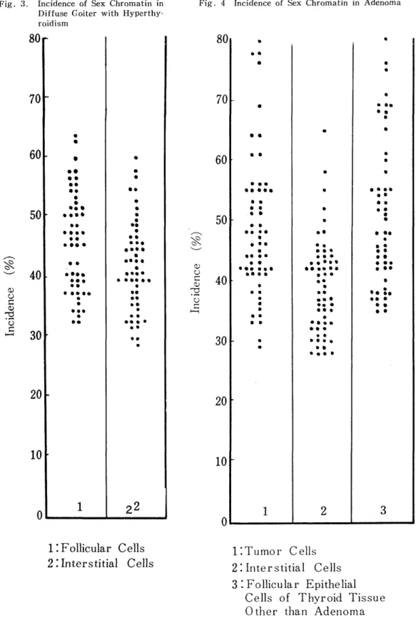
In the 60 cases of diffuse goiter with hyperthyroidism, the inci- dence of sex chromatin in the follicular epithelial cells ranged from 31% to 63%, the mean value being 45%. The incidence in the interstitial cells ranged from 28% to 60% with the mean of 39%. No significant difference was noted between the two incidences nor was observed any specific change due to age. (Fig. 3)
The incidence of sex chromatin in the 16 cases of chronic thyroi- ditis was calculated only in the follicular epithelial cells and it ranged from 26% to 46% with the mean of W/o.
The incidence of sex chromatin in the 8 cases of adenomatous goiter was calculated in the follicular epithelial cells and it ranged from 29% to 52% with the mean of 41%. Calculation in the interstitial cells was not performed. (Table 2)
Fig. 2. Variation in Incidence of Sex Chromatin by Age
Fig. 3. Incidence of Sex Chromatin in Diffuse Goiter with Hyperthy-
roidism
Fig. 4 Incidence of Sex Chromatin in Adenoma
1: Follicular Cells 2:Interstitial Cells
1:Tumor Cells 2: Interstitial Cells 3 : Follicular Epithelial
Cells of Thyroid Tissue
Other than Adenoma
In the 60 cases of adenoma of the thyroid, sex chromatins were calculated in three constitu- ent parts of thyroid, namely, tumor cells, interstitial cells of the capsule of adenoma and folli- cular epithelial cells of normal thyroid tissue other than adeno- ma, and the incidence was com- pared among the three parts.
Table 2.
Tissue Type and Sex Chromatin in Thyroid Cancer
Tissue Type No. of Range Mean Cases (/
o) (/o) Papillary Adenocarcioma 63 53-32 19 Follicular Aeenocarcinoma 49 10-42 22 Undifferentiated Carcinoma 8 10-20 15
The incidence in the tumor cells of adenoma ranged from 29% to 78%
but in most of the cases from 40% to 60% indicating the mean value of 44%. The incidence of sex chromatin in the interstitial cells of the capsule of adenoma showed the mean value of 39% ranging from 28%
to 65%. In the follicular epithelial cells of normal thyroid tissue other than adenoma, the mean value was 49% ranging from 34% to 80%. No significant difference (P= 0.05) was noted between the incidence of sex chromatin in the tumor cells of adenoma and that in the follicular epithelial cells of the normal part. However, the inci- dence in the interstitial cells of the capsule was significantly lower
(P=0.05) than the incidence in the other two parts. (Fig. 4)
Fig. 5. Schema of So-Called Malignant Adenoma
1 : Tumor Cells with Capsular Invasion 2 Tumor Cells without Capsular Invasion
3 : Follicular Epithelial Cells of Normal Parts outside the Capsule
The author has heretofore described the ranges and mean values of incidence of sex chromatin in the follicular epithelial cells and interstitial cells of the normal thyroid and the thyroid tissue of benign
Fig. 6. Incidence of Sex Chromatin in So- Called Malignant A- enoma
1 : Tumor Cells with Capsular Invasion
2 : Tumor Cells without Capsular Invasion
3 : Follicular Epithelial Cells of Normal Parts outside the Capsule
thyroid diseases such as diffuse goiter with hyper- thyroidism, chronic thy- roiditis, adenoma and adenomatous goiter. The mean value of the inci- dence of sex chromatin in the follicular epithelial cells was 50% in normal thyroid, 45% in diffuse goiter with hyperthyroidi- sm, 44% in adenoma, 41
% in adenomatous goiter and 34% in chronic thy- roiditis; the value was the highest in the normal thyroid and the lawest in chrodic thyroiditis. The relationship between the age and incidence of sex chromatin is as stated previously in this paper.
In the. follicular epithelial cells of normal thyroid, the incidence of sex chro- matin gradually decreased as the age advanced but afer age 40 it remained almost constant indicating the value of approximate-
ly 40% . No specific
rleation between age and
incidence of sex chroma-
tin was noted in the
follicular epithelial cells
or tumor cells as well
as in the interstitial cells
of benign thyroid diseas-
es. Anyhow, the inciden-
ce `of sex chromatin in
the follicular epithelial
cells and the interstitial
cells of benign thyroid diseases was over 30% in most of the cases.
(Table 1, Fig. 2)
In 60 cases of so-called "malignant adenoma of the thyroid" which was diagnosed because of the evidence of capsular invasion, the incidence of sex chromatin in the follicular epithelial cells of seeming- ly normal parts outside the capsule showed the mean value of 48%
ranging from 30% to 67%.
The incidence of sex chromatin in the tumor cells of the region without capsular invasion within so-called malignant adenoma of the thyroid was 37% in mean value ranging from 20% to 58%. There
Photo. 1 This picture shows a capsular invasion (arrow) of so-called Maliguant Adenoma. (x 100. H-E stain)
Photo. 3 Sex Chromatin is seldom seen in the nuclei of papillary adenocarcinoma
cells of thyroid gland (X1400. Feulgen s stain)
were only 9 cases out of 60 that showed the value less than 30% . The incidence in the tumor cells with capsular invasion in the cases of so-called malignant adenoma of the thyroid ranged widely from 11%
to 68'% but the incidence less than 30% was noted in 39 cases out of 60 or in 2/3 of the cases,
The difference in inci- dence of sex chromatin among three regions in the follicular epithelial cells of so-called malignant adenoma was significant.
The incidence was lower in the region of malig- nant adenoma than in the seemingly normal region.
Within the regions of malignant adenoma, the incidence was the lowest in the region of capsular invasion. (Fig. 5, 6 ) (Photo. 1)
For calculation of sex chromatins in the cases of thyroid cancer, which was classified by UICC (International Union against Cancer) classifica- tion into papillary adeno- carcinoma, follicular ade- nocarcinoma and undiffe- rentiated carcinoma, sex chromatins were counted in cancer cells and inter- stitial cells respectively.
Sex chromatins were also counted in the follicular epithelial cells of normal regions without cancer invasion. For the cases of metastasized thyroid cancer, calculation of sex chromatins was made in the primary lesions and metastasized lesion in the lymph nodes and the
Fig. 7 Incidence of Sex Chromati in Thyroid Cancer
1: Cancer Cells 2:Interstitial Cells 3 : Follicular Epithelial
Cells outside Thyroid
Cancer
incidence in the two lesions were compared.
The incidence of sex chromatin in the cancer cells in 63 cases of papillary adenocarcinoma was 19% in mean ranging from 5% to 32%.
The incidence in the cancer cells in 49 cases of follicular adenocarcino- ma ranged from 10% to 42% with the mean of 22% which was some- what lower than that in the cases of papillary adenocarcinoma.
However, there was no significant difference (P=0.05) between two
Fig. 8. Incidence of Sex Chromatin in Occult Sclerosing Carcinoma
Present in Diffuse Coiter with
Hyperthyroidism