Acta med. Nagsaki . 12 : 142-157
Comparative Biochemistry of Hemoglobins
X. Amino Acid Sequences in Soluble Tryptic Peptides, βT9,βT13,βT14, and βT15 from β‑polypeptide
Chain in Macaca mulatta Monkey Hemoglobin
Masaharu MIGITA*
Department of Biochemistry, Nagasaki University School of Medicine, Nagasaki, Japan
Received for publication, March 1.0, 1968
F rom the part soluble at pH 6.4 of tryptic hydrolyzate of β‑chain in
Macaca mulatta monkey hemoglobin, the so-called `soluble tryptic peptides' were isolated and purified by column and paper chromatography. Four of
these peptides, which were called βT9, βT13, βT14, and βT15, were hyd‑
rolyzed with pepsin and isolated by column and paper chromatography.
Then, amino acid compositions of the four peptides were analyzed and sequ- ences were determined by the DNP and PTC methods.
The results were compared respectively with the amino acid sequences of the corresponding peptides in human hemoglobin. Differences between the
two hemoglobins were found in βT9 and βT13, namely, human hemoglobin has alanine at the tenth position from the N‑terminus of βT9, whereas
macaca mulatta monkey hemoglobin has asparagine. Similarly, the former
has proline at the fifth position from the N‑terminus of βT13, whereas the latter, glutamine. The sequence of the remaining forty amino acid residues were all the same with each other.
INTRODUCTION
It is said that amino acid sequences of proteins which are produced in living things are under the control of gene, presumably DNA.
However, at the present stage that the structure of DNA has not been wholly elucidated, investigations on specificities in the primary struct- ures of proteins will give some valuable information to evolutional problems from the viewpoint of molecular level. In this respect, hemo- globin is one of good materials owing to its wide distribution and com- paratively easy isolation and purification.
The primary structure of human adult hemoglobin has been already
*右 田方 春
determined by BRAUNITZER et al.') and KONIGSBERG et al. '1 It seems very interesting to inquire into differences in the primary structure between
human adult and monkey adult hemoglobins from the evolutional point of view.
It was reported that macaca mulatta monkey hemoglobin closely re- sembled human hemoglobin immunologically,191 chromatographically, 18) elect rophoretically'81 and in alkali-denaturation.3' However, ZUCKERKA- NDEL et al.25j compared the tryptic peptides of these two hemoglobins by the fingerprint method and pointed out the slight differences between them.
In our laboratory, various kinds of investigations have been carried out in order to discover differences in the primary structure between human and macca mulatta monkey hemoglobin. 9)14)15)16)20)22) According to the results obtained so far, these two adult hemoglobins differ in their amino acid compositions by having five differences in the three tryptic peptides soluble at pH 6.4 in (/-chain, and six differences in n-chain.
In order to demonstrate these differences clearer, amino acid sequence of each tryptic peptides must be determined.
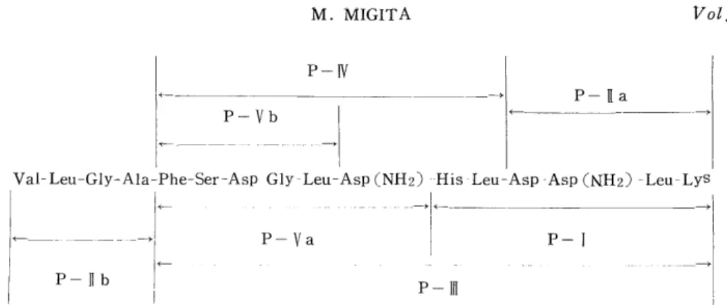
The present paper reports the determination of amino acid sequences of 9T9, 9T13, 8T14, and 9T15 among tryptic peptides from 9-chain in macaca mulatta monkey hemoglobin.
MATERIALS AND METHODS
1) Preparation of Tryptic Peptides
Isolation and purification of the tryptic peptides from 9-chain in macaca mulatta monkey hemoglobin were performed by the methods reported previously by TAKEI22)
According to DRABKIN' S method, 4) hemoglobin was obtained from blood of macaca mulatta monkeys. It was dehemed to obtain globin by TEALE' S method. 23) The globin was separated into a- and ,9-chains by countercurrent distribution methods" by using a system of sec.-butanol containing 0.08% trichloroacetic acid, propionic acid, and water (8.7:
1.8: 11.0). 9-chain was denatured in 8M urea at 60°C for 45 minutes.
After it was dialysed against water to remove urea, it was hydrolyzed
with trypsin at 37°C at pH 8.0 for four hours. This hydrolyzate was
adjusted to pH 6.4. Consequently, the so-called `core' was precipitated,
and then removed by centrifugation. From the remaining soluble
part, the so-called `soluble tryptic peptides' were isolated and purified
by column and paper chromatography. The column chromatography
was developed on Dowex 1 x 2 with acetate buffer containing some
organic bases such as pyridine, collidine, and lutidine. Descending
paper chromatography was employed with the system of n-butanol,
acetic acid, and water (4 : 1 : 5). A portion of each tryptic peptide
thus isolated was subjected to amino acid analysis to assess its quantity and purity.
2) Digestion of the Peptides with Pepsin
A sample of each tryptic peptide (15-20 umole) was dissolved in 15 ml of deionized water and adjusted to pH 2.0 with 1.0 N HC1. To this solution was added 4 mg of pepsin ( Thrice- crystallyzed, Sigma) which was dissolved in 2 ml of 1/16 N HC1. Digestion was performed at 37 °C for the appropriate time and stopped by adding 1.0 N NaOH to the reaction mixture which was consequently brought to pH 9.0.
3) Resolution of the Peptic Peptides by Column Chromatography
The resin (Dowex 1 x 2, 200-400 mesh) was washed with 1N NH4OH, water, acetic acid, and water. It was suspended with the starting buffer, evacuated thoroughly, and filled up in a column (1.0cm x 60cm) .
The column was fully equilibrated again with the starting buffer. The peptic digest of each tryptic peptide was put on the column. The acetate buffer containing some organic bases such as pyridine, collidine, and picoline was used as the starting buffer of the developer.
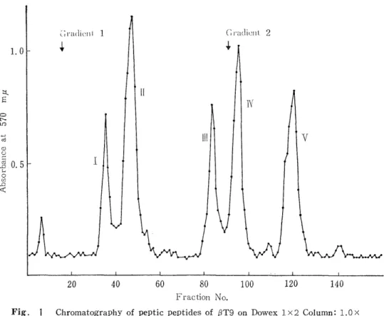
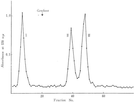
Next, pH gradient was carried out with acetic acid. (see Fig 1, 3, 5) The development was performed at 37°C by keeping a flow rate of 100 ml/h. The effluent was collected into 8 ml fractions by a fraction -collector . A portion of each fraction (0.3 ml) was subjected to ninhy- drin reaction after alkali-hydrolysis, according to YEMM and COCKING'S method.24) The fractions positive to this reaction were combined toge- ther, evaporated to dryness under reduced pressure below 30 C. It was dissolved in deionized water.
4) Identification and Purification of the Peptides by Paper Chromatography
Descending paper chromatography was employed by using Toyo filter paper No. 50. The upper phase of the system of n-butanol, acetic acid, and water (4: 1 : 5) was used as a developer. For the detection of the peptides on the paper chromatogram, 0.2 % ninhydrin-n-butanol solution was sprayed on the paper and heated with an iron to color.
In addition, PAULI's reactionzl) and a-nitrosonaphtol reactions' were employed in order to locate the peptides containing histidine and tyrosine. In case of purification of the peptides with the above-menti- oned system, elution of the peptide from the paper was done by using 5 % acetic acid.
5) Paper Electrophoresis of the da(Jppas
Electrophoresis was carried out at 2 KV for two hours on an electro-
phoretic apparatus (Ishidai-type) by usiu2 Toyo filter paper No. 51 (10
cm x 60cm) and pyridine acetate buffer, pH 6.4 (pyridine : acetic acid :
water= 100: 4 : 900). The peptides were detected by ninhydrin reaction
as in case of the paper chromatography described previously.
6) DNP-Method for the N-Terminal Amino Acid Analysis of the Peptides
Each peptide (0.2-0.5 pmole) was dissolved in 3 ml of 1 % NaHCO3i and to this was added 0.05 ml of dinitrofluorobenzene (DNFB). After it was dinitrophenylated at 40°C for two hours, the excess of DNFB was extracted with ether. The DNP-peptide in the aqueous phase was concentrated to dryness under reduced pressure and redissolved in 4 ml of constant boiling point HC1 and hydrolyzed in sealed tubes at 105°C for 24 hours. The N-terminal DNP-amino acid was extracted three times from the hydrolyzate with 10 ml of ether. This ether extract was transferred into a heart-shaped flask (MILLS' apparatus)10' in order to evaporate to dryness under reduced pressure. Then the flask was connected to the coldfinger, and the content was evacuated by a vacuum pump in order to remove as much dinitrophenol as possible.
Identification of the DNP-amino acids was carried out by ascending paper chromatography on Toyo filter paper No. 51 (40cm x4Ocm). For the first dimension, the developer was the upper phae of an n-butanol- 1 N NH4OH (1 : 1) mixture; the second dimension, 0.5 M phosphate buffer. Yellow spots on the paper were cut out and and eluted with 5 ml of warm water. The quantitative determination was performed
by measurement of the optical density at 360 mu of this eluate.
7) PTC Method for Amino Acid Sequence Analysis of the Peptides
EDMAN'S original method' '7' was modified for phenylisothiocyanation of the peptides. The ring-formation was performed with trifluoroacetic
acid. 11) Analysis was carried out by the elimination methods' by comparing the amino acid compositions of the PTC-peptidos with the original peptides.
A sample of each peptide (0.5-2.0 umole) was dissolved in 2.5 ml of 66 % pyridine in a heart-shaped flask and to this was added 0.05 ml of phenylisothiocyanate (PTC). The PTC reaction was performed at 37°C for three hours after adjusting the reaction mixture ti pH 7.5. The reaction mixture was concentrated to dryness under reduced pressure.
The excess PTC was removed by 30 minute aspiration with a vacuum pump by connecting the flask to the cold finger. To this was added 1 ml of trifluoroacetic acid in order to occur the ring-formation. This was allowed to stand at room temperature for four hours. After the reaction, trifluoroacetic acid was removed under reduced pressure.
The dry residue was dissolvod in 3 ml of deionized water and extracted with 3 ml of benzene three times. Consequently, the PTH-amino acids were extracted in the benzene phase.
The original peptide lost its N-terminal residue and remained in the aqueous phase. An aliquot of the aqueous phase was concentrated to dryness after HCl-hydrolysis and subjected to amino acid analysis.
The remainder of the aqueous phase after the concentration to dryness
was forr the following stage of the PTC method.
8) Amino Acid Analysis of the Peptides
Each peptide was dissolved in 4 ml of constant boiling point HC1 (twice distilled)and was hydrolyzed in sealed tubes at 105°C for 24 hours.
The HCl was removed from the hydrolyzate by repeating dissolution in water and concentration to dryness. The amino acid analysis was carried out on Hitachi KLA-2 amino acid analyzer. The losses of the amino acids during the hydrolysis were not corrected and the composi- tions were indicated by molecular ratios of the amino acids.
RESULTS AND DISCUSSION
1) Preparation of the tryptic peptides 1T9, 8T13, 1T 14, and 1 T J5
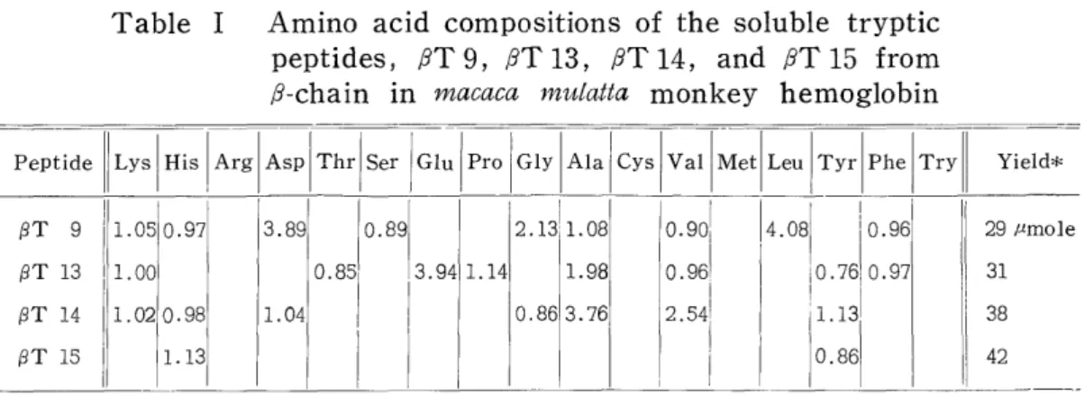
Table I shows the amino acid compositions and. the yields of i T9, /9T 13, PST 14, and PPT 15 which were obtained by paper and column chromatographies from the tryptic hydrolyzate of 1 g of p-chain in macaca mulatta monkey hemoglobin.
Table I Amino acid compositions of the soluble tryptic peptides, j9T 9, iT 13, iT 14, and RT 15 from
,8-chain in macaca mulatta monkey hemoglobin
Peptide Lys His Arg Asp Thr Ser I Glu Pro Gly Ala Cys Val Met Leu Tyr Phe Try Yield*
(3T 9 1.05 0.97 3.89 0.89 2.1311. 08 0.901 4.08 j 0.96 29 mmole
PT 13 1.00 0.85' 3.94 1.14 1.98 0.961 I0.761 0.97 31
i


![Fig. 6 Paper chromatography of peptic peptides of (3T]4 n-butanol : acetic acid: water (4 : 1 : 5)](https://thumb-ap.123doks.com/thumbv2/123deta/10140017.1973887/13.766.224.551.91.572/fig-paper-chromatography-peptic-peptides-butanol-acetic-water.webp)
