N
- ac et yl gal ac t os am
i nyl t r ans f er as e- 1 and - 2
著者
Shi m
bo M
i ki , Suz uki Ri ku, Fus eya Sayaka, Sat o
Takas hi , Ki yohar a Kat s ue, H
agi w
ar a Koz ue,
O
kada Ri s a, W
akui H
i r om
as a, Ts unakaw
a Yuki ,
W
at anabe H
i det o, Ki m
at a Koj i , N
ar i m
at s u
H
i s as hi , Kudo Takas hi , Takahas hi Sat or u
j our nal or
publ i c at i on t i t l e
PLO
S O
N
E
vol um
e
12
num
ber
12
page r ange
e0190333
year
2017- 12
権利
( C) 2017 Shi m
bo et al .
Thi s i s an open ac c es s ar t i c l e di s t r i but ed
under t he t er m
s of t he Cr eat i ve Com
m
ons
At t r i but i on Li c ens e, w
hi c h per m
i t s unr es t r i c t e
d us e, di s t r i bu t i on, and r epr oduc t i on i n any
m
edi um
, pr ovi ded t he or i gi nal aut hor and
s our c e ar e c r edi t ed.
U
RL
ht t p: / / hdl . handl e. net / 2241/ 00151185
Postnatal lethality and chondrodysplasia in
mice lacking both chondroitin sulfate
N
-acetylgalactosaminyltransferase-1 and -2
Miki Shimbo1,2☯, Riku Suzuki1,3☯, Sayaka Fuseya1,4☯, Takashi Sato5, Katsue Kiyohara5, Kozue Hagiwara5, Risa Okada1, Hiromasa Wakui1,4, Yuki Tsunakawa1,3, Hideto Watanabe6, Koji Kimata7, Hisashi Narimatsu5, Takashi Kudo1,8*, Satoru Takahashi1,8,9*
1Department of Anatomy and Embryology, Faculty of Medicine, University of Tsukuba, Tsukuba, Ibaraki, Japan,2Doctoral Program in Biomedical Sciences, Graduate School of Comprehensive Human Sciences, University of Tsukuba, Tsukuba, Ibaraki, Japan,3Ph.D. Program in Human Biology, School of Integrative and Global Majors, University of Tsukuba, Tsukuba, Ibaraki, Japan,4Master’s Program in Medical Sciences, Graduate School of Comprehensive Human Sciences, University of Tsukuba, Tsukuba, Ibaraki, Japan,
5Glycoscience and Glycotechnology Research Group, Biotechnology Research Institute for Drug Discovery, National Institute of Advanced Industrial Science and Technology (AIST), Tsukuba, Ibaraki, Japan,6Institute for Molecular Science of Medicine, Aichi, Japan,7Multidisciplinary Pain Center, Aichi Medical University, Aichi, Japan,8Laboratory Animal Resource Center (LARC), University of Tsukuba, Tsukuba, Ibaraki, Japan,
9Transborder Medical Research Center, Faculty of Medicine, University of Tsukuba, Tsukuba, Ibaraki, Japan
☯These authors contributed equally to this work.
*[email protected](TK);[email protected](ST)
Abstract
Chondroitin sulfate (CS) is a sulfated glycosaminoglycan (GAG) chain. In cartilage, CS plays important roles as the main component of the extracellular matrix (ECM), existing as side chains of the major cartilage proteoglycan, aggrecan. Six glycosyltransferases are known to coordinately synthesize the backbone structure of CS; however, theirin vivo syn-thetic mechanism remains unknown. Previous studies have suggested that two glycosyl-transferases, Csgalnact1 (t1) and Csgalnact2 (t2), are critical for initiation of CS synthesisin vitro. Indeed,t1single knockout mice (t1KO) exhibit slight dwarfism and a reduction in CS content in cartilage compared with wild-type (WT) mice. To reveal the synergetic roles of t1 and t2 in CS synthesisin vivo, we generated systemic single and double knockout (DKO) mice and cartilage-specifict1andt2double knockout (Col2-DKO) mice. DKO mice exhibited postnatal lethality, whereast2KO mice showed normal size and skeletal development. Col2-DKO mice survived to adulthood and showed severe dwarfism compared witht1KO mice. Histological analysis of epiphyseal cartilage from Col2-DKO mice revealed disrupted endochondral ossification, characterized by drastic GAG reduction in the ECM. Moreover, DKO cartilage had reduced chondrocyte proliferation and an increased number of apoptotic chondrocytes compared with WT cartilage. Conversely, primary chondrocyte cultures from Col2-DKO knee cartilage had the same proliferation rate as WT chondrocytes and low GAG expression levels, indicating that the chondrocytes themselves had an intact proliferative ability. Quantitative RT-PCR analysis of E18.5 cartilage showed that the expression levels ofCol2a1 andPtch1transcripts tended to decrease in DKO compared with those in WT mice. The CS content in DKO cartilage was decreased compared with that int1KO cartilage
a1111111111 a1111111111 a1111111111 a1111111111 a1111111111 OPEN ACCESS
Citation:Shimbo M, Suzuki R, Fuseya S, Sato T, Kiyohara K, Hagiwara K, et al. (2017) Postnatal lethality and chondrodysplasia in mice lacking both chondroitin sulfateN
-acetylgalactosaminyltransferase-1 and -2. PLoS ONE 12(12): e0190333.https://doi.org/10.1371/ journal.pone.0190333
Editor:Jung-Eun Kim, Kyungpook National University School of Medicine, REPUBLIC OF KOREA
Received:September 29, 2017
Accepted:December 12, 2017
Published:December 29, 2017
Copyright:©2017 Shimbo et al. This is an open access article distributed under the terms of the
Creative Commons Attribution License, which permits unrestricted use, distribution, and reproduction in any medium, provided the original author and source are credited.
Data Availability Statement:All relevant data are within the paper and its Supporting Information files.
Funding:This work was performed as a part of the “Medical Glycomics (MG) Project” supported by the New Energy and Industrial Technology Development Organization (NEDO).
but was not completely absent. These results suggest that aberrant ECM caused by CS reduction disrupted endochondral ossification. Overall, we propose that both t1 and t2 are necessary for CS synthesis and normal chondrocyte differentiation but are not sufficient for all CS synthesis in cartilage.
Introduction
Chondroitin sulfate (CS) is a long-linear glycosaminoglycan (GAG) chain consisting of repeat-ing sulfated disaccharide units ofN-acetylgalactosamine (GalNAc) and glucuronic acid (GlcUA) that is covalently attached to core proteins via a linkage region to form a proteoglycan (PG). Depending on the type of core proteins, chondroitin sulfate proteoglycans (CSPGs) can be cell membrane-bound or part of the extracellular matrix (ECM), are particularly abundant in cartilage and the brain, and play an important role in development and homeostasis. Aggre-can is a large CSPG comprising approximately 100 CS chains and residing in the ECM. It con-tributes to water retention and the compression resistance of cartilage by aggregating with hyaluronan and link protein 1 [1]. In brain ECM, various CSPGs, including aggrecan, versican, and neurocan, form a structure called a perineuronal net that surrounds neuronal cell bodies to regulate their plasticity and activity [2–4]. Not only core proteins but also CS chains have indispensable roles in CSPG function. Among the functions, glycosylation prevents the core protein from degrading [5], and we previously reported that reduced CS chains accelerate core protein degradation of cartilage aggrecan [6]. Furthermore, CS shows diversity in sulfation patterning, exerting effects on cortical layer formation and axon guidance of retinal growth cones [7,8]. Although the mechanism of CS recognition by other proteins remains unknown, previous studies revealed that cytokines and growth factors, such as midkine and pleiotrophin [9], as well as receptors, including transmembrane protein tyrosine phosphatase (PTPσ) and contactin-1 [10–12], bind to CS chains.
Although the CS glycosyltransferase activities have been well studiedin vitro, their function in vivoremains unknown. To examine this, CS glycosyltransferase knockout (KO) mice were generated and analyzed.Css2KO mice showed no morphological phenotypes; however, their
Fig 1. Phenotype oft2null mice.(A) Schematic showing the CS biosynthetic pathway and relevant glycosyltransferases. Arrows indicate the catalytic activity of each glycosyltransferase. Half-filled diamonds, open squares, open circles, and stars refer to GlcA, GalNAc, Gal, and Xyl, respectively. (B) Quantitative analysis oft1andt2gene transcription in humeral cartilage of WT mice (n = 3) using real-time RT-PCR. The expression of each transcript was normalized to that ofβ-actin. (C) Targeting strategy for conditional deletion of thet2gene. The exon containing the initiation codon and transmembrane domain was flanked by loxP elements. This region was deleted via mating with Ayu1-Cre mice, to generate systemict2KO mice. Probe position for Southern hybridization is indicated by the bold line. (D) Southern blot analysis ofBglII-digested genomic DNA and PCR-based genotyping of progeny from intercrossing heterozygotes. (E) Postnatal growth kinetics of WT mice (male, n = 3; female, n = 6) andt2KO littermates (male, n = 11; female, n = 5).F-H, Double whole body staining with Alizarin red and Alcian blue (F), upper (G) and lower limbs (H) of the WT and
t2KO littermates at E18.5. Scale bars: 1 cm (F) and 1 mm (G,H).
CS chain length in cartilage was significantly shorter than that of wild-type (WT) mice [21], suggesting that Css2 has elongation activityin vivo, similar to that observed within vitro stud-ies. Conversely,Css1KO mice displayed skeletal phenotypes including abnormal digit pattern-ing and endochondral ossification, and their CS sulfation patterns in cartilage were altered [21], suggesting that Css1 may regulate sulfationin vivo, which was not shownin vitro. With regard to this,t1KO mice also showed abnormal endochondral ossification, which resulted in slight dwarfism and impaired aggrecan metabolism [6,22]. Their CS showed no difference in chain length or sulfation patterns, but the number of CS chains decreased by approximately half. These results not only replicated thein vitroresults of t1 initiation activityin vivobut also suggested the possibility that CS glycosyltransferases other than t1 have initiation activity, which raises the hypothesis that t1 and t2 both regulate the initiation activityin vivo.
In the current study, we generated and characterizedCsgalnact1::Csgalnact2double KO (DKO) mice. DKO mice exhibited postnatal lethality due to respiratory failure and mild dwarfism and their cartilage showed more severe abnormality of endochondral ossification due to reduction of CS than cartilage fromt1KO mice. These observations suggest that CS synthesis is not enough for t1 and t2 and the decreased CS in cartilage causes abnormal chondrocyte function.
Materials and methods
Animals
Mice were maintained under specific pathogen-free conditions at the Laboratory Animal Resource Center of the University of Tsukuba. All experiments were approved by the institutional animal care and use committees of the University of Tsukuba (No. 16–102 and 17–106) and were con-ducted according to related guidelines and the applicable laws of Japan. Mouse cages were placed in an air-conditioned room (average temperature; 24.1˚C, average relative humidity; 42.8%RH) with a 12:12-h light-dark cycle. The mice were fed with CRF-1 (Oriental Yeast Co., Ltd., Tokyo, Japan) and given waterad libitum. The drinking water was autoclaved tap water. We checked the health condition of the mice every day, and unexpected deaths were very rare. The humane end-points that we used were behavioral changes and 20% weight loss. All mice were euthanized via the inhalation of lethal doses of isoflurane and then subjected to dissection to collect tissue samples.
Generation of mice with genetically modified
t1
and
t2
genes
The generation oft1KO mice and mice carrying the floxedt1gene was described in a previous report by our group [6]. To distinguish thet1+,t1-, andt1floxalleles, the genotypes of the mice were confirmed via PCR using the following 3 primers:5’-TAGATGAACTGTCCATCCTAC
AG-3’,5’-GAGACGGCTCTCTTGCTTCCAAGG-3’, and5’-CCTTTACTAAAATGGCGAC
CTGCC-3’.
To generate chondrocyte-specific conditionalt1andt2KO mice,t1flox/-::t2flox/-mice were further crossed with transgenic mice expressing Cre recombinase under the collagen2a1 pro-moter (Col2-cre (B6;SJL-Tg(Col2a1-cre)1Bhr/J), Jackson Laboratory, Bar Harbor, ME, USA). The primers used for the genotyping of Col2-cre were5’-GGACATGTTCAGGGATCGCCA
GGCGT-3’and5’-GCATAACCAGTGAAACAGCATTGCTG-3’.
Quantitative analysis of six CS glycosyltransferase transcripts using
real-time RT-PCR
Total RNA was isolated from the humeral cartilage of embryonic day 18.5 (E18.5) embryos using Isogen (Nippon Gene, Tokyo, Japan), and cDNA templates were synthesized from the total RNA with a QuantiTect Reverse Transcription Kit (QIAGEN, Venlo, the Netherlands). The primers and probes selected from TaqMan Gene Expression Assays (Applied Biosystems, Foster City, CA, USA) and the primers used with the THUNDERBIRD SYBR qPCR system (Toyobo, Osaka, Japan) are listed inS3 Table. PCR products were continuously measured with a 7500 Fast Real-Time PCR System (Applied Biosystems). Relative transcript levels were nor-malized to the amount of theβ-actintranscript in the same cDNA sample.
Skeletal histology
The whole skeleton of E18.5 embryos was fixed in 95% ethanol and stained overnight in a solu-tion containing Alcian blue 8GX (Sigma-Aldrich, St. Louis, MO, USA). Samples were placed in 1% KOH (vol/vol)/Alizarin red S (Sigma-Aldrich) for 2 h and then cleared with 2%-0.2% KOH (vol/vol)/20-80% glycerol for several days. To measure bone length, isolated humeri and tibiae were photographed with a digital microscope (VH-8000; KEYENCE, Osaka, Japan) and then measured with ImageJ software (freeware; National Institute of Health, USA).
Histological and immunohistochemical analyses
Structural analysis of CS chains
Rib cartilage was obtained from E18.5 mice after the administration of general anesthesia, and the cartilage of 4 to 5 mice of each genotype was then mixed. Unsaturated disaccharides in the filtrates were analyzed according to Toyoda’s method [25]. Briefly, freeze-dried rib cartilage was digested with 1 mg/ml Pronase (Calbiochem, CA, USA) for 3 h at 60˚C and then filtered through a 0.45-μm membrane. The obtained filtrates were digested with ChaseABC and Chas-eAC-II for 2 h at 37˚C, and the mixture was then ultrafiltered using a centrifugal ultrafiltration tube. The unsaturated disaccharides derived from CS in rib cartilage were analyzed via HPLC.
Ki67 and TUNEL staining
For antigen retrieval, sections of the proximal tibial epiphysis were heated in 10 mM citrate buffer (pH 6.0) in an autoclave at 115˚C for 10 min. After blocking with 10% normal goat serum/0.1% Tween 20 in PBS for 1 h at room temperature, the sections were stained with an anti-Ki67 antibody (cat. no. NCL-Ki67p, 1:100; Novacastra, UK) overnight at 4˚C. Detection was achieved using an Alexa Fluor 488-conjugated secondary antibody (Molecular Probes). The sections were subsequently incubated for 5 min with 0.1% Hoechst 33258 to stain nuclei. The number of Ki67-positive cells was calculated using Dynamic Cell Count BZ-H1C software (KEYENCE). Fluorescent TUNEL assays were performed with a DeadEndTMFluorometric TUNEL System (Promega, Fitchburg, WI, USA) according to the manufacturer’s protocol. Images were captured using a BIOREVO BZ-9000 microscope.
Chondrocyte isolation and primary chondrocyte culture
Chondrocytes from mouse cartilage were isolated and cultured according to a protocol described in a previous report [26]. Briefly, knee cartilage was isolated from E18.5 embryos and digested via two incubations in 3 mg/ml collagenase D (Clostridium histolyticum; Roche, Switzerland) in low-glucose Dulbecco’s modified Eagle’s medium (DMEM). Incubation was conducted under a 5% CO2atmosphere in a humidified incubator at 37˚C for 45 min. After the cartilage was additionally incubated overnight with 0.5 mg/ml collagenase D/DMEM, the digested chondrocytes were completely separated via pipetting, followed by filtration through a cell strainer with a 0.4-μm pore size (Greiner Bio-One, Austria). Primary chondrocytes were seeded in a 96-well plate, with 8×10³cells per well, and cultured in DMEM containing 10% fetal bovine serum. Cell proliferation was assessed using a Cell Counting Kit-8 (Dojindo, Kumamoto, Japan) according to the manufacturer’s instructions, and absorbance values at 450 nm were read with a microplate reader. To quantify GAG production, Alcian blue staining was performed after seven days of culture as described in a previous study [26]. Primary chon-drocytes were fixed in Mildform 10 N at room temperature for 15 min, followed by washes with 0.1 N HCl. Sulfated GAG was detected with 1% (w/v) Alcian Blue 8GX in 0.1 N HCl at room temperature for 1 h. After two rinses with 0.1 N HCl, Alcian blue dye was extracted with 6 M guanidine hydrochloride, and absorbance values were quantified at 595 nm using a micro-plate reader.
In situ
hybridization
mix (Roche). Hind limbs were fixed in 4% PFA/PBS at 4˚C overnight, then maintained in 0.5 M sucrose/PBS at 4˚C for cytoprotection and subsequently embedded in O.C.T. compound. Sections with a size of 20μm treated with 0.5μg/ml proteinase K (Wako)/0.1% Tween 20 in PBS at 37˚C for 10 min and then post-fixed in 4% PFA/PBS for 10 min and rinsed in PBS. Hybridization with DIG-labeled riboprobes was conducted overnight at 55˚C in 50% formam-ide, 5×Denhardt’s solution, 5×SSC, and 50μg/ml yeast tRNA and then washed with 5×SSC and 2×SSC at 55˚C for 15 min each. After washing, the slides were digested with RNase A (20μg/ml; Sigma-Aldrich) in 0.5 M NaCl/10 mM Tris-HCl pH 7.5/0.1% Tween 20 for 30 min at 37˚C. Next, the slides were washed twice with 2×SSC at 42˚C for 20 min. After incubation with an alkaline phosphatase-conjugated anti-DIG antibody (Roche), DIG-labeled RNA duplexes were detected with BM purple (Roche).
Statistical analysis
The results are reported as the mean±SEM. Statistical analyses were performed using ANOVA or Student’st-test.Pvalues are provided in the figure legends and are indicated by asterisks within the figures.
Results
t2
KO mice show normal skeletal development
Previous reports, including those from our group, have shown thatt1KO mice exhibit slight dwarfism and an approximately 50% decrease in the CS content of cartilage compared with WT mice [6,22]. The gene expression level oft2, which is another initiation enzymein vitro, was several times higher than that oft1in mouse humeral cartilage at E18.5 (Fig 1B). To fur-ther expand this finding, we hypothesized that t2 may have initiation activityin vivoas well. To investigate this, we generatedt2KO mice (Fig 1C), which intriguingly showed normal development, fertility, and growth rates (Fig 1E) compared with WT mice. Analysis of the off-spring resulting from crosses amongt2heterozygotes revealed a Mendelian distribution of WT, heterozygous, and homozygous offspring (S1 Table). In addition, whole body skeletal preparation double-stained with Alizarin red and Alcian blue showed no size differences or skeletal deformities (Fig 1F and 1H).
t1
::
t2
double KO mice show severe dwarfism and postnatal lethality
humeral and tibial lengths of DKO mice were significantly reduced by 22% compared with those oft2KO mice (humerus;t2KO: 3.93±0.04 mm, n = 5, DKO: 3.07±0.02 mm, n = 10, tibia;t2KO: 3.64±0.04 mm, n = 5, DKO: 2.81±0.02 mm, n = 10) (Fig 2I). Previous data indi-cated there was no difference in bone length between WT andt1KO embryos. Therefore, our DKO embryo showed a severe phenotype compared witht1KO embryos, signifying the neces-sity of t2 in skeletal development processes.
Chondrocyte-specific
t1
::
t2
double KO mice show phenotypes similar to
DKO mice, with a number of surviving progeny
Normal cartilage development is indispensable for bone elongation, which was abnormal in DKO mice. CS is widely known to be expressed in cartilage andt1KO andCss1KO mice were
Fig 2. Phenotype of DKO mice.(A) Representative photograph oft2KO pups and cyanotic DKO littermates at E18.5. Scale bar: 1 cm. (B) Body weight oft2KO (n = 68) and DKO (n = 51) embryos at E18.5.*P<0.05. (C) Lung sections fromt2KO embryos and DKO littermates were stained with HE. Scale bar: 100μm. (D-H) Skeletal whole body (D), upper limb (E), lower limb (F), lumber spine (G), and cranial bone (H) preparation. (I) Humeral and tibial bone length at E18.5 oft2KO (n = 5) and DKO (n = 10) mice.**P<0.01.
reported to exhibit a deformed skeletal system as a result of abnormal cartilage development [6,22,27]. To investigate the cartilage-specific function of t1 and t2, we generated chondrocyte-specifict1::t2double KO mice (Col2-DKO) usingCollagen2a1-Cre mice [28]. Since heterozy-gous KO oft1ort2showed the same phenotype, WT,t1+/-::t2+/+,t1+/+::t2+/-, andt1+/-::t2 +/-mice were used as control groups (Control). E18.5 embryos of Control +/-mice started spontane-ous respiration after cesarean section (Fig 3A). Forty-three Col2-DKO mice among 53 imme-diately died of respiratory failure after birth. However, the remaining 10 Col2-DKO mice started spontaneous respiration. Col2-DKO mice rarely survived to adulthood and died by P14 in most cases. Although Col2-DKO mice showed the same body weight as Control mice at E18.5, they showed significantly (50%) lower body weight than Control mice and severe dwarf-ism at P7 (Control: 3.80±0.15 g, n = 4, Col2-DKO: 2.40±0.22 g, n = 4) and P14 (Control: 7.70±0.30 g, n = 4, Col2-DKO: 3.80±0.15 g, n = 4) (Fig 3B). At E18.5, when body weight showed no difference between Col2-DKO and Control mice, skeletal preparation of humeri and tibiae from Col2-DKO mice were shorter than those of Control mice (humerus; Control: 4.06±0.09 mm, n = 6, Col2-DKO: 3.28±0.08 mm, n = 7, tibia; Control: 3.72±0.09 mm, n = 6, Col2-DKO: 2.97±0.08 mm, n = 7) (Fig 3C). The quantitative RT-PCR analysis of knee cartilage showed no or slightt1andt2gene expression in Col2-DKO mice compared with
Fig 3. Phenotype of Col2-DKO mice.(A) Representative photograph of Control pups and Col2-DKO littermates at E18.5, P7, and P14. Scale bar: 1 cm. (B) Body weight of Control (E18.5; n = 16, P7; n = 4, P14; n = 4) and Col2-DKO (E18.5; n = 12, P7; n = 4, P14; n = 4) mice at various stages.*P<0.05,**P<0.01. (C) Humeral and tibial bone length at E18.5 of Control (n = 6) and Col2-DKO (n = 7) mice. (D) Quantitative analysis oft1andt2transcripts in the knee cartilage of WT (n = 3) and Col2-DKO (n = 3) mice at E18.5 using real-time RT-PCR. The expression of each transcript was normalized to that ofβ-actin.**P<0.01.
those of WT mice, and thus, chondrocyte-specifict1::t2KO was confirmed (Fig 3D). These results indicate that deficiency of both t1 and t2 in chondrocytes not only affects endochondral ossification during the fetal period but also influences bone growth after birth.
Impaired CS content in cartilage and induction of abnormal
endochondral ossification by
t1
::
t2
double deficiency
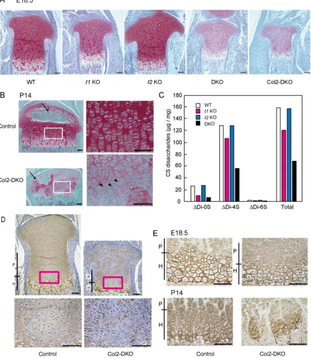
Since t1 and t2 function in cartilage is crucial for skeletal development, we compared CS content in each genotype of mice including WT,t1KO,t2KO, and DKO at E18.5 by performing histo-logical analysis of the proximal tibial growth plate with Safranin-O staining. Safranin-O is a red stain that detects the GAG content of cartilage ECM. To verify the GAG that we are detecting, we treated the samples with chondroitinase ABC, a CS- and hyaluronic acid (HA)-specific diges-tive enzyme, because it is possible that Safranin-O staining detects GAGs other than CS, such as heparan sulfate. Safranin-O staining of chondroitinase ABC-treated samples diminished, indi-cating that the Safranin-O staining in this condition was certainly CS and HA derived from car-tilage ECM (S1 Fig). Once the specificity of the Safranin-O staining was confirmed in WT mice, other genotypes were also stained with Safranin-O, and the staining intensity was similar in both t2KO and WT mice (Fig 4A). For the case of t1KO mice, a slight decrease in intensity compared with that in WT mice was observed. The staining intensity revealed that the largest difference when comparing DKO mice to WT mice was a weaker intensity compared witht1KO mice. Structural organization of the proliferative and hypertrophic zones of the DKO mice did not show any significant changes compared with WT mice; however, the size of their epiphysis was smaller than that of WT mice (Fig 4B). A drastic decrease in Safranin-O staining was also noticed in Col2-DKO mice, which exhibit phenotypes similar to those of the DKO mice men-tioned above. These results indicate that the existence of GAG is correlated with the skeletal phe-notype, where a lower amount of CS leads to a more severe defect in skeletal development.
Fig 4. Safranin-O staining, CS content, and immunohistochemical analyses in cartilage.(A) Safranin-O staining in E18.5 proximal tibial cartilage of WT,t1KO,t2KO, DKO, and Col2-DKO mice. (B) Safranin-O staining in P14 tibial cartilage of Control and Col2-DKO mice. Arrows indicate secondary ossification centers. Each right panel is a higher magnification image of the regions labeled with white squares in the left panel. Black arrowheads indicate abnormal shape and cell layer structure of the growth plate in Col2-DKO mice. White arrowheads indicate ectopic localization of proliferating chondrocytes in the hypertrophic zone. (C) The total amount and disaccharide analysis of the rib cartilage in WT,t1KO,t2
KO and DKO mice.Δdi-0S,ΔHexAα1-3GalNAc;Δdi-4S,ΔHexAα1-3GalNAc(4S);Δdi-6S,ΔHexAα1-3GalNAc(6S). (D) Immunohistochemical aggrecan staining using 1C6 monoclonal antibody. The sections from Control and Col2-DKO mice were used for staining after chondroitinase ABC treatment. Each lower panel shows a higher magnification image of the regions labeled by magenta squares in the upper panel. Scale bar: 100μm. (E) Immunostaining with an antibody against type X collagen in Control and Col2-DKO cartilage at E18.5 and P14. P, Proliferative chondrocyte; H, Hypertrophic chondrocyte. Scale bar: 100μm.
showed uniform aggrecan expression in cartilage ECM in growth plates. In contrast, most por-tions of the Col2-DKO ECM were revealed to be aggrecan-negative, and scarce aggrecan-posi-tive regions were restricted to the periphery of the cartilage lacunae (Fig 4D). These findings indicate that CS loss induced byt1andt2KO disturbs aggrecan core protein distribution throughout the cartilage ECM. To assess the differentiation stages of chondrocytes, we next performed immunohistochemical staining of collagen X, a marker for hypertrophic chon-drocytes, using tibial growth plates at E18.5 and P14. No difference was observed between Col2-DKO and WT mice at E18.5. At P14, collagen X expression was distributed irregularly in Col2-DKO mice, whereas a uniformly stained hypertrophic zone was seen in WT mice (Fig 4E). These results suggest that both t1 and t2 function in chondrocytes are required for CS syn-thesisin vivo, and their loss induces abnormal endochondral ossification, including disrupted growth plate structure and differentiation stages.
Cartilage-specific
t1
::
t2
double deficiency induces reduced proliferation
and increased apoptosis of chondrocytes
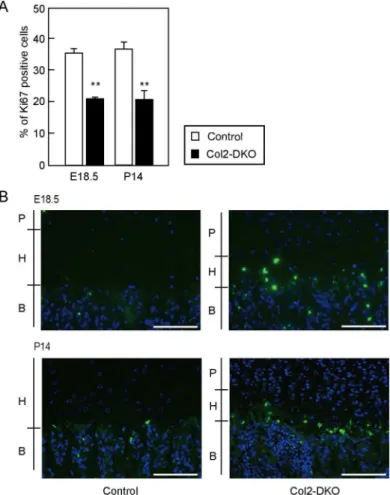
Abnormal endochondral ossification and reduced epiphyseal size, which were found in Col2-DKO mice, often reflect alterations in the balance of proliferation versus apoptosis. To test this, we performed immunohistochemical staining of Ki67, a marker of proliferating cells, and terminal dUTP nick-end labeling (TUNEL) staining using tibial growth plates at E18.5 and P14. Col2-DKO mice displayed an approximately 50% reduction in the percentage of Ki67-positive cells in the proliferative zone compared with WT mice at both E18.5 (Control: 35.4±1.3%, n = 4, Col2-DKO: 19.7±1.6%, n = 3,P<0.001) and P14 (Control: 36.6±2.5%, n = 3, Col2-DKO: 21.1±2.7%, n = 6,P<0.001) (Fig 5A). When apoptosis was examined with TUNEL staining, Col2-DKO mice displayed increased apoptosis in the hypertrophic zone at both E18.5 and P14, while Control mice exhibited moderate apoptosis (Fig 5B). These results indicate that the CS reduction caused byt1andt2KO leads to decreased proliferation and accelerated apoptosis in chondrocytes, contributing to the disrupted endochondral ossification and morphology of the growth plate.
Characterization of primary chondrocyte culture from Col2-DKO mice
The above findings further led us to the hypothesis that disrupted aggrecan causes a disordered ECM environment, resulting in abnormal endochondral ossification in Col2-DKO mice. To test this, primary chondrocytes from knee and femur cartilage at E18.5 were cultured, and their ability to respond to exogenous factors was examined. The accumulation of sulfated GAG in primary chondrocyte cultures was quantified through Alcian blue staining, and the amount of sulfated GAG was found to show a significant decrease of approximately 60% in Col2-DKO primary chondrocyte cultures compared with WT mouse chondrocytes (WT: 0.047±0.004, n = 3, Col2-DKO: 0.028±0.004, n = 3) (Fig 6A and 6B). Growth rate measure-ments of these chondrocyte cultures revealed no difference between Col2-DKO and WT cells (Fig 6C). These results show that although the CS amount is reduced in Col2-DKO mice, their chondrocytes can respond to extracellular cues.
Quantitative RT-PCR and
in situ
hybridization analyses of humeral
cartilage
collagen type IIα1 chain (Col2a1) int1KO andt2KO mice was found to be similar to that in WT mice (Fig 7A), but the expression ofCol2a1was prone to decrease in DKO cartilage com-pared with WT cartilage (Acan(F[3,23]= 0.75,P= 0.53),Col2a1(F[3,23]= 2.79,P= 0.06)). However,in situhybridization showed that the distribution ofAcanandCol2a1transcripts in t1KO,t2KO or DKO tibial cartilage was also similar to that in WT cartilage (Fig 7B). The transcripts of Indian hedgehog (Ihh) and one of the hedgehog target genes,Ptch1, were also expressed at low levels in DKO cartilage compared with WT cartilage, but these differences were not statistically significant (Ihh(F[3,23]= 1.38,P= 0.27),Ptch1(F[3,23]= 2.75,P= 0.07)). Moreover, the distribution ofIhhandPtch1transcripts was normal in DKO cartilage. When comparisons between WT and DKO were performed using Student’st-test, the expression of Col2a1andPtch1was found to be significantly decreased in DKO mice. To clarify whether other CS synthases can compensate for a loss int1,t2or both genes, the expression of six CS synthase transcripts in WT,t1KO,t2KO, and DKO humeral cartilage at E18.5 was measured using real-time RT-PCR (S2 Fig). While complete loss was observed int1expression int1KO and DKO ort2expression int2KO and DKO,t1expression int2KO ort2expression int1 KO did not change compared with that in WT. No change was observedCss1,Css2,Css3and Csglcatgene expression between each mouse genotype.
Fig 5. Cell proliferation and apoptosis in proximal tibial epiphyseal cartilage.(A) Quantification of cell proliferation assessed by Ki67 immunostaining in tibia sections from Control (E18.5; n = 4, P14; n = 3) and Col2-DKO (E18.5; n = 3, P14; n = 6) mice at P14.**P<0.01. (B) TUNEL staining of tibia sections from Control (n = 3) and Col2-DKO (n = 3) mice at E18.5 and P14. P, Proliferative chondrocyte; H, Hypertrophic chondrocyte; B, Bone. Scale bar: 100μm.
Discussion
In this study, we generated and analyzed mice lackingt2or botht1::t2gene expression to eluci-date the role of CS in cartilage. Previously,t1KO mice were found to exhibit slight dwarfism due to minor impairment of endochondral ossification and reduction of cartilaginous CS content [6]. According to anin vitroenzymatic characterization, among the six CS glycosyl-transferases, only t1 and t2 possessed the ability to independently initiate CS synthesis. We previously reported that t1 efficiently transfers GalNAc onto the linkage tetrasaccharidein vitro, which is common to both CS and heparin sulfate/heparin, and t1 initiation activity is stronger than that of t2 [15,17].t1KO mice demonstrated a similar or slightly increased CS chain length, although the CS levels of CS int1KO cartilage decreased to ~50% those in nor-mal mouse cartilage [6]. The remaining CS contents int1KO cartilage are predicted to be syn-thesized by other CS glycosyltransferases. t2 is most likely to possess initiation activity for CS synthesisin vivoand predicted to affect endochondral ossification as well as t1. However,t2
Fig 6. GAG accumulation and cell proliferation in primary chondrocyte cultures.(A) Proliferation of WT (n = 3) and Col2-DKO (n = 3) chondrocytes. (B) Primary chondrocytes from knee cartilage of WT and Col2-DKO mice at E18.5 were stained for GAG accumulation using Alcian blue at culture day 7. (C) The absorption intensity at 595 nm of Alcian blue-stained cell extracts in WT (n = 3) and Col2-DKO (n = 3) mice was measured to quantify sulfated GAG accumulation.*P<0.05.
KO mice showed no apparent structural cartilage abnormality. Although Izumikawaet al. [29] demonstrated the proportion of linkage region saccharide int1KO cartilage was changed compared with that of WT, it was not changed between WT andt2KO mice. These results suggest that t1 has higher specific activity toward initiation of CS synthesis than t2in vivo. Therefore, the CS synthesis ability of t2 might almost be compensated for by t1, but t1 has suf-ficient CS synthetic ability even if t2 is absent. Takeuchi et al. reported that theirt1KO exhib-ited reduced scar formation after spinal cord compression injury [30]. They also generatedt2 KO mice, which showed normal CS production in areas with glial scars. Their study described a spinal cord-specifict2KO phenotype but did not mention cartilage development. These data suggest that t2 does not individually influence CS biosynthesis in cartilagein vivo. On the other hand, t2 has elongation activity and regulates CS chain length [31]. However, our data showed that although the CS content was exactly the same between WT andt2KO cartilage, the CS content in DKO cartilage was less than that int1KO cartilage. These data suggest that a synergistic effect on CS synthesis might be caused by coordination of t1 and t2. In WT carti-lage, there might be another initiation enzyme to generate a heterodimer with t1 preferentially. This other initiation enzyme may function by combining with t2 when t1 is lost, but its CS syn-thesis ability is lower than that of the t1 heterodimer. If the other initiation enzyme fails to form a complex with t1 or t2, initiation activity might be lost.
DKO mice exhibited smaller body size thant1KO mice and postnatal lethality. The Safra-nin-O staining intensities and CS content in DKO cartilage were lower than those int1KO cartilage, whereas those int2KO cartilage were similar to the levels in WT cartilage.Slc35d1 null mice, which encode an endoplasmic reticulum nucleotide-sugar transporter that trans-ports UDP-GlcA and UDP-GalNAc, show a drastic decrease in CS content, extremely short limbs, and no survival in the neonatal period [32]. The skeletal formation ofSlc35d1KO mice showed a more severe defect than our DKO mice, suggesting that the CS content in DKO car-tilage was not completely lacking. These observations indicate that residual CS content in DKO cartilage is synthesized by other CS glycosyltransferases as initiation enzymes. There are only two CS glycosyltransferases inC.elegans, cChSy and PAR2.4, which are orthologous to CSS1 and CSS2, respectively.cChSy-RNAi andPAR2.4-RNAi worms showed decreases in chondroitin of 73% and 54%, respectively [33], indicating that CSS1 or CSS2 might be essential for CS biosynthesis. The heterodimers among four enzymes, CSS1, CSS3, CSGlcAT and CSS2, synergistically increased CS elongation activity [34]. Moreover, the heterodimer of the CSGlcAT/CSS2 complex has initiation activityin vitro[19]. However, the formation of a t1 and t2 heterodimeric complex for hyperactivity of CS initiationin vitrohas not been reported.
Some researchers have reported the phenotype of cartilage in CS glycosyltransferase null mice.Css1KO mice display multiple skeletal defects including chondrodysplasia and decreased bone density due to an imbalance in chondroitin sulfation [27]. However,Css2KO mice exhibited no overt abnormalities, while the CS content was slightly decreased in cartilage to 80% that of WT mice [21]. Biochemical analysis ofCss2KO primary chondrocyte cultures showed thatCss1regulates both initiation and elongation activity.
All DKO and almost all Col2-DKO mice died just after birth due to respiratory failure, and they exhibited severe dwarfism due to insufficient endochondral ossification. Degradation of aggrecan core protein was observed int1KO cartilage [6], suggesting that aggrecan
Fig 7. Quantitative gene expression andin situhybridization of genes associated with endochondral ossification.(A) Quantitative analysis of transcription of ECM and Ihh signaling molecules in E18.5 humeral cartilage using real-time RT-PCR. The expression of each transcript was normalized to that ofHprt. The amount of each transcript in WT cartilage was set to a value of 1.0.*P<0.05. (B) The expression of the indicated probes was examined byin situhybridization in tibial sections at E18.5. The black dotted lines indicate the contour of the proximal epiphysis of the tibia. Scale bar: 100μm.
metabolism was further changed in DKO cartilage.Cartilage matrix deficiency(cmd/cmd) mice, with a null mutation of the aggrecan gene, exhibited perinatal lethal dwarfism and cra-niofacial abnormalities [35,36]. Recently, Lauing et al. reported that the cause of lethality was severe tracheomalacia and tracheal stenosis [37]. However, DKO and Col2-DKO mice exhib-ited slight tracheal obstruction but normal tracheal development (S3 Fig). The brachymorphic mouse, with spontaneous mutation in phosphoadenosine phosphosulfate (PAPS) synthase 2, which synthesizes the sulfate donor PAPS for GAG sulfation, showed postnatal chondrodys-plasia, reduction in sulfation level on CS in cartilage, and abnormal Indian hedgehog (Ihh) dis-tribution in cartilage ECM [38]. Moreover, recombinant Ihh protein was bound to aggrecan via its CS chains, indicating that Ihh signaling plays an important role in growth plate develop-ment. We observed thatAcan,Col2a1,Ihh, andPtch1transcripts were prone to be downregu-lated in DKO cartilage. Our results suggest that the abnormal endochondral ossification observed in DKO mice may also play a role in the reduction of Ihh signaling for cartilage development, in addition to the decreased expression of ECM molecules.
CS and HS synthesis utilizes an identical tetrasaccharide linkage region, and each initiation enzyme competes for accepter substrate. HS production was increased in injured spinal cord t1KO mice, replacing CS reduction [30]. HS synthases are also expressed in cartilage, and HS is indispensable for normal limb formation [39]. Our Safranin-O staining after chondroitinase ABC treatment showed no increase in staining signals in Col2-DKO cartilage at E18.5 com-pared with that in WT, indicating that GAG on CSPGs in DKO cartilage is not replaced by HS chains to substitute for CS chains. This result suggests that either CS or HS transfer to the link-age region is not due to competition of both initiation enzymes in cartillink-age.
At P14, columnar proliferating chondrocytes were scattered in Col2-cre cartilage, and the ECM around it was strongly stained with Safranin-O. The Safranin-O staining intensity was similar to that of ECM in WT proliferating chondrocytes. The cause of aberrant GAG expres-sion is thought to be due to mosaic ablation of t1 and t2 in chondrocytes by Col2-cre recombi-nase driver mice.
We observed that chondrocyte proliferation was reduced in Col2-DKO mice, as demon-strated by the reduction in Ki67-positive chondrocytes. Because cell apoptosis is increased in the hypertrophic zone of Col2-DKO mice, the reduced epiphyseal size of Col2-DKO mice might be due to the combined effect of a decreased number of chondrocytes entering the hypertrophic zone and increased chondrocyte apoptosis.
We have shown for the first time that CS synthases other than t1 and t2 may exhibit initia-tion activityin vivo. There are inherited diseases in humans associated with mutations in the CSGALNACT1 gene [40], and a decrease in chondroitin sulfate is associated with osteoarthri-tis in humans [41].In vitroexperiments have demonstrated that overexpression of thet1gene enhances chondroitin sulfate synthesis [20]; thus, identification of the combination of enzymes with the best ability to synthesize chondroitin sulfate may make it possible to induce chondroi-tin sulfate synthesisin vivo. The application of these findings is expected to lead to the develop-ment of therapeutic and preventive methods for these diseases. Therefore, the demonstration of the CS synthetic abilities of t1 and t2in vivois important for complete elucidation of the mechanism of chondroitin sulfate biosynthesis.
Supporting information
S1 Fig. Quantitative analysis of the expression of six CS synthases in E18.5 humeral carti-lage.The amount of each transcript in WT cartilage was set to a value of 1.0. WT (n = 6),t1 KO (n = 5),t2KO (n = 7), DKO (n = 9). ND; Not detected.
S2 Fig. Safranin-O staining of WT tibial cartilage at E18.5 without and with chondroiti-nase ABC pretreatment.
(TIF)
S3 Fig. Safranin-O staining of tracheal cartilage at E18.5.
(TIF)
S1 Table. Genotypic analysis of offspring fromCsgalnact-1orCsgalnact-2 heterozygous intercrosses.
(XLSX)
S2 Table.Csgalnact-1::Csgalnact-2double-deficient embryos die at birth.
(XLSX)
S3 Table. Primer information for real-time PCR.
(XLSX)
S4 Table. Primer sequences forin situhybridization.
(XLSX)
Author Contributions
Conceptualization:Miki Shimbo, Takashi Sato, Takashi Kudo, Satoru Takahashi.
Data curation:Miki Shimbo, Riku Suzuki, Sayaka Fuseya, Takashi Sato, Kozue Hagiwara, Yuki Tsunakawa, Takashi Kudo.
Formal analysis:Miki Shimbo, Riku Suzuki, Sayaka Fuseya, Takashi Sato, Katsue Kiyohara, Risa Okada, Hiromasa Wakui, Takashi Kudo.
Funding acquisition:Hisashi Narimatsu, Takashi Kudo, Satoru Takahashi.
Investigation:Miki Shimbo, Katsue Kiyohara, Kozue Hagiwara, Takashi Kudo.
Methodology:Miki Shimbo.
Supervision:Hideto Watanabe, Koji Kimata, Hisashi Narimatsu.
Writing – original draft:Miki Shimbo, Yuki Tsunakawa, Takashi Kudo.
References
1. Watanabe H, Yamada Y, Kimata K. Roles of aggrecan, a large chondroitin sulfate proteoglycan, in carti-lage structure and function. J Biochem. 1998; 124: 687–693. PMID:9756610
2. Dityatev A, Bruckner G, Dityateva G, Grosche J, Kleene R, Schachner M. Activity-dependent formation and functions of chondroitin sulfate-rich extracellular matrix of perineuronal nets. Dev Neurobiol. 2007; 67: 570–588.https://doi.org/10.1002/dneu.20361PMID:17443809
3. Karetko M, Skangiel-Kramska J. Diverse functions of perineuronal nets. Acta Neurobiol Exp (Wars). 200; 69: 564–577.
4. Beurdeley M, Spatazza J, Lee HH, Sugiyama S, Bernard C, Di Nardo AA, et al. Otx2 binding to peri-neuronal nets persistently regulates plasticity in the mature visual cortex. J Neurosci. 2012; 32: 9429– 9437.https://doi.org/10.1523/JNEUROSCI.0394-12.2012PMID:22764251
5. Kudo T, Sato T, Hagiwara K, Kozuma Y, Yamaguchi T, Ikehara Y, et al. C1galt1-deficient mice exhibit thrombocytopenia due to abnormal terminal differentiation of megakaryocytes. Blood. 2013; 122: 1649–1657.https://doi.org/10.1182/blood-2012-12-471102PMID:23794065
7. Ichijo H, Kawabata I. Roles of the telencephalic cells and their chondroitin sulfate proteoglycans in delimiting an anterior border of the retinal pathway. J Neurosci. 2001; 21: 9304–9314. PMID:11717364
8. Ishii M, Maeda N. Oversulfated chondroitin sulfate plays critical roles in the neuronal migration in the cerebral cortex. J Biol Chem. 2008; 283: 32610–32620.https://doi.org/10.1074/jbc.M806331200
PMID:18819920
9. Muramatsu T. Midkine and pleiotrophin: two related proteins involved in development, survival, inflam-mation and tumorigenesis. J Biochem. 2002; 132: 359–371. PMID:12204104
10. Shen Y, Tenney AP, Busch SA, Horn KP, Cuascut FX, Liu K, et al. PTPsigma is a receptor for chondroi-tin sulfate proteoglycan, an inhibitor of neural regeneration. Science. 2009; 326: 592–596.https://doi. org/10.1126/science.1178310PMID:19833921
11. Mikami T, Yasunaga D, Kitagawa H. Contactin-1 is a functional receptor for neuroregulatory chondroitin sulfate-E. J Biol Chem. 2009; 284: 4494–4499.https://doi.org/10.1074/jbc.M809227200PMID:
19075012
12. Coles CH, Shen Y, Tenney AP, Siebold C, Sutton GC, Lu W, et al. Proteoglycan-specific molecular switch for RPTPsigma clustering and neuronal extension. Science. 2011; 332: 484–488.https://doi. org/10.1126/science.1200840PMID:21454754
13. Kitagawa H, Uyama T, Sugahara K. Molecular cloning and expression of a human chondroitin synthase. J Biol Chem. 2001; 276: 38721–38726.https://doi.org/10.1074/jbc.M106871200PMID:11514575
14. Gotoh M, Yada T, Sato T, Akashima T, Iwasaki H, Mochizuki H, et al. Molecular cloning and characteri-zation of a novel chondroitin sulfate glucuronyltransferase that transfers glucuronic acid to N-acetylga-lactosamine. J Biol Chem. 2002; 277: 38179–38188.https://doi.org/10.1074/jbc.M202601200PMID:
12145278
15. Gotoh M, Sato T, Akashima T, Iwasaki H, Kameyama A, Mochizuki H, et al. Enzymatic synthesis of chondroitin with a novel chondroitin sulfate N-acetylgalactosaminyltransferase that transfers N-acetyl-galactosamine to glucuronic acid in initiation and elongation of chondroitin sulfate synthesis. J Biol Chem. 2002; 277: 38189–38196.https://doi.org/10.1074/jbc.M203619200PMID:12163485
16. Uyama T, Kitagawa H, Tamura Ji J, Sugahara K. Molecular cloning and expression of human chondroi-tin N-acetylgalactosaminyltransferase: the key enzyme for chain initiation and elongation of chondroichondroi-tin/ dermatan sulfate on the protein linkage region tetrasaccharide shared by heparin/heparan sulfate. J Biol Chem. 2002; 277: 8841–8846.https://doi.org/10.1074/jbc.M111434200PMID:11788602
17. Sato T, Gotoh M, Kiyohara K, Akashima T, Iwasaki H, Kameyama A, et al. Differential roles of two N-acetylgalactosaminyltransferases, CSGalNAcT-1, and a novel enzyme, CSGalNAcT-2. Initiation and elongation in synthesis of chondroitin sulfate. J Biol Chem. 2003; 278: 3063–3071.https://doi.org/10. 1074/jbc.M208886200PMID:12446672
18. Uyama T, Kitagawa H, Tanaka J, Tamura J, Ogawa T, Sugahara K. Molecular cloning and expression of a second chondroitin N-acetylgalactosaminyltransferase involved in the initiation and elongation of chondroitin/dermatan sulfate. J Biol Chem. 2003; 278: 3072–3078.https://doi.org/10.1074/jbc. M209446200PMID:12433924
19. Kitagawa H, Izumikawa T, Uyama T, Sugahara K. Molecular cloning of a chondroitin polymerizing factor that cooperates with chondroitin synthase for chondroitin polymerization. J Biol Chem. 2003; 278: 23666–23671.https://doi.org/10.1074/jbc.M302493200PMID:12716890
20. Sakai K, Kimata K, Sato T, Gotoh M, Narimatsu H, Shinomiya K, et al. Chondroitin sulfate N-acetylga-lactosaminyltransferase-1 plays a critical role in chondroitin sulfate synthesis in cartilage. J Biol Chem. 2007; 282: 4152–4161.https://doi.org/10.1074/jbc.M606870200PMID:17145758
21. Ogawa H, Hatano S, Sugiura N, Nagai N, Sato T, Shimizu K, et al. Chondroitin sulfate synthase-2 is necessary for chain extension of chondroitin sulfate but not critical for skeletal development. PLoS One. 2012; 7: e43806.https://doi.org/10.1371/journal.pone.0043806PMID:22952769
22. Watanabe Y, Takeuchi K, Higa Onaga S, Sato M, Tsujita M, Abe M, et al. Chondroitin sulfate N-acetyl-galactosaminyltransferase-1 is required for normal cartilage development. Biochem J. 2010; 432: 47– 55.https://doi.org/10.1042/BJ20100847PMID:20812917
23. Tanimoto Y, Iijima S, Hasegawa Y, Suzuki Y, Daitoku Y, Mizuno S, et al. Embryonic stem cells derived from C57BL/6J and C57BL/6N mice. Comp Med. 2008; 58: 347–352. PMID:18724776
24. Niwa H, Araki K, Kimura S, Taniguchi S, Wakasugi S, Yamamura K. An efficient gene-trap method using poly A trap vectors and characterization of gene-trap events. J Biochem. 1993; 113: 343–349. PMID:8387481
26. Gosset M, Berenbaum F, Thirion S, Jacques C. Primary culture and phenotyping of murine chondro-cytes. Nat Protoc. 2008; 3: 1253–1260.https://doi.org/10.1038/nprot.2008.95PMID:18714293
27. Wilson DG, Phamluong K, Lin WY, Barck K, Carano RA, Diehl L, et al. Chondroitin sulfate synthase 1 (Chsy1) is required for bone development and digit patterning. Dev Biol. 2012; 363: 413–425.https:// doi.org/10.1016/j.ydbio.2012.01.005PMID:22280990
28. Ovchinnikov DA, Deng JM, Ogunrinu G, Behringer RR. Col2a1-directed expression of Cre recombinase in differentiating chondrocytes in transgenic mice. Genesis. 2000; 26: 145–146. PMID:10686612
29. Izumikawa T, Sato B, Mikami T, Tamura J, Igarashi M, Kitagawa H. GlcUAbeta1-3Galbeta1-3Galbeta1-4Xyl(2-O-phosphate) is the preferred substrate for chondroitin N-acetylgalactosaminyltransferase-1. J Biol Chem. 2015; 290: 5438–5448.https://doi.org/10.1074/jbc.M114.603266PMID:25568321
30. Takeuchi K, Yoshioka N, Higa Onaga S, Watanabe Y, Miyata S, Wada Y, et al. Chondroitin sulphate N-acetylgalactosaminyl-transferase-1 inhibits recovery from neural injury. Nat Commun. 2013; 4: 2740.
https://doi.org/10.1038/ncomms3740PMID:24220492
31. Izumikawa T, Okuura Y, Koike T, Sakoda N, Kitagawa H. Chondroitin 4-O-sulfotransferase-1 regulates the chain length of chondroitin sulfate in co-operation with chondroitin N-acetylgalactosaminyltransfer-ase-2. Biochem J. 2011; 434: 321–331.https://doi.org/10.1042/BJ20101456PMID:21138417
32. Hiraoka S, Furuichi T, Nishimura G, Shibata S, Yanagishita M, Rimoin DL, et al. Nucleotide-sugar trans-porter SLC35D1 is critical to chondroitin sulfate synthesis in cartilage and skeletal development in mouse and human. Nat Med. 2007; 13: 1363–1367.https://doi.org/10.1038/nm1655PMID:17952091
33. Izumikawa T, Kitagawa H, Mizuguchi S, Nomura KH, Nomura K, Tamura J, et al. Nematode chondroitin polymerizing factor showing cell-/organ-specific expression is indispensable for chondroitin synthesis and embryonic cell division. J Biol Chem. 2004; 279: 53755–53761.https://doi.org/10.1074/jbc. M409615200PMID:15485872
34. Izumikawa T, Koike T, Shiozawa S, Sugahara K, Tamura J, Kitagawa H. Identification of chondroitin sul-fate glucuronyltransferase as chondroitin synthase-3 involved in chondroitin polymerization: chondroitin polymerization is achieved by multiple enzyme complexes consisting of chondroitin synthase family members. J Biol Chem. 2008; 283: 11396–11406.https://doi.org/10.1074/jbc.M707549200PMID:
18316376
35. Kimata K, Barrach HJ, Brown KS, Pennypacker JP. Absence of proteoglycan core protein in cartilage from the cmd/cmd (cartilage matrix deficiency) mouse. J Biol Chem. 1981; 256: 6961–6968. PMID:
7240256
36. Watanabe H, Kimata K, Line S, Strong D, Gao LY, Kozak CA, et al. Mouse cartilage matrix deficiency (cmd) caused by a 7 bp deletion in the aggrecan gene. Nat Genet. 1994; 7: 154–157.https://doi.org/10. 1038/ng0694-154PMID:7920633
37. Lauing KL, Cortes M, Domowicz MS, Henry JG, Baria AT, Schwartz NB. Aggrecan is required for growth plate cytoarchitecture and differentiation. Dev Biol. 2014; 396: 224–236.https://doi.org/10. 1016/j.ydbio.2014.10.005PMID:25446537
38. Cortes M, Baria AT, Schwartz NB. Sulfation of chondroitin sulfate proteoglycans is necessary for proper Indian hedgehog signaling in the developing growth plate. Development. 2009; 136: 1697–1706.
https://doi.org/10.1242/dev.030742PMID:19369399
39. Matsumoto Y, Matsumoto K, Irie F, Fukushi J, Stallcup WB, Yamaguchi Y. Conditional ablation of the heparan sulfate-synthesizing enzyme Ext1 leads to dysregulation of bone morphogenic protein signal-ing and severe skeletal defects. J Biol Chem. 2010; 285: 19227–19234.https://doi.org/10.1074/jbc. M110.105338PMID:20404326
40. Vodopiutz J, Mizumoto S, Lausch E, Rossi A, Unger S, Janocha N, et al. Chondroitin Sulfate N-acetyl-galactosaminyltransferase-1 (CSGalNAcT-1) Deficiency Results in a Mild Skeletal Dysplasia and Joint Laxity. Hum Mutat. 2017; 38: 34–38.https://doi.org/10.1002/humu.23070PMID:27599773