1
The comparison of clinical findings and treatment between unilateral and bilateral
1
vertebral artery dissection
2 3
Masaki Takahara, MD1, Toshiyasu Ogata, MD, PhD2, Hiroshi Abe, MD, PhD1, Toshio
4
Higashi, MD, PhD1, Takashi Morishita, MD, PhD1, Koichi Takano, MD, PhD3, Tooru
5
Inoue, MD, PhD1
6 7
1Department of Neurosurgery, Fukuoka University, Fukuoka Japan
8
2Department of Neurology, Fukuoka University, Fukuoka Japan
9
3Department of Radiology, Fukuoka University, Fukuoka Japan
10 11
Degree for each author: M.T., drafting the manuscript for content, study concept, and
12
analysis of data; T.O., drafting the manuscript for content, study design, statistical
13
analysis, and study supervision; H.A., T.H., critical revision of the manuscript for
14
intellectual content; T.M., acquisition of data and obtaining funding; K.T., critical
15
revision of the manuscript for important intellectual content; and T.I., study supervision
16
and obtaining funding.
17 18
2
Corresponding author: Toshiyasu Ogata, MD
19
Department of Neurology, Faculty of Medicine, Fukuoka University, 7-45-1,
20
Namakuma, Jonan-ku, Fukuoka 814-0180, Japan
21
Tel: 092-801-1011 (ext. 3445); E-mail: [email protected]
22
https://doi.org/10.1016/j.jstrokecerebrovasdis.2019.01.009
23 24
The information of our study
25
The Fukuoka Dissection Registry is a hospital-based study from our affiliated hospitals
26
in which patients with cerebral artery dissections are prospectively enrolled. This study
27
was approved by the human subject ethics committee at Fukuoka University hospital
28
(IRB No.: 2016M062).
29 30
Acknowledgements
31
The authors thank Ms. Asuka Ikezaki for support in data collection, and Edanz Group
32
(www.edanzediting.com/ac) for editing a draft of this manuscript.
33 34
Funding
35
This study was partially supported by a grant from the Clinical Research Foundation in
36
3
Japan. Dr. Morishita has received grant support from the Japan Society for Promotion of
37
Science, St. Luke Life Science Institute, Nakatomi Foundation, Takeda Science
38
Foundation, the Uehara Memorial Foundation, and the Central Research Institute of
39
Fukuoka University.
40 41
Conflict of interest
42
None
43 44
Disclosure
45
Dr. Morishita has received an honoraria from Boston Scientific and Medtronic as a
46
consultant within the past 12 months.
47 48
4 49
Abstract
50
Background: There are limited clinical studies of bilateral vertebral artery dissection
51
(VAD).
52
Objective: To compare the characteristics, imaging findings, and treatments between
53
patients with bilateral and unilateral VAD.
54
Methods: Between February 2007 and May 2017, 31 (mean age: 53.0 years; 23 men,
55
eight women) out of 171 VAD patients were hospitalized because of bilateral VAD.
56
Onset type, dissection site, dominant side of the VA, imaging features, treatments, and
57
outcomes were investigated based on medical records. The dominant side of the VA was
58
determined by basi-parallel anatomical scanning.
59
Results: Twenty (64.5%) of 31 patients exhibited bilateral VAD on both sides of V4.
60
The dominant side of the VA was right in 16 patients and left in 15 patients. The pearl
61
and string sign (an angiographical finding with both dilatation and stenosis) was
62
frequently observed on the dominant VAD side, while a tapered occlusion and string
63
sign were most common on the non-dominant side. For clinical subtype of VAD, six
64
(19.4%) patients had subarachnoid hemorrhage, 10 (32.3%) ischemic stroke, three
65
(9.7%) infarction plus subarachnoid hemorrhage, and 12 (38.7%) only headache. The
66
5
frequency of infarction was increased in bilateral VAD compared with unilateral
67
(P<0.05). Surgical intervention was performed in three cases, while 14 patients received
68
endovascular intervention.
69
Conclusions: Infarction occurred frequently in bilateral VAD patients, and 17 patients
70
required an intervention (mainly endovascular) for VA. The treatment strategy varied
71
depending on the clinical subtype, imaging findings of VAD, and morphology of the
72
dominant VAD side.
73 74
Key words: Vertebral Artery Dissections; Clinical features; Unilateral and Bilateral;
75
Morphology; Interventions
76 77
Running head: Features of bilateral VAD
78 79
Word count: 2331 words
80 81
Itemized list of tables and figures: 4 tables and 2 figures
82
83
6
Introduction
84
Vertebral artery dissection (VAD) has an estimated incidence of 1–1.5 per 100,000
85
individuals annually, and is frequently seen in the Japanese population, which contrasts
86
with the Western population in which carotid dissection predominates 1, 2. Although
87
neck injury is a known cause of VAD, spontaneous VAD is increasingly reported 3. VAD
88
is typically diagnosed based on both clinical symptoms (e.g., stroke and headache) and
89
imaging findings, especially magnetic resonance imaging (MRI). We previously
90
reported the importance of unilateral headache and neck pain for diagnosis of VAD 4,
91
the use of new MRI methods to confirm VAD 5, and the comparison between
92
intracranial and extracranial VADs 6.
93
Bilateral VAD is rare. As a potential mechanism of bilateral VAD, it was recently
94
suggested that VAD may continue via the VA union to the contralateral side of the VA.
95
Alternatively, occlusion or severe stenosis on either side of the VA may increase
96
perfusion of the contralateral side of the VA, and thus cause contralateral VAD 7.
97
Several therapeutic options have been proposed for bilateral VAD to preserve flow in
98
both VAs, and to prevent enlargement or rebleeding of the aneurysm 7-9.
99
However, many of the reports concerning bilateral VAD are case reports 7-15, in
100
which the risk factors and mechanisms of bilateral VAD were not reported. Thus, the
101
7
frequency of the two potential mechanisms of bilateral VAD remains unclear, and the
102
treatment strategies of bilateral VAD are not fully described. The aim of the present
103
study was to examine the risk factors and morphological findings of bilateral VAD, and
104
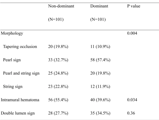
describe current treatment strategies and outcomes.
105 106
Methods
107
The patients who are diagnosed to have cerebral artery dissections are prospectively
108
enrolled as previously reported6, 16. Among the patients in our study between February
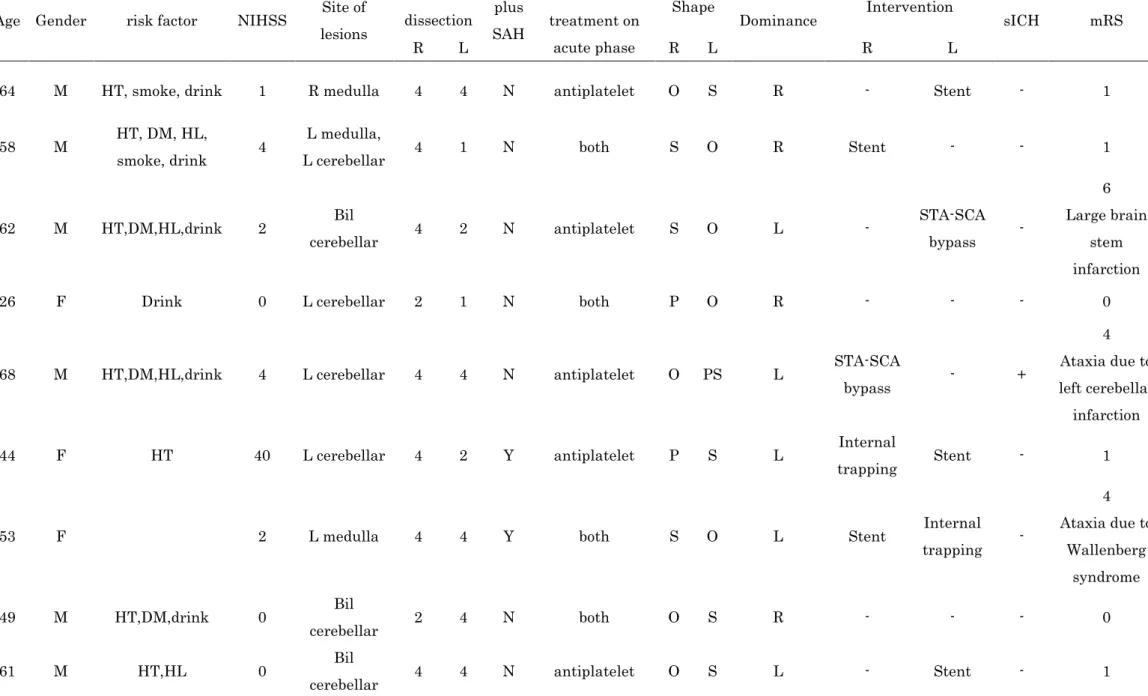
109
2007 and May 2017, we used a dataset of patients with VAD in the present study. VAD
110
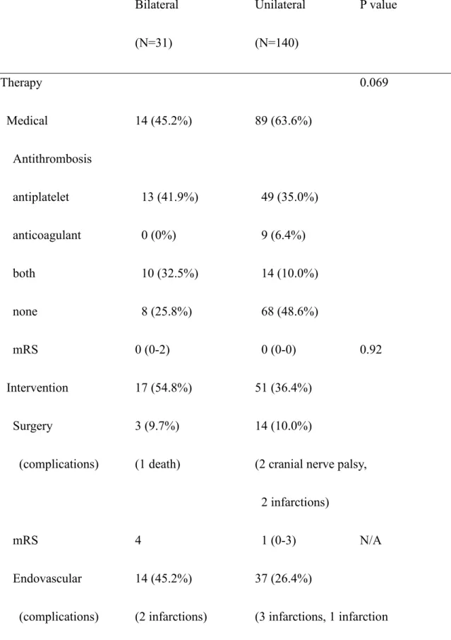
was diagnosed as previously reported 4, 6. In brief, if a patient who complained of
111
sudden onset of headache with or without neurological deficit was suspected to have
112
VAD, then MRI and/or digital subtraction angiography (DSA) was performed after
113
written informed consent was obtained. This study was approved by the human subject
114
ethics committee in our hospital. VAD was diagnosed according to the criteria from a
115
multicenter observational study (SCADS-Japan) 6.
116
When we applied the recent diagnostic criteria of dissection to the patients with
117
intracranial VAD 3, the numbers of each degree of certainty were 117 in definite, nine in
118
probable, and 23 in possible dissection. Atherosclerotic risk factors were determined by
119
8
medical records. The clinical subtype of the patients was categorized as subarachnoid
120
hemorrhage (SAH), infarction, SAH and infarction, and headache. Neurological
121
severity at admission was estimated using the National Institute of Health and Stroke
122
Scale score, while the clinical outcome was determined by the modified Rankin Scale
123
(mRS) at discharge.
124
VAD was diagnosed by a high signal intensity on T1-weighted 3D turbo spin echo
125
(VISTA) MRI within the VA wall, which indicates intramural hematoma 5, or by
126
morphological findings of the pearl and string sign, string sign, pearl sign, or tapered
127
occlusion on DSA. We obtained MRI around 7 days after the symptom onset for the
128
evaluation of intramural hematoma. The morphology of VAD was determined and
129
analyzed vessel-by-vessel. The site of VAD was defined as previously reported 17. For
130
the mechanism of bilateral VAD, we reviewed whether the bilateral VAD was connected
131
through the basilar artery union or existed separately. The dominant side of the VA was
132
defined as the side with a diameter ≥0.3 mm wider than the contralateral side when
133
measured at the bilateral V4 portion within 3 cm from the union of the basilar artery.
134
The dominant side was determined by basi-parallel anatomical scanning. When the
135
difference was <0.3 mm, the VA side that was linearly connected to the BA was
136
considered dominant. A similar method was recently reported for deciding the dominant
137
9
side of the VA 18.
138
We mainly used medical therapy such as antithrombotic and/or antihypertensive
139
agents, while a surgical or endovascular intervention was used in some patients if
140
required. Our policy was that medical therapy was first considered as a treatment
141
option, although endovascular intervention was frequently performed in VAD patients
142
with SAH. In cases with infarction in which it was difficult to continue only medical
143
treatment, endovascular or surgical interventions were considered. Endovascular
144
intervention for SAH was usually conducted acutely, while intervention on patients with
145
infarction was performed after a delay. The indications of superficial temporal artery-
146
superior cerebellar artery bypass surgery (STA-SCA) were as follows: severe steno-
147
occlusive lesions existed on bilateral VA; collateral formation of anterior circulation into
148
the brain stem was insufficient; neurological symptoms were progressed regardless of
149
the medications; and stent insertion was difficult technically. By contrast, the indication
150
of surgery in patients with unilateral VAD was the presence of aneurysmal formation of
151
VAD with a risk of rupture. If patients had a large dissecting VA aneurysm that involved
152
the origin of the posterior inferior cerebellar artery (PICA), clipping or trapping of the
153
VA aneurysm with or without occipital artery (OA) -PICA bypass was considered. The
154
treatment strategy was decided based on the age, clinical subtype, morphology, the
155
10
degree of morphological changes, and personal choice of each patient.
156
Patients were divided into bilateral and unilateral VAD groups for statistical
157
comparisons. The Fisher exact test was used to assess differences in clinical background
158
treatment and imaging data between the groups, the unpaired t-test used to compare age,
159
and the Mann–Whitney U-test used to compared the National Institute of Health and
160
Stroke Scale scores and mRS. Chi square test was used for the statistical analyses of
161
clinical subtype, morphology and therapy. All statistical analyses were performed with
162
statistical software (SPSS version 22.0; IBM, Armonk, New York, USA).
163 164
Results
165
There were 140 patients admitted to our Neuro-center with unilateral VAD, and 31
166
patients with bilateral VAD. The background and characteristics of the patients are
167
summarized in Table 1. There were no differences between the unilateral and bilateral
168
VAD groups, except for the frequency of diabetes (unilateral 11.4% vs. bilateral 25.8%).
169
Infarction and/or SAH was observed in approximately 60% of bilateral VAD patients
170
and 30% of unilateral VAD patients (P<0.001). Three patients with bilateral VAD
171
exhibited both SAH and infarction. No patients were treated with intravenous
172
thrombolysis. Two patients with unilateral VAD had mild trauma as a potential trigger
173
11
19. The numbers of segments of the right VAD were three in V1, two in V2, one in V3,
174
and 25 in V4, while the numbers of segments of the left VAD were two in V1, three in
175
V2, two in V3, and 24 in V4 portion.
176
In bilateral VAD, three (9.7%) of 31 patients had the right and left sides connected
177
via a basilar artery union, while the remaining patients had bilateral VAD that existed
178
separately. V4 was the main site of dissection in the dominant and non-dominant sides
179
in bilateral VAD. The number of patients with unilateral and bilateral VAD was 140 and
180
31, respectively. Thus, a total of 202 vessels were analyzed. No patient had VAD
181
pseudo-aneurysm formation. For imaging findings in the 62 vessels of 31 patients with
182
bilateral VAD, the pearl and string sign was observed in 21.0% of patients, string sign in
183
25.8%, pearl sign in 27.4%, and tapered occlusion in 25.8%. However, the frequency of
184
the VAD morphologies differed between VAD in the dominant and non-dominant sides.
185
The pearl sign (35.5%) occurred most frequently in the dominant side of VAD, while
186
the string sign (32.3%) and tapered occlusion (32.3%) were mainly observed in the non-
187
dominant side. This pattern was consistently observed in the 202 vessels analyzed,
188
indicating that the string sign and tapered occlusion were more frequent in the non-
189
dominant side of VAD, while the pearl sign was the main morphology in the dominant
190
side of VAD (Table 2). Intramural hematoma was more frequent in the non-dominant
191
12
than the dominant side of the VA. The features of the bilateral VAD patients with
192
ischemic stroke are summarized in Table 3. Seven of 13 patients with ischemic stroke
193
showed a string sign or tapered occlusion on both sides of the VAD.
194
Endovascular intervention was frequently used in bilateral VAD patients,
195
especially those with ischemic stroke (eight of 13 patients), while >50% of unilateral
196
VAD patients were treated medically (Table 4). STA-SCA was performed in the two
197
bilateral VAD patients with stenosis and/or occlusion, while surgical intervention was
198
conducted in 10% of patients with unilateral VAD. One patient with bilateral VAD died
199
after STA-SCA. Although many patients with unilateral VAD exhibited a favorable
200
mRS outcome of 0–1, bilateral VAD patients were significantly more severe.
201 202
Representative case
203
A 53-year-old woman with a chief complaint of headache, dysphagia and vertigo had
204
ischemic stroke in the left lateral medullary and hospitalized immediately. Although the
205
stenotic sites did not reveal high intensity on VISTA, the second VISTA acquisition
206
indicated existence of intramural hematoma in the bilateral VA, which confirmed
207
bilateral VAD (Figure 1). Right VA angiography revealed the string sign in the
208
intracranial VA, while left VA angiography indicated the tapered occlusion in the
209
13
intracranial VA (Figure 2). The patient complained of headache on the 16th day of
210
hospitalization, and computed tomography showed SAH. A left VAD was the cause of
211
SAH based on recanalization on a follow-up DSA study. Endovascular coil trapping was
212
performed in the left VA during right VA stent placement. Her mRS at discharge was 4.
213 214
Discussion
215
The main finding of the present study was that bilateral VAD was caused ischemic
216
stroke more frequently than unilateral VAD. There was also a lower frequency of VAD
217
in which both sides were connected via a union. Further, the morphology of VAD was
218
significantly different between the dominant and non-dominant sides of VAD, with the
219
pearl sign the most common on the dominant side, and the string sign or tapered
220
occlusion the most common on the non-dominant side. There were no differences in the
221
frequency of intracranial VAD between unilateral and bilateral VAD. However, medical
222
therapy was used most frequently in unilateral VAD, while endovascular intervention
223
was more common in bilateral VAD. Finally, the outcome of bilateral VAD was more
224
severe at discharge
225
In previous case reports of bilateral VAD, SAH was the main stroke subtype 8, 9, 20-
226
23, while ischemic stroke caused by bilateral VAD was relatively uncommon 10, 11. By
227
14
contrast, in the present study we found higher rates of ischemic stroke than SAH, which
228
is likely because many of the bilateral VAD patients with ischemic stroke had a steno-
229
occlusive lesion on either side. We also found that diabetes was a factor associated with
230
bilateral VAD, which may have contributed to the increased rates of ischemic stroke.
231
There are two main hypotheses for the mechanism of bilateral VAD: type 1, where
232
the dissection progresses from one side to the other through the basilar union 11, 24, and
233
type 2, where the dissection on one side of the VA causes hemodynamic stress on the
234
other side, which leads to VA dissection 7. Type 1 is considered the main mechanism of
235
bilateral VAD, which contrasts with the findings in the present study. The presence of
236
separated dissection sites can be difficult to diagnose, although recent imaging
237
technologies may provide more accurate assessment of bilateral VAD. The mechanism
238
of bilateral VAD can be determined by examining the initial side of the dissection. We
239
suggest that occlusion of one side of the VA causes an increase in contralateral VA flow.
240
In turn, this may increase stress on the contralateral VA and cause dissection. Our data
241
suggest that the initial symptoms of many patients with bilateral VAD were less severe,
242
and that prevention of a second episode of SAH or ischemic stroke is critical. Our
243
cohort included three patients with the right and left VA connected via a basilar artery
244
union, all of whom developed severe ischemic stroke. The risks and prevention of type
245
15
1 VAD should be examined in future studies.
246
In the present study, patients with bilateral VAD received endovascular
247
interventions more often than those with unilateral VAD. Endovascular intervention was
248
frequently performed in VAD patients with SAH. However, in the present study,
249
endovascular intervention was also used for treatment of bilateral VAD with ischemic
250
stroke. Bilateral VAD often shows imaging findings of a string sign and/or tapered
251
occlusion on either side. When one side requires treatment because of severe stenosis,
252
then an endovascular intervention, particularly stent insertion, is used to prevent further
253
ischemic stroke. However, we were concerned that using a stent to treat one side of a
254
bilateral VAD with severe bilateral stenoses may cause occlusion of the treated side, and
255
thus induce further ischemic stroke. To prevent this, we occasionally performed surgical
256
interventions such as STA-SCA anastomosis in cases with severe bilateral stenoses, to
257
maintain basilar arterial flow 25, 26. Although STA-SCA anastomosis can preserve the
258
perfusion of the brainstem in patients with bilateral severe VA stenoses, there are some
259
reports of technical difficulties and potential safety concerns. Further studies are
260
required to elucidate whether and which interventions should be chosen in cases with
261
bilateral VAD with severe stenoses.
262
There are some potential limitations of our study. First, the number of patients
263
16
participating was small. Second, this was a single center study, and thus various
264
treatment options were not attempted. Third, although our treatment strategy was not
265
changed, advances in endovascular intervention equipment and techniques may have
266
influenced our treatment decision. Fourth, antithrombotic agents were used in many
267
cases with bilateral VAD, and it is difficult to prove the association of these medications
268
with the incidence of SAH. Fifth, univariate analysis suggested that intramural
269
hematoma was more frequent in the non-dominant compared with the dominant side of
270
the VA, which should be examined in future studies. Finally, we did not collect follow-
271
up data from our patients.
272 273
Conclusion
274
We found that infarction was relatively frequent in bilateral VAD patients. Endovascular
275
intervention was common in patients with bilateral VAD with SAH or infarction. The
276
treatment strategy varied depending on the clinical subtype, imaging findings of VAD,
277
and dominant or non-dominant side of VAD.
278 279
17
References
280
[1] Schievink WI. Spontaneous dissection of the carotid and vertebral arteries. N Engl J Med.
281
2001;344:898-906.
282
[2] Redekop GJ. Extracranial carotid and vertebral artery dissection: a review. The Canadian 283
journal of neurological sciences Le journal canadien des sciences neurologiques. 2008;35:146- 284
285 52.
[3] Debette S, Compter A, Labeyrie M-A, et al. Epidemiology, pathophysiology, diagnosis, and 286
management of intracranial artery dissection. The Lancet Neurology. 2015;14:640-54.
287
[4] Fukuhara K, Ogata T, Ouma S, et al. Impact of initial symptom for accurate diagnosis of 288
vertebral artery dissection. International journal of stroke : official journal of the 289
International Stroke Society. 2015;10 Suppl A100:30-3.
290
[5] Takemoto K, Takano K, Abe H, et al. The new MRI modalities "BPAS and VISTA" for the 291
diagnosis of VA dissection. Acta neurochirurgica Supplement. 2011;112:59-65.
292
[6] Kobayashi H, Morishita T, Ogata T, et al. Extracranial and intracranial vertebral artery 293
dissections: A comparison of clinical findings. Journal of the neurological sciences.
294
2016;362:244-50.
295
[7] Wilkinson DA, Wilson TJ, Stetler WR, Jr., Pandey AS. Subarachnoid haemorrhage with 296
bilateral intracranial vertebral artery dissecting aneurysms treated by staged endovascular 297
stenting. BMJ case reports. 2013;2013.
298
[8] Ishikawa T, Yamaguchi K, Anami H, Ishiguro T, Matsuoka G, Kawamata T. Stent-assisted 299
coil embolisation for bilateral vertebral artery dissecting aneurysms presenting with 300
subarachnoid haemorrhage. The neuroradiology journal. 2016;29:473-8.
301
[9] Kono K, Shintani A, Fujimoto T, Terada T. Stent-assisted coil embolization and 302
computational fluid dynamics simulations of bilateral vertebral artery dissecting aneurysms 303
18
presenting with subarachnoid hemorrhage: case report. Neurosurgery. 2012;71:E1192-200;
304
discussion E200-1.
305
[10] Fukuda M, Aiba T, Takahashi S. Bilateral medial medullary infarction due to bilateral 306
vertebral artery dissection. Clinical neurology and neurosurgery. 2004;106:132-5.
307
[11] Funaki T, Oshimoto T, Wataya T, et al. [Bilateral vertebral artery dissection and its 308
chronological changes detected by MR angiography: a case report]. No to shinkei = Brain and 309
nerve. 2004;56:247-50.
310
[12] Garcia-Monco JC, Fernandez Canton G, Gomez Beldarrain M. Bilateral vertebral artery 311
dissection in a patient with afibrinogenemia. Stroke; a journal of cerebral circulation.
312
1996;27:2325-7.
313
[13] Kato Y, Nagoya H, Abe T, et al. Progressive Bilateral Vertebral Artery Dissection in a 314
Case of Osteogenesis Imperfecta. Journal of stroke and cerebrovascular diseases : the official 315
journal of National Stroke Association. 2017;26:e43-e6.
316
[14] Frankowska E, Brzozowski K, Staszewski J, Kolmaga N, Stepien A, Boguslawska- 317
Walecka R. Combined thrombolysis in posterior circulation stroke caused by bilateral 318
vertebral artery dissection in squash player. Neurologia i neurochirurgia polska.
319
2014;48:299-304.
320
[15] Fukunaga A, Tabuse M, Naritaka H, Nakamura T, Akiyama T. Spontaneous resolution 321
of nontraumatic bilateral intracranial vertebral artery dissections. Neurologia medico- 322
chirurgica. 2002;42:491-5.
323
[16] Matsumoto J, Ogata T, Abe H, Higashi T, Takano K, Inoue T. Do characteristics of 324
dissection differ between the posterior inferior cerebellar artery and the vertebral artery?
325
Journal of stroke and cerebrovascular diseases : the official journal of National Stroke 326
Association. 2014;23:2857-61.
327
19
[17] Subclavian system of arteries. In: PL W, editor. Gray's anatomy. New York: Churchill 328
Livingstone; 1995. p. 1529-34.
329
[18] Hong JM, Chung CS, Bang OY, Yong SW, Joo IS, Huh K. Vertebral artery dominance 330
contributes to basilar artery curvature and peri-vertebrobasilar junctional infarcts. Journal 331
of neurology, neurosurgery, and psychiatry. 2009;80:1087-92.
332
[19] Engelter ST, Grond-Ginsbach C, Metso TM, et al. Cervical artery dissection: trauma and 333
other potential mechanical trigger events. Neurology. 2013;80:1950-7.
334
[20] Otawara Y, Ogasawara K, Ogawa A, Kogure T. Dissecting aneurysms of the bilateral 335
vertebral arteries with subarachnoid hemorrhage: report of three cases. Neurosurgery.
336
2002;50:1372-4; discussion 4-5.
337
[21] Shin YS, Kim BM, Kim SH, et al. Endovascular treatment of bilateral intracranial 338
vertebral artery dissecting aneurysms presenting with subarachnoid hemorrhage.
339
Neurosurgery. 2012;70:75-81; discussion 340
[22] Yoon SM, Shim JJ, Kim SH, Chang JC. Bilateral vertebral artery dissecting aneurysms 341
presenting with subarachnoid hemorrhage treated by staged coil trapping and covered stents 342
graft. Journal of Korean Neurosurgical Society. 2012;51:155-9.
343
[23] Zhao WY, Zhao KJ, Huang QH, Xu Y, Hong B, Liu JM. Single-stage endovascular 344
treatment of subarachnoid hemorrhage related to bilateral vertebral artery dissecting 345
aneurysms. Interventional neuroradiology : journal of peritherapeutic neuroradiology, 346
surgical procedures and related neurosciences. 2016;22:138-42.
347
[24] Hosoya T, Adachi M, Yamaguchi K, Haku T, Kayama T, Kato T. Clinical and 348
neuroradiological features of intracranial vertebrobasilar artery dissection. Stroke; a journal 349
of cerebral circulation. 1999;30:1083-90.
350
[25] Ota T, Usami K, Iijima A, Saito N. Staged surgical treatment for symptomatic 351
20
vertebrobasilar artery stenosis: combined treatment with endovascular angioplasty and 352
bypass surgery. World neurosurgery. 2012;78:90-4.
353
[26] Ausman JI, Diaz FG, Vacca DF, Sadasivan B. Superficial temporal and occipital artery 354
bypass pedicles to superior, anterior inferior, and posterior inferior cerebellar arteries for 355
vertebrobasilar insufficiency. Journal of neurosurgery. 1990;72:554-8.
356 357 358
Figure 1: T1-weighted 3D turbo spin echo (VISTA) findings. Left: VISTA finding on the 5th day of hospitalization. High intensity signals in the bilateral vertebral artery (VA) were not visible. Right: VISTA finding on the 17th day. High intensity signals were detected in the bilateral VA, indicating the existence of VA dissection.
A
B
Figure 2: Digital subtraction angiographical (DSA) findings in a representative case.
Left: DSA on the right side of the patient, with evidence of a string sign. Right: DSA on the left side of the patient, with evidence of a tapered occlusion.
Table 1: Comparison of background characteristics between patients with unilateral and bilateral vertebral artery dissection (VAD).
Unilateral (N=140) Bilateral (N=31) P value
Age 55.3±13.6 53.0±12.5 0.39
Gender (male) 86 (61.4%) 23 (74.2%) 0.22
Hypertension 87 (62.1%) 20 (64.5%) 0.84
Diabetes 16 (11.4%) 8 (25.8%) 0.047
Hyperlipidemia 41 (29.3%) 8 (25.8%) 0.83
Smoking 53 (37.9%) 10 (32.3%) 0.68
Drinking 95 (67.9%) 19 (61.3%) 0.53
NIHSS score 0 (0-1) 0 (0-4) 0.016
Clinical subtype <0.001
non-stroke 98 (70%) 12 (38.7%)
infarction 23 (16.4%) 10 (32.3%)
SAH 19 (13.6%) 6 (19.4%)
intracranial bilaterally: 6
infarction+SAH 0 (0%) 3 (9.7%)
extra/intracranial: 2
intracranial bilaterally: 1
NIHSS: National Institute of Health and Stroke Scale; SAH: subarachnoid hemorrhage.
extra/intracranial: intracranial on one side, and extracranial on the other.
Table 2: Relationship of VA dominancy with VAD morphology.
Non-dominant Dominant P value
(N=101) (N=101)
Morphology 0.004
Tapering occlusion 20 (19.8%) 11 (10.9%)
Pearl sign 33 (32.7%) 58 (57.4%)
Pearl and string sign 25 (24.8%) 20 (19.8%)
String sign 23 (22.8%) 12 (11.9%)
Intramural hematoma 56 (55.4%) 40 (39.6%) 0.034
Double lumen sign 28 (27.7%) 35 (34.5%) 0.36
Table 3: The features of bilateral VAD patients with ischemic stroke.
Age Gender risk factor NIHSS Site of lesions
Site of
dissection plus SAH
Antithrombotic treatment on
acute phase
Shape
Dominance Intervention
sICH mRS
R L R L R L
64 M HT, smoke, drink 1 R medulla 4 4 N antiplatelet O S R - Stent - 1
58 M HT, DM, HL,
smoke, drink 4 L medulla,
L cerebellar 4 1 N both S O R Stent - - 1
62 M HT,DM,HL,drink 2 Bil
cerebellar 4 2 N antiplatelet S O L - STA-SCA
bypass -
6 Large brain
stem infarction
26 F Drink 0 L cerebellar 2 1 N both P O R - - - 0
68 M HT,DM,HL,drink 4 L cerebellar 4 4 N antiplatelet O PS L STA-SCA
bypass - +
4 Ataxia due to left cerebellar infarction
44 F HT 40 L cerebellar 4 2 Y antiplatelet P S L Internal
trapping Stent - 1
53 F 2 L medulla 4 4 Y both S O L Stent Internal
trapping -
4 Ataxia due to
Wallenberg syndrome
49 M HT,DM,drink 0 Bil
cerebellar 2 4 N both O S R - - - 0
61 M HT,HL 0 Bil
cerebellar 4 4 N antiplatelet O S L - Stent - 1
66 M HT,HL,drink 4 R medulla 1 4 N both O S R - - -
4 Ataxia due to
Wallenberg syndrome
58 M DM,Smoke,drink 1 L cerebellar 4 4 N both PS O L - Internal
trapping - 1
35 M HT,HL,Smoke 2 L medulla 4 4 N both P PS R Stent+coil - - 1
34 M 11
R medulla, bil cerebellar
4 2 Y both S P L Internal
trapping Stent+coil -
5 Sever left hemiparesis
due to medulla infarction M: male; F: female; SAH; subarachnoid hemorrhage; mRS; modified Rankin Scale at discharge; HT: hypertension; DM: diabetes mellitus; HL: hyperlipidemia; O: tapered occlusion;
S: string sign; P: pearl sign; PS: pearl and string sign; STA-SCA: superficial temporal artery-superior cerebellar artery; internal trapping: a treatment to insert coils into the dissecting aneurysm including proximal artery occlusion; Stent+coil: stent assisted coil embolization.
Table 4: Treatment differences between patients with unilateral and bilateral VAD.
Bilateral Unilateral P value
(N=31) (N=140)
Therapy 0.069
Medical 14 (45.2%) 89 (63.6%)
Antithrombosis
antiplatelet 13 (41.9%) 49 (35.0%) anticoagulant 0 (0%) 9 (6.4%)
both 10 (32.5%) 14 (10.0%)
none 8 (25.8%) 68 (48.6%)
mRS 0 (0-2) 0 (0-0) 0.92
Intervention 17 (54.8%) 51 (36.4%)
Surgery 3 (9.7%) 14 (10.0%)
(complications) (1 death) (2 cranial nerve palsy, 2 infarctions)
mRS 4 1 (0-3) N/A
Endovascular 14 (45.2%) 37 (26.4%)
(complications) (2 infarctions) (3 infarctions, 1 infarction
with sICH, 1 death)
mRS 1 (1-1) 0 (0-2) 0.21
mRS of all patients 1 (0-1) 0 (0-1) 0.021
sICH: symptomatic intracerebral hemorrhage, N/A: not available