九州大学学術情報リポジトリ
Kyushu University Institutional Repository
植物ポリフェノールの抗菌および抗毒素活性に関す る研究
ファム, ティ, ヴィン
http://hdl.handle.net/2324/4110566
出版情報:Kyushu University, 2020, 博士(農学), 課程博士 バージョン:
権利関係:
Study on antibacterial and antitoxin activities of plant polyphenols
PHAM THI VINH
2020
i
Study on antibacterial and antitoxin activities of plant polyphenols
A thesis submitted by
PHAM THI VINH
in the Partial Fulfillment of the Requirements for the Degree of Doctor of Philosophy
at the Department of Bioscience and Biotechnology,
Graduate School of Bioresource and Bioenvironmental Sciences, Kyushu University, Japan.
September, 2020
i
TABLE OF CONTENTS
Abbreviations ... iv
Chapter 1. Introduction ...1
1.1 Foodbrone illness ...1
1.2 Enterohaemorrhagic Escherichia coli ...2
1.2.1 Introduction enterohaemorrhagic Escherichia coli ...2
1.2.2 Virulence factors of EHEC ...4
1.2.2.1 The adhesion of EHEC ...4
1.2.2.2 Shiga toxin ...5
1.2.2.3 Other virulence factors of EHEC ...7
1.3. Plant polyphenols ...9
1.3.1 Flavonoids ... 10
1.3.2 Phenolic acids ... 13
1.3.3 Stilbenes ... 14
1.3.4 Lignans... 14
1.4 Antibacterial activity of polyphenols ... 14
1.5 Antitoxin activity of polyphenols ... 16
1.6 Purpose of study ... 18
Chapter 2. The combqined effects of tea polyphenols and food additives against enterohaemorrhagic Escherichia coli ... 19
2.1 Introduction ... 19
2.2 Materials and methods ... 21
2.2.1 Preparation of bacteria ... 21
2.2.2 Preparation of tea polyphenols and food additives solution ... 21
2.2.3 Determination of the minimum inhibitory concentration (MIC)... 22
ii
2.2.4 Synergy testing of tea polyphenols with food additives using broth dilution
method in test tube ... 22
2.2.5 Time-kill assays ... 23
2.2.6 Viable cell count ... 23
2.3 Results... 23
2.3.1 Single antibacterial effect ... 23
2.3.2 Combined antibacterial effect ... 24
2.3.3 Time kill assay of the combination ... 26
2.4 Discussion ... 34
2.5 Summary ... 37
Chapter 3. Combined effects of epigallocatechin gallate and heat treatment against enterohaemorrhagic Escherichia coli ... 39
3.1 Intruduction ... 39
3.2 Methods ... 41
3.2.1 Bacterial strains and culture condition ... 41
3.2.2 Heat treatment and recovery ... 41
3.2.3 Determination of viable counts... 42
3.2.4 Measurement of protein leakage ... 42
3.2.5 Measurement of nucleic acid leakage ... 43
3.2.6 Statistical analysis ... 43
3.3 Results... 43
3.3.1 Combined effect of EGCg and heat treatment on the growth of E.coli ... 43
3.3.2 Combined effects of EGCg and heat treatment on protein leakage from E. coli ... 46
3.3.3 Combined effects of EGCg and heat treatment on nucleic acid leakage from E. coli ... 47
iii
3.4 Discussion ... 48
3.5 Summary ... 51
Chapter 4. Effects of baicalein on the cytotoxicity, production and secretion of Shiga toxins of enterohaemorrhagic Escherichia coli ... 52
4.1 Introduction ... 52
4.2 Materials and methods ... 53
4.2.1 Analysis of interaction between StxB pentamers and natural products... 53
4.2.2 Preparation of Stx1 and Stx2 ... 54
4.2.3 Determination of Stx-inhibitory activity of baicalein ... 55
4.2.4. Determination of protective effect of baicalein on Vero cells against Stx .... 56
4.2.5 Determination of transcription of stx, production and secretion of Stx... 56
4.3 Results... 59
4.3.1 Effects of baicalein on the cytotoxicity of Stx ... 59
4.3.2 Effects protective of baicalein on Vero cells against Stx ... 61
4.3.3 Effects of baicalein on productivity of Stx by EHEC... 62
4.3.4 Interaction of baicalein with Stx1B and Stx2B pentamers ... 65
4.4 Discussion ... 66
4.5 Summary ... 70
Chapter 5: Conclussions ... 71
Acknowledgement ... 74
References ... 75
iv
ABBREVIATIONS
AEEC: Attaching and effacing Escherichia coli BSA: Bovine serum albumin
CDC: Center for Disease Control and Prevention DHL: Deoxycholate Hydrogen Sulfide-lactose E. coli: Escherichia coli
EHEC: Enterohaemorrhagic Escherichia coli ETEC: Enterotoxigenic Escherichia coli FDA: Food and Drug Administration
Gb3: Glycosphingolipid globotriaosylceramide HUS: Hemolytic uremic syndrome
LB: Luria broth
PCR: Polymerase Chain Reaction S. dysenteriae: Shigella dysenteriae Shiga toxin: Stx
STEC: Shiga toxin producing Escherichia coli TSA: Tryptic Soy Agar
TSB: Tryptic Soy Broth VT: verotoxin
WHO: World Health Organization
1
Chapter 1. Introduction
1.1 Foodborne illness
Foodborne illness is caused by consuming contaminated foods. There are many causes of foodborne illness, including bacteria, viruses, fungi, protozoa and parasites.
Among all causes of foodborne illness, bacterial pathogens are the most common cause of foodborne illness outbreaks. According to World Health Organization (WHO) assessment, there are about 600 million cases of foodborne illness and 420,000 deaths every year worldwide (World Health Organization, 2019). Center for Disease Control and Prevention (CDC) also estimates Salmonella infection cause 1.35 million foodborne illnesses, with 26,500 hospitalizations, and 420 deaths each year in United States (CDC, 2013). In 2019 in Indiana States, the number of cases of Salmonella infection was estimated approximately 137 foodborne illnesses, with 38 hospitalizations (Food and Drug Administration (FDA), 2019). Early 2020 in Republic of Korea, 36 foodborne illnesses, resulting in 36 hospitalizations and 4 deaths is thought to be associated with Listeria monocytogenes in the mushrooms (CDC, 2020).
Among all bacterial pathogens causing food poisoning, the prevalence of pathogenic Escherichia coli (E. coli) strains in foods are the most common cause of foodborne illness outbreaks. Most outbreaks of E. coli infection have been found to be associated with consumption of various foods, such as meat and meat products, dairy products, vegetables and fruits. E. coli O157:H7-contaminated undercooked meat as cause of several outbreaks of hemorrhagic colitis was investigated involving 47 cases of illness occurred in Oregon and Michigan in 1982 (Riley et al., 1983). The foodborne outbreaks of E. coli O157:H7 occurred in Washington and California in 1994 have been linked to eating dry fermented salami (Tilden et al., 1996). After this outbreak, the requiring least a 5-log10 unit reduction of E. coli O157:H7 during processing dry fermented salami was established
2
by the United States (US) Department of Agriculture Food Safety Inspection Service to ensure food safety (Chacon et al., 2006). Each year in US, the number of cases of E. coli O157:H7 infection was estimated to be approximately 73,500 cases (Lahmer et al., 2017).
Of all multistate incidents of food poisoning by E. coli associated with fresh products occurred in US from 2010 to 2017, more than half of them were due to E. coli O157:H7 infections (Carstens et al., 2019). The outbreak of 167 cases of foodborne diseases and 85 hospitalizations in US and Canada in 2019 found to be linked to E. coli O157:H7 infection in salad has also been reported by FDA on January, 2020 (FDA, 2020). In the Isfahan province, Iran, the prevalence of E. coli strains in ready-to-eat food is 5%, among them the prevalence of Shiga toxin producing E. coli (STEC) strains is 2.63%, in which the prevalence of enterohaemorrhagic E. coli (EHEC) and attaching and effacing E. coli (AEEC) were 36.84%
and 52.63%, respectively (Shahreza et al., 2017). In 1995, 70% of 40 patients were infected by E. coli O157:H7 linked to leaf lettuce in western Montana (Ackers et al., 1998). In the Piedmont region of Italy, the ratio of STEC contamination in dairy and meat products was 42% (Rantsiou et al., 2012). From April 2000 to July 2002, the prevalence of E. coli O157:H7 is 1.7% in beef cattle, 6.7% in dairy cattle, 0.3% in pig and none in chicken in Korea (Jo et al., 2004). Although prevalence of EHEC is less than that of enterotoxigenic E.
coli (ETEC), there is significant risk for the E. coli O157:H7 infection by contaminated meat products from livestock animals (Kang et al., 2004; Cho et al., 2014).
1.2 Enterohaemorrhagic Escherichia coli
1.2.1 Introduction enterohaemorrhagic Escherichia coli
Escherichia coli is a bacterium found in the food, environment, and in the intestines of human and animals. Most strains of E. coli are vital part in the healthy human intestinal and harmless to humans. However, some strains such as STEC, ETEC and diarrheagenic E.
coli can cause some foodborne disease. The categorization of pathogenic E. coli is
3
associated with their pathogenicity profiles such as clinical syndromes, and virulence factors and phylogenetic description. Six intestinal pathotypes of pathogenic E. coli are ETEC, EHEC, enteropathogenic E. coli, enteroaggregative E. coli, enteroinvasive E.
coli and diffusely adherent E. coli (Kaper et al., 2004).
EHEC is known cause of severe intestinal infection in humans, including abdominal pain, diarrhea and bloody diarrhea. It may be transferred to humans by the consumption of contaminated food such as undercooked meat products, contaminated fresh vegetables and fruits (Berger et al., 2010). EHEC can grow in a wide range of temperature from 7oC to 50oC, in acidic food with pH of 4.4 and at minimum water activity of 0.95 (Balamurugan et al., 2015).
Shiga toxin (Stx), a strong toxin produced by EHEC, can give rise to hemorrhagic colitis and hemolytic uremic syndrome (HUS). EHEC was first recognized in 1982 after two outbreaks of hemorrhagic colitis and was identified to have resulted from eating undercooked ground beef contaminated with E. coli O157:H7 (CDC, 1982; Riley et al., 1983). Since then, the other serotypes of E. coli also have been associated with the outbreaks of hemorrhagic colitis in humans. Meng et al. reported that more than 600 different serotypes of STEC including 160 O serogroups and 50 H types have been isolated from patients with STEC infection (Meng et al., 2007). EHEC O157:H7 strain was reported to cause outbreaks of hemorrhagic colitis in many countries in the world such as Japan (Trofa et al., 1999), the United States (O’Brien et al., 1983), and Australia (McPherson et al., 2011). EHEC infections not only affect on public health but also impose a significant economic cost on society. The estimated total cost for each case of EHEC infection is AUD$3,132 in South Australia (McPherson et al., 2011)), $5,515 in United States (Frenzen et al., 2005).
4 1.2.2 Virulence factors of EHEC
An early event in EHEC infection is hemorrhagic colitis that is thought to be caused by various virulence factors including fimbrial and nonfimbrial adhesion, flagella, Stx, and the Type III secretion system (Foster, 2013), of which the adhesion and the production of Stx are the most important factors for the pathogenicity of EHEC.
1.2.2.1 The adhesion of EHEC
Some studies have described the adhesion mechanisms of EHEC. Jeter and Matthysse showed the characteristics of the binding of both non-pathogenic and pathogenic strains to various plant surfaces (Jeter & Matthysse, 2005). Contrary to non- pathogenic E. coli, EHEC O157:H7 strains showed strong adherence to spinach leaves, roofs of alfalfa sprout and tomato skin. The attachment to these plant surfaces is mediated by curli (Jeter & Matthysse, 2005). However, the production of curli on the surface of non- pathogenic strains has been shown to be sufficient to allow bacterial adhesion to roofs of alfalfa sprouts, but curli are not required for the attachment of pathogenic strains to plant surfaces, suggesting that other adhesion factors were involved together.
E. coli O157 and O26 strains use the filamentous type III secretion system (fT3SS) comprising EspA filaments for binding to salad leaves (Shaw et al., 2008). In addition, fT3SS-dependent protein secretion in E. coli O157 plays an important role in the adhesion of bacteria to mammalian cells (Garmendia et al., 2005). In contrast to attachment of mammalian hosts, the adhesion of E. coli O157 to salad leaves was independent of translocation of effector protein (Shaw et al., 2008). Flagella also play a role for binding of E. coli O157 to baby spinach and lettuce leaves as deletion of the fliC flagellin gene in EHEC significantly reduced the adhesion to the surface of leaves (Xicohtencatl-Cortes et al., 2009). The mutation of escN (ATPase) gene that linked to the function of fT3SS also significantly reduced the level of adhesion. This suggested that E. coli O157 used more than one mechanism for binding to host cells.
5
It has also been reported that both the fimbrial and non-fimbrial EHEC adhesins have been related to adhesion of EHEC to host cells. F9 fimbriae, a long polar fimbriae of EHEC O157:H7, can prevent or disrupt the adhesion of EHEC to human cells because mutants defective in F9 fimbriae increased the adhesion of EHEC to cultured epithelial cells (Low et al., 2006). E. coli common pilus, a pilus fimbrial structure produced by both pathogenic and non-pathogenic E. coli, promoted intercellular interactions in biofilms communities (Rendón et al., 2007; Martínez-Santos et al., 2012). The non-fimbrial EHEC adhesins that have been implicated in adhesion include the chromosomal genetic locus efa1 (EHEC factor for adherence 1), plasmid-encoded toxB, calcium-binding antigen 43 homolog, outer membrane protein A, and the chromosomally-encoded adhesins (Tatsuno et al., 2001; Johnson et al., 2005; Torres et al. 2005)
1.2.2.2 Shiga toxin
Major virulence factor of EHEC strains is Shiga toxin (Stx) that causes some diseases in humans such as hemolytic uremic syndrome and hemorrhagic colitis. Stx was known as verotoxin (VT) because of its similarities to VT (Johnson et al., 1983) and the words "Shiga toxin" or "vero toxin" refer to the same toxin (Kaper & O’Brien, 2014).
Based on the antigenic characteristics of the prototypical Stx produced by Shigella dysenteriae (S. dysenteriae) I, Stxs produced by STEC are subdivided into two major types, Stx1 and Stx2 (Strockbine et al., 1986; Scheutz et al., 2012). STEC strains can produce both Stx1 and Stx2 or either one. While Stx1 is a similar cytotoxin to Stx of S.
dysenteriae I or differs by only one amino acid, Stx2 shares 56% amino acid sequence similarity with Stx1 (Kaper & O’Brien, 2014). Purified Stx2 has higher toxic activity than Stx1, and strains of STEC O157:H7 that express Stx2 are more likely to cause HUS than strains expressing Stx1 (Scotland et al., 1987; Louise & Obrig, 1995; Boerlin et al., 1999).
6
Stxs are the members of AB5-toxin family, consisting of an enzymatically active A subunit linked with a pentameric B subunits and structure of Stx is shown in Figure 1-1.
Figure 1-1. Structure of Stx from E. coli O157:H7 (Sandvig, 2001)
The A subunit comprised of two main domains, A1 (27.5 kDa) and A2 (4.5 kDa), that are linked together by disulfide bond between cysteines 242 and 261 (Garred et al., 1995). The A1 domain had an N-glycosidase activity responsible for the toxicity to the host cell. The activity of N-glycosidase on 28S rRNA of the 60S ribosomal subunit causes inhibition of protein synthesis and results in endothelial cell death. The A2 domain is essential for binding of the remainder of A subunit to B pentamer to create the holotoxin which is responsible for binding to glycosphingolipid globotriaosylceramide (Gb3) found on the surface of vascular endothelial cells and kidney epithelial cells. The chemical structure of Gb3 is shown in Figure 1-2.
Figure 1-2. Chemical structure of glycosphingolipid globotriaosylceramide (Paton &
Paton, 1998) B subunit
A subunit
7
Stxs are encoded by stx genes found on lambdoid bacteriophages. At low iron concentrations, stx1 can be expressed in the absence of lytic phage replication, whereas the expression of stx2 depends mainly on prophage induction (Wagner & Waldor, 2002; Tyler et al., 2004). Stx production of STEC strains is closely correlated with stx-encoding phages and could be significantly enhanced by DNA-damaging, antimicrobial and SOS-inducing agents such as mitomycin C, quinolones, furazolidone and trimethoprim (Al-Jumaili et al., 1992; Kimmitt et al. 2000; Wagner et al., 2001b). It has also been reported that Stx promoted EHEC adhesion to host epithelial cells by enhancing surface expression of two receptor candidates, nucleolin and phosphatidylethanolamine (Foster et al., 2000; Robinson et al., 2006).
1.2.2.3 Other virulence factors of EHEC
Other virulence factors of EHEC include the locus of enterocyte effacement (LEE) and the non-LEE encoded effectors. The LEE consists of five major polycistronic operons (LEE1-5) that encode the components of type III secretion system (TT3SS) and effector proteins (Mcdaniel et al., 1995). EHEC colonization was promoted by the LEE-encoded proteins, including Map, EspF, EspH, EspA ans EspG (Ritchie & Waldor, 2005; Campellone et al., 2007). In addition to the LEE, the non-LEE encoded effector proteins are also translocated into the host cell through the TT3SS (Connolly et al., 2015). Both the LEE and the non-LEE contribute to the modulation of host cell signaling to assist the replication and survival of bacteria, as well as host colonization and disease development.
1.2.3 Prevention of EHEC contamination in food
EHEC strains can survive and grow in many food products. Thus, the minimizing initial contamination and subsequent growth of EHEC in foods are essential to prevent foodborne disease. Some EHEC strains can induce stress responses to enhance their
8
resistance and growth. It has been reported that acid tolerance of E. coli O157:H7 enable the pathogen to survive in acidic conditions of gastrointestinal tract and finally, cause disease (Arnold & Kaspar, 1995; Diez-Gonzalez et al., 1998).
Furthermore, pathogenic E. coli can also survive all production stages of smear- ripened cheese for up to 70 days after manufacture (Maher et al., 2001). Hence, control steps in the food supply chain are necessary for the prevention of bacterial infection leading to the risk reduction to public health. In the industry, heat treatment is an effective method to eliminate EHEC from food products because of the sensitivity of EHEC to heat (Juneja et al., 1997; Li & Gänzle, 2016). Cooking at a high temperature can prevent E. coli O157:H7 infection in minced meat (Abong’o & Momba, 2009). The treatment of spinaches inoculated with E. coli O157:H7 and Salmonella enterica with the antimicrobial from lactic acid bacteria (LAB) was effective to reduce E. coli O157:H7 and Salmonella viable cells (Cálix-Lara et al., 2014). To reduce contamination with E. coli O157:H7 in apple cider, sodium benzoate was used in the some processes, including washing, brushing and preservation (Zhao et al., 1993). Some recent research demonstrated the effects of the treatment by bacteriophage to control E. coli O157:H7 in chicken, pork, tomato, spinach, beef and in both cooked and raw beef (Flynn et al., 2004; Abuladze et al., 2008; Hudson et al., 2013; Seo et al., 2016).
In addition, multiple hurdles may be applied during food processing to ensure safety of the processed food. For example, the combination of treatments with chloride and LAB before packaging and the packaging in modified atmosphere such as nitrogen (80%
N2, 20% O2) or oxygen (80% O2, 20% N2) significantly reduced the survival of E. coli O157:H7 and Clostridium sporogenes in spinach (Brown et al., 2011). Combination of the lytic activity of bacteriophage BPECO19 and low temperature (4oC) was effective to control E. coli O157:H7 in meat (Cho et al., 2011). Although many methods were used in the control of EHEC in food products, the use of natural antimicrobial compounds in food
9
industry is increasing due to consumer concerns about the adverse effects of chemically synthesized food preservatives.
1.3. Plant polyphenols
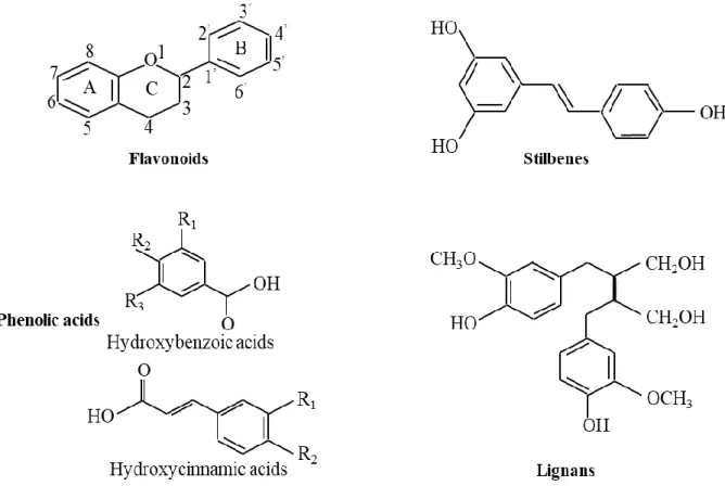
Polyphenols are organic compounds found in plant such as fruits, vegetables, cereals, tea, wine, beverages and dark chocolate. Their beneficial effect on human health is believed to be due to their antioxidant activity (Sato et al., 2002). Meta-epidemiological studies suggested that long term consumption of plant polyphenol-rich foods had an important role in human health, helps to protect against many disease such as cancers, lung damage, diabetes, osteoporosis, type 2 diabetes, cardiovascular and neurodegenerative illness (Pandey & Rizvi, 2009; Martín-Peláez et al., 2013; Xiao & Hogger, 2014; Fujiki et al., 2015). Over 8,000 types of polyphenolic compounds have been identified in many plants. Based on the number of phenol ring in polyphenol structure and the structural elements that bind these rings to each other, polyphenols were categorized into 4 main groups, including flavonoids, phenolic acids, stilbenes and lignans (Pandey & Rizvi, 2009).
The basic structure of main groups of polyphenols is shown in Figure 1-3.
10
Figure 1-3. Basic structure of the different classes of polyphenols
1.3.1 Flavonoids
Flavonoids account for about 60% of all polyphenols and are found in various foods such as fruits, tea, vegetables, flowers and grains. They have a common basic structure consisting of two benzene rings (A and B) and a heterocyclic pyrane ring (C) (Fig.
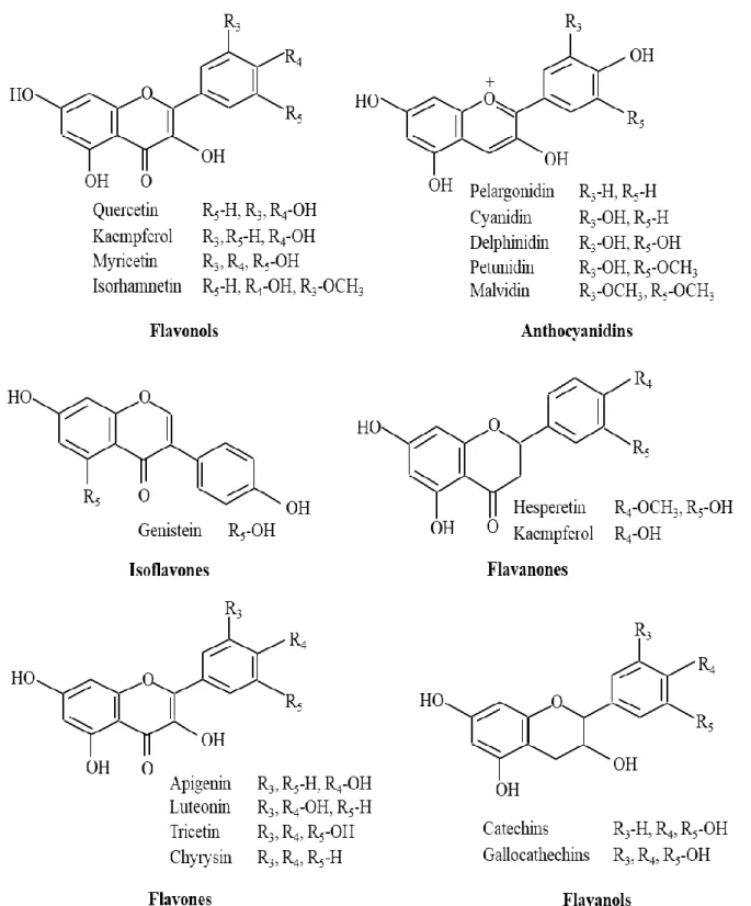
1-3) (Brown, 1980). Flavonoids may be subdivided into six subgroups: flavones, flavonols, flavanones, anthocyanidins, flavanols and chalcones (Fig. 1-4).
11
Figure 1-4. Structures of different classes of flavonoids
The health benefits of flavonoids in humans are associated with their antioxidant, anticarcinogenic anti-inflammatory and antibacterial activities. Many flavonoids show antioxidative activity, anticancer activity, preventing coronary heart disease and free radical scavenging activity, while some other flavonoids are shown to have functions of
12
anti-human immunodeficiency virus (Brown et al., 1998; Mishra et al., 2013). The antioxidant activity of flavonoids includes suppression of ROS generation, scavenging ROS, and upregulation of antioxidant defenses (Mishra et al., 2013). Hepatoprotective activities were also shown in several flavonoids such as rutin, catechin, naringenin, apigenin, quercetin, and venoruton (Tapas et al., 2008). It has been reported that liver damage was improved by treatment with flavonoids in histopathological examinations (Wu et al., 2006). According to the report of Cushnie and Lamb, some flavonoids (such as isoflavones, flavonol glycosides, apigenin, quercetin, flavone, chalcones and galangin) have been shown to have potent antibacterial activity (Cushnie & Lamb, 2005a). Catechin was reported to have antibacterial activity against Vibrio cholerae, Shigella, Steptococcus mutans and some other bacteria (Moerman, 1996). In addition, the anti-inflammatory activity of flavonoids was shown through their inhibitory effects on enzymes which are responsible for the inflammatory responses. For example, cyclooxygenase-2 that is responsible for the production of inflammatory mediators was inhibited by luteolin and quercetin (Kimata et al., 2000). Rutin effectively inhibited phospholipases A2 that can increase inflammatory response (Lindahl & Tagesson, 1997)). Hesperidin, a flavanone glycoside, inhibited induction of mammary and colon cancers in rats by azoxymethanol (Ren et al., 2003). The antiviral activity of various flavonoids has been reported, such as the inhibitory effect of baicalin on HIV-1 infection and replication (Li et al., 2000), and inhibition of DNA polymerases of HIV-1 by catechin (Cushnie & Lamb, 2005a). Furthermore, several flavonoids also showed the antitoxin activities. For example, the mixture of catechins inhibited the production and activity of Stx2 (Okubo et al., 1998). The extracellular release of both Stx1 and Stx2 from EHEC were inhibited by gallocatechin gallate (GCg) and epigallocatechin gallate (EGCg) (Sugita-Konishi et al., 1999).
13 1.3.2 Phenolic acids
Phenolic acids account for around 30% of all polyphenolic compounds. They are mostly found in vegetables and fruits and play a vital role in the formation of unique taste and flavor of food quality. Some common phenolic acids include caffeic acid, ferulic acid, sinapic acid and gallic acid. The basic structure of phenolic acids contains an aromatic ring and an organic carboxylic acid function (Fig. 1-3) (Brown, 1980). They were divided into two groups: hydroxycinnamic acid and hydroxybenzoic acid (Daglia et al., 2014). Phenolic acids have beneficial effects on human health because they act as antioxidants that significantly delay cellular damage by scavenging hydroxyl radical (Kukić et al., 2006).
Some studies showed that phenolic acids have a major role in the prevention of many chronic illnesses because of their contribution to the intake of natural antioxidants in human diets (Balasundram et al., 2006; El-Seedi et al., 2012; Upadhyay and Rao, 2013). It has been reported that the antioxidant activity of phenolic acids is more effective than that of vitamin E or vitamin C (Tsao & Deng, 2004). Furthermore, phenolic acids are responsible for anticancer activity. Several phenolic acids (such as gallic acid, ferulic acid, and caffeic acid) showed high anticancer activities (Hwang et al., 2006; Madlener et al., 2007). The inhibitory effect of caffeic acid on human colon cancer cells was also reported (Jaganathan, 2012). The possible role of phenolic acid in inhibiting the toxin-production by microorganism has been reported. The toxin produced by Clostridium botulinum was inhibited by gallic acid (Reddy et al., 1982). Phenolic acids including chlorogenic, ferulic and p-coumaric acids showed the inhibitory effects on the growth and production of HT-2 and T-2 toxins by Fusarium sporotrichioides and Fusarium langsethiae (Ferruz et al., 2016a, 2016b).
14 1.3.3 Stilbenes
Stilbenes are found in many plant species such as sorghum, grapes, and peanut (Chong et al., 2009). They contain two phenyl moieties connected by a two-carbon methylene bridge (Fig. 1-3) (Brown, 1980). Most stilbenes work as antifungal phytoalexins that are synthesized in response to injury, infection, fungal attacks and UV irradiation (Reinisalo et al., 2015). Resveratrol, a stilbenoid polyphenol found largely in grapes, is a phytoalexin that acts against bacterial and fungal pathogens (Beekwilder et al., 2006; Wang et al., 2011; Li et al., 2015; Nawaz et al., 2017). Resveratrol was also shown strong inhibitory effects on VacA toxin producing by Helicobacter pylori (Tombola et al., 2003).
1.3.4 Lignans
Lignans are diphenolic compounds found in many plants and food, including wine, tea, nuts, grains, seeds and vegetables (Suzuki & Umezawa, 2007). The concentration of lignans found in sesame and flax seeds is higher than that in other foods (Landete, 2012).
Lignans comprise of two propylbenzene unit linked by a β, β’-bond (Fig. 1-3) (Brown, 1980). Based on the cyclization pattern and the oxygen incorporation into the molecule, lignans may be subdivided into 8 subgroups: arynaphthalenes, dibenzylbutanes, furofurans, dibenzylbutyrolactones, aryltetralins, dibenzocylcooctadienes, and dibenzylbutyrolactols (Pan et al., 2009). Lignans express various biological characteristics, including antioxidant, antitumor and anti-inflammatory activity (Ionkova, 2011).
1.4 Antibacterial activity of polyphenols
Polyphenols are an important compound for the finding and the development of novel antibacterial drugs because of their strong antibacterial activities. In vitro, many studies reported that polyphenols have antibacterial activities against Gram-negative and Gram-positive bacteria. The mechanisms of the antibacterial activity of polyphenols are
15
being increasingly recorded. Due to the structural variety of polyphenol groups, it is impossible to predict the interaction between bacteria and polyphenol which are responsible for the antibacterial activities of polyphenol.
Based on the structural differences of some flavonoids (such as dihydrobiochanin A, darbergioidin and ferreirin) to evaluate their antibacterial activities by using paper disk agar diffusion assay, Osawa et al. (1992) suggested that 5,2,4-trihydroxyl-7- methoxyisoflavanone inhibited the growth of Streptococcus cricetus, S. mutans, S.
sobrinus, and S. rattus, in which 5-hydroxyisoflavanones and 5-hydroxyflavanones with hydroxyl group at position 2’ are important for their antibacterial activity. Tsuchiya et al.
(1996) indicated that 5,7-dihydroxylation of the A ring and 2’,4’- or 2’,6’-dihydroxylation of the B ring in flavanone structures are essential to antibacterial activity against methicillin resistant Staphylococcus aureus, while substitution at the 6 or 8 position with a certain aliphatic group also enhanced this activity.
Cowan (1999) suggested that the binding of OH group or OH groups to both the aromatic rings and the oxygen substituted ring linked to the antimicrobial activities of polyphenols. It has been reported that the antibacterial activities of flavonoids are closely related to the number, positions and alkylation of OH groups, the backbone structure, and the presence of glycoside linkages (Liu et al., 2010; Wu et al., 2013b). Mori et al. (1987) suggested that myricetin, (−)-epigallocatechin and robinetin significantly inhibited DNA, protein and lipid syntheses in Proteus vulgaris. The inhibitory effects on these syntheses were attributed to the role of ring B of flavonoids in intercalation or hydrogen bonding with the stacking of nucleic acid bases (Mori et al., 1987).
Some flavonoids, including isoflavones, flavonol glycosides, apigenin, chalcones, quercetin, flavone, and galangin have been shown to have strong antibacterial activity (Cushnie & Lamb, 2005a). Taguri et al. (2004) demonstrated that 10 types of polyphenols including proanthocyanidins, hydrolyzable tannins, and catechins and their oxidation products,
16
inhibited the growth of four groups of foodborne bacteria such as Staphylococcus aureus, Escherichia coli, Salmonella, and Vibrio. The susceptibility of bacteria to polyphenol is related to the structure of both polyphenol and bacterial strain. Taguri et al. (2004) also suggested that 3,4,5-trihydroxyphenyl groups play an important role in the antibacterial activities of epigallocatechin, epigallocatechin-3-O-gallate, prodelphinidin and castalagin which show relatively strong antimicrobial activities.
Several studies proposed that EGCg inhibited the growth of bacteria by inhibiting the function of cytoplasmic membrane (Cushnie & Lamb, 2005b; Nakayama et al., 2008, 2013).
Cushnie and Lamb (2005b) showed that galangin (3,5,7-trihydroxyflavone) induced a significant increase in potassium leakage in Staphylococcus aureus cells, which may be associated with the cytoplasmic membrane damage of the bacterium.
1.5 Antitoxin activity of polyphenols
Various bacterial toxins were inhibited by polyphenols. Polyphenols could directly inhibit the activities of toxin, or could block the binding of the toxins to the receptor.
Resveratrol inhibited cholera toxin (CT) activity against Vero cell by suppressing the toxin activity and toxin internalization (Morinaga et al., 2010). Several plant polyphenols have been shown the inhibitory effects on CTA1 by directly inhibiting the catalytic activity of the toxin (Oi et al., 2002). Some polyphenol extracts inactivated heat-labile enterotoxin (LT) produced by enterotoxigenic E. coli by inhibiting the binding of LT to its intestinal receptor GM1(Verhelst et al., 2013). The inhibitory activity of EGCg on cholera toxin appear to specifically disrupt CT-GM1 interactions by its binding with toxin (Cherubin et al., 2016), in contrast to the inhibition of LT-GM1 interaction by toxin precipitation with EGCg (Verhelst et al., 2013).
Several polyphenols could aggregate bacterial toxins by sharing a common structural organization that contains catalytic A subunit and a cell binding B subunit
17
(Friedman, 2007). It has been reported that the inhibitory activity of EGCg against the cytotoxicity of Stx2 is due to forming a binding structure at the pocket of Stx2B subunit pentamer (Miyamoto et al., 2014). Caffeic acid protected Vero cells from the cytotoxicity of ochratoxin A by altering principally the lysosomal function in Vero cells (Cariddi et al., 2015). Yahiro et al. (2005) reported that the inhibitory activity of polyphenols against VacA toxin produced by Helicobacter pylori might be due to forming of a complex between polyphenol and toxin or inhibiting binding of toxin to cell receptors in the digestive tract.
Bacterial toxins are proteins and their virulence was determined by 3-D conformation. Thus, the change of the native structural integrity of these proteins could inactivate the activities of bacterial toxin by preventing molecular interactions with cell membrane receptors of host cells (Friedman, 2007). EGCg strong inhibited anthrax toxin produced by Bacillus anthracis by binding between phenolic OH groups of EGCg to the zinc atom linked to the metalloproteinase of toxins (Friedman et al., 1986; Benelli et al., 2002). Some polyphenols isolated from black tea also inhibited the botulinum neurotoxin produced by Clostridium botulinum by binding with the metalloproteinase part of toxin (Satoh et al., 2002). The inhibitory effects of catechins on the production and extracellular release of Vero toxins produced by E. coli O157:H7 appears to be caused by interference of the transfer of periplasmic proteins through the outer membrane of bacterial cells by catechins (Sugita-Konishi et al., 1999).
In addition, the specific structure of each polyphenol is also responsible for its antitoxin activity. EGCg inhibited CT, exotoxin A, diphtheria toxin and ricin, whereas epicatechin gallate which differs from EGCg by a single OH group only inhibited exotoxin A (Cherubin et al., 2016). Verhelst et al. (2013) suggested that the presence of at least two galloyl moieties in polyphenol structure is essential for the inhibitory effects of polyphenol on enterotoxin. Pentagalloyl glucose which contains more galloyl moieties than EGCg and GCg has stronger inhibitory activity on LT than EGCg and GCg (Verhelst et al., 2013).
18 1.6 Purpose of this study
EHEC causes foodborne illness, and if it becomes severe, it causes hemorrhagic colitis and potentially fatal hemolytic uremic syndrome due to the action of Stx produced by the bacterium. The beneficial effect of polyphenols on human health is believed to be due to their various properties such as antibacterial, antioxidant and antitoxin activities.
They have been reported to inhibit the growth of various bacteria and the activities of toxins produced by bacteria. The combinations of polyphenols with other antimicrobial agents have also shown synergistic effects against many bacteria. In this study, to obtain basic evidences for the use of plant polyphenols for controlling EHEC, antibacterial activities of the tea polyphenol formulations were used to investigate the combined effects with some food additives and heat treatment. It has been shown that the cytotoxicity of Stx1 decreased after pre-incubation with GCg and EGCg. However, the cytotoxicity of Stx2 was not inhibited by preincubation with catechins and theaflavins. To find out plant polyphenols effective against both Stx1 and 2, phenolic compounds were screened by docking simulation based on the analysis of interaction between Stx B subunit pentamers and natural products from the natural compounds database. The effects of selected compounds on the cytotoxicity of Stxs were investigated. Since baicalein inhibited the cytotoxicity of both Stx1 and 2 among compounds selected by in silico screening, effects of baicalein on cytotoxicity and productivity of Stxs were investigated in detail.
19
Chapter 2. The combined effects of tea polyphenols and food additives against enterohaemorrhagic Escherichia coli
2.1 Introduction
Enterohaemorrhagic Escherichia coli (EHEC) is an important foodborne pathogen causing many diseases, and it can lead to hemolytic uremic syndrome (HUS) in humans (Kaper et al., 2004). Escherichia coli O157:H7, a serotype in EHEC, is one of the most important foodborne pathogens. E. coli O157:H7 can survive in acidic conditions (as low as pH 2.0) (Leyer et al., 1995; Diez-Gonzalez et al., 1998) and grow in TSB containing high concentration of salt up to 6.5% NaCl or at pH from 4.5 to 9.0 (Glass et al., 1992). Many antibiotics are contraindicated in treatment for E. coli O157:H7-infected patients because they might increase the risk of the hemolytic uremic syndrome (Wong et al., 2000).
The beneficial effect of tea polyphenols on health is believed to be due to their strong antioxidant activity (Forester & Lambert, 2011). Moreover, tea polyphenols also have anti-inflammatory, antiviral and anticancer activities (Serafini et al., 2011).
Epigallocatechin gallate (EGCg), one of the green tea catechins, has been reported to inhibit the growth of both Gram-positive and Gram-negative bacteria, including E. coli O157:H7 (Nakayama et al., 2008). EGCg inhibited the growth of E. coli by direct interaction with proteins in the outer membrane of E. coli (Nakayama et al., 2013).
Nakayama et al. (2008) also indicated that the antibacterial activity of green tea extract increased in the presence of 4% NaCl and have also shown its combined effects with various food additives against both E. coli O157:H7 and Listeria monocytogenes.
Theaflavins (TF), a group of polyphenols, are formed from polymerization of catechins during the fermentation of black tea. Theaflavin (TF1), theaflavin-3-gallate, theaflavin-3- gallate (TF2A, TF2B), and theaflavin-3,3-digallate (TF3) are the major TF, specifically TF3
20
has strongest antioxidant activity among all catechins and TF derivatives (Leung et al., 2001).
Food additives are substances added to food for various purposes such as inhibiting the growth of bacteria, preserving fresh attributes and nutritional quality etc. Some food additives have antibacterial activities against various foodborne pathogens. For example, sodium citrate (Na citrate) and sodium acetate (Na acetate) can inhibit the growth of several bacteria such as Staphylococcus epidermidis, S. aureus and E. coli (Lee et al., 2002; McWilliam & Stewart, 2002), sodium chloride (NaCl) inhibits the growth of Pediococcus acidilactici (Aina, 2017), S. aureus and E.coli (Omotoyinbo, 2016) by lowering the water activity. Moreover, ethylenediaminetetraacetic acid (EDTA) and ethanol are also capable of inhibiting the growth of bacteria (Farca et al., 1997; Brewer et al., 2002; Gill & Holley, 2003). Although widely used in food industry, food additives were viewed as a major risk to health because they could generate harmful chemical changes and should be considered as potentially toxic materials. Trasande et al. (2008) have been reported that certain food additives could cause endocrine disruption and other potential adverse effects on consumer health, especially children’s health. In food industry, food additives can be used alone or in combination. The combination of food additives has been increasing interest because the combination of two or more food additives could increase their antibacterial effects and produce synergistic effects. For example, ethanol enhanced the sensitivity of Listeria monocytogenes to nisin, NaCl and organic acids (Barker & Park, 2001; Brewer et al., 2002), the combination nisin and EDTA also reduced the growth of Salmonella (Ukuku & Fett, 2004), and E. coli (Field et al., 2017). However, the antibacterial activity of food additives in combination with tea polyphenols on the growth of E. coli O157:H7 have been rarely reported.
In this study, the combined effects of EGCg and theaflavin with some food additives including EDTA, ethanol, Na acetate, Na citrate and NaCl on the growth of E.
21
coli O157:H7 were evaluated by determining minimum inhibitory concentration (MIC).
The fractional inhibitory concentrations (FIC) index were further determined through the study on the modes of actions of the compounds using time kill assays. The synergistic effects of these combinations could be a way to overcome the risk of using food additives.
2.2 Materials and methods 2.2.1 Preparation of bacteria
E. coli O157:H7 No.28 strain was used in this study. The bacterium was inoculated into 5 mL of Tryptic Soy Broth (TSB) medium (Becton, Dickinson & Co., Franklin Lakes, NJ, USA) and cultured overnight at 37oC with shaking at 130 rpm to obtain cells in the stationary phase of growth. This culture was then adjusted by dilution in sterilized water to a final OD660 of 0.10 (bacterial concentration, ca. 108 CFU/mL). The bacterial cells were harvested by centrifugation (6,000 g, 5 min, 25oC), and then were suspended in sterile water to attain a final concentration of ca. 108 CFU/mL for use in subsequent experiments.
2.2.2 Preparation of tea polyphenol and food additive solutions
The tea polyphenol products used in this study were food additive formulation including EGCg or theaflavins. Teavigo containing 95%(w/w) EGCg was purchased from DSM Nutrition Japan, Tokyo, Japan. TF40, a theaflavins extract containing 40%(w/w) theaflavins, was purchased from Yaizu Seisakusho Kogyo Co., Japan. To prepare tea polyphenol solutions, Teavigo and TF40 powders were dissolved in sterilized water and in 0.1 % dimethyl sulfoxide (DMSO), respectively. These solutions were sterilized by filtration through a Millex-GP 0.22 μm filter (Merck Millipore, Billerica, MA, USA).
Food additives used in this study were EDTA, NaCl, Na acetate, Na citrate and ethanol. All the reagents were of analytical grade. To prepare working solutions, reagents were dissolved in sterilized water and then autoclaved at 121oC for 15 min.
22
After autoclaving, these solutions were stored at room temperature to use in the following experiments.
2.2.3 Determination of the minimum inhibitory concentration (MIC)
Minimum inhibitory concentration (MIC) determinations were performed using broth dilution method in test tube as described previously (Wiegand et al., 2008). To 3.8 mL of glucose added ammonium (GA) medium (0.909 g/L (NH4)2HPO4, 0.182 g/L KCl, 0.182 g/L MgSO4/7H2O, 0.182 g/L yeast extract and 9.091 g/L glucose, pH 6.5) in separate test tube, 100 µL of each compound with various concentration were added. And then, 100 µl of bacterial suspension with a final concentration of 2 x 10⁵ CFU/mL was inoculated to these tubes. The inoculated tubes were incubated at 37oC for 24 h. The MIC was defined as the lowest concentration of compounds which showed no visible bacterial growth.
Final concentrations of each compound were used as follows: 2000, 1500, 1000, 500 and 250 μg/mL for Teavigo, 1000, 750, 500 and 250 μg/mL for TF40, 0.1, 0.05, 0.025, 0.0125 and 0.00625 (%) for EDTA, 0.5, 0.25, 0.125, 0.0625 and 0.03125 (%) for Na acetate, 12, 8, 4, 2, 1, 0.5 (%) for Na citrate, and 25, 20, 15, 10, 5 and 1 (%) for ethanol.
2.2.4 Synergy testing of tea polyphenols with food additives using broth dilution method in test tube
To determine the synergistic interaction between tea polyphenols and food additives, the fractional inhibitory concentrations (FIC) index was calculated using formula:
FIC = ([A]/MICA) + ([B]/MICB)
[A] and [B] are the MIC of each compound when used together.
MICA and MICB are the MIC of each compound when used alone.
The interaction effects were determined as follows: FIC ≤ 0.5, synergy; 0.5 < FIC ≤ 0.75, partial synergy; 0.75 < FIC ≤ 1.0, additive effect; 1.0 < FIC ≤ 4.0, indifference, and 4.0 < FIC, antagonism (Berenbaum, 1981).
23 2.2.5 Time-kill assays
The time-kill assays were performed with a final concentration of bacterial inoculation about 2 x 10⁵ CFU/mL in a final volume of 4 mL in a test tube. The samples and the control (a bacterial suspension in GA) were incubated at 37oC. Viable cell count was determined at 0, 3, 6, 9 and 24 h incubation.
2.2.6 Viable cell count
Each sample was divided into 1 mL aliquots, and the sample was 10-times serially diluted with phosphate buffered saline (PBS). One hundred microliters of each dilution were spreaded onto tryptic soy agar (TSA, Becton, Dickinson & Co., Franklin Lakes, NJ, USA) plate. After cultivation of the plates for 48 h at 37oC, the number of formed colonies was counted, and then the viable cell counts were calculated.
2.2.7 Statistical analysis
Viable cell counts were determined for three separate experiments and analyzed in Microsoft Excel 2010 (Microsoft, Seattle, WA, USA). Statistical significance of the viable counts was determined by Student’s T-test. Multiple comparisons of the viable cell count between differently treated sample groups were performed using the Tukey- Kramer’s multiple comparison post hoc test followed by the one-way ANOVA by Statcel 3 (Yanai, 2011), which is an add-in application in Microsoft Excel.
2.3 Results
2.3.1 Antibacterial effect of tea polyphenols and food additives alone
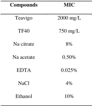
The antibacterial effect of the test compounds against E. coli O157:H7 was evaluated by MIC determinations. The MIC values of 2 tea polyphenol formulations and 5 food additives against E. coli O157:H7 are summarized in Table 2-1. For 2 tea polyphenol
24
formulations, TF40 showed the stronger inhibitory effect with MIC value about 2.7-fold lower compared to that of Teavigo. Among 5 food additives, EDTA exhibited the highest inhibitory effect against E. coli O157:H7 (MIC: 0.025%), whereas ethanol was the weakest inhibitor (MIC: 10%). The order of strong antibacterial activity was EDTA > Na acetate >
NaCl > Na citrate > ethanol.
Table 2-1. Minimum inhibitory concentration (MIC) of tea polyphenols and food additives against E. coli O157:H7
Compounds MIC
Teavigo 2000 mg/L
TF40 750 mg/L
Na citrate 8%
Na acetate 0.50%
EDTA 0.025%
NaCl 4%
Ethanol 10%
2.3.2 Combined antibacterial effect of tea polyphenols and food additives
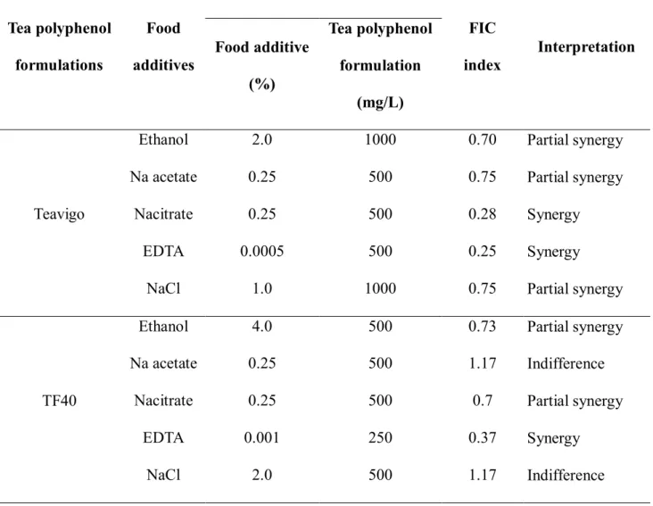
To evaluate whether the antibacterial activities of tea polyphenol formulations can be improved by the combined use with food additives, FIC determinations were carried out.
When 5 food additives used in this study were combined with 2 tea polyphenols formulations, synergistic effects on the inhibition of growth of E. coli O157:H7 were observed. The combined effects of food additives and Teavigo or TF40 against E. coli O157:H7 were shown in Table 2-2. In general, there were 2 synergistic effects that observed in the combination of Teavigo with EDTA or Na citrate, whereas the synergistic effect was only observed in the combination of TF40 with EDTA. The partial synergistic
25
effect was observed in the combination of Teavigo and ethanol, NaCl or Na acetate against E. coli O157:H7. However, the combination of TF40 with ethanol or Na citrate also showed the partial synergistic effect. There was no combined effect of TF40 and Na acetate or NaCl in this study.
Table 2-2. Minimum inhibitory concentration (MIC) and fractional inhibitory concentration (FIC) index of tea polyphenol formulations and food additives in
combination against E. coli O157:H7
Tea polyphenol formulations
Food additives
MIC
FIC
index Interpretation Food additive
(%)
Tea polyphenol formulation
(mg/L)
Teavigo
Ethanol 2.0 1000 0.70 Partial synergy
Na acetate 0.25 500 0.75 Partial synergy
Nacitrate 0.25 500 0.28 Synergy
EDTA 0.0005 500 0.25 Synergy
NaCl 1.0 1000 0.75 Partial synergy
TF40
Ethanol 4.0 500 0.73 Partial synergy
Na acetate 0.25 500 1.17 Indifference
Nacitrate 0.25 500 0.7 Partial synergy
EDTA 0.001 250 0.37 Synergy
NaCl 2.0 500 1.17 Indifference
The concentration of Teavigo required for inhibition of the growth of E. coli O157:H7 in the presence of food additives decreased as follows: 1/2 MIC in the presence of ethanol or NaCl, and 1/4 MIC in the presence of Na acetate or Na citrate or EDTA. The combination of TF40 with food additives, the concentration of TF40 required for inhibition
26
of the growth of E. coli O157:H7 decreased to 2/3 MIC in the presence of EDTA and 1/3 MIC in the presence of other 4 food additives (NaCl, ethanol, Na citrate or Na acetate).
2.3.3 Time kill assay of the combined use
To examine the mode of combined effect of tea polyphenol formulations and food additives in more detail, effects of tea polyphenol formulations and food additives on the growth of E. coli O157:H7 were determined.
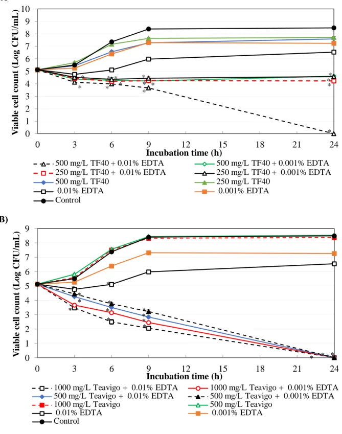
For TF40, the combination of 500 mg/L TF40 and 0.01% EDTA reduced the viable cell counts of E. coli O157:H7 to below detection limit after 24 h incubation. The antibacterial activity of combinations of 500 mg/L TF40 and 0.001 % EDTA, 250 mg/L TF40 and 0.01 and 0.001% EDTA were more effective than that of single use (Fig. 2-1A).
When Teavigo (500 and 1000 mg/L) was used in combination with EDTA (either at 0.01%
or at 0.001%), the viable cell counts decreased to below lower detection limit after 24 h incubation (Fig. 2-1B). Teavigo and TF40 reduced EDTA concentration to inhibit the growth of E. coli O157:H7 to 1/50 and 1/25, respectively (Table 2-1 and 2-2).
27
Figure 2-1. Combined effect of tea polyphenol formulations and EDTA on the growth of E. coli O157:H7. The mixtures of EDTA and TF40 (A) or Teavigo (B) were added to E. coli O157:H7 culture and incubated at 37oC. Viable cell count was determined at 0, 3, 6, 9 and 24 h incubation. Values are average ± SD for 3 separate experiments. *, P < 0.05.
B) A)
0 1 2 3 4 5 6 7 8 9
0 3 6 9 12 15 18 21 24
Viable cell count (Log CFU/mL)
Incubation time (h)
1000 mg/L Teavigo + 0.01% EDTA 1000 mg/L Teavigo + 0.001% EDTA 500 mg/L Teavigo + 0.01% EDTA 500 mg/L Teavigo + 0.001% EDTA
1000 mg/L Teavigo 500 mg/L Teavigo
0.01% EDTA 0.001% EDTA
Control
*
* *
* *
* *
*
*
* *
*
* * * *
0 1 2 3 4 5 6 7 8 9 10
0 3 6 9 12 15 18 21 24
Viable cell count (Log CFU/mL)
Incubation time (h)
500 mg/L TF40 + 0.01% EDTA 500 mg/L TF40 + 0.001% EDTA 250 mg/L TF40 + 0.01% EDTA 250 mg/L TF40 + 0.001% EDTA
500 mg/L TF40 250 mg/L TF40
0.01% EDTA 0.001% EDTA
Control
* *
*
*
*
* *
* * * *
* *
* *
28
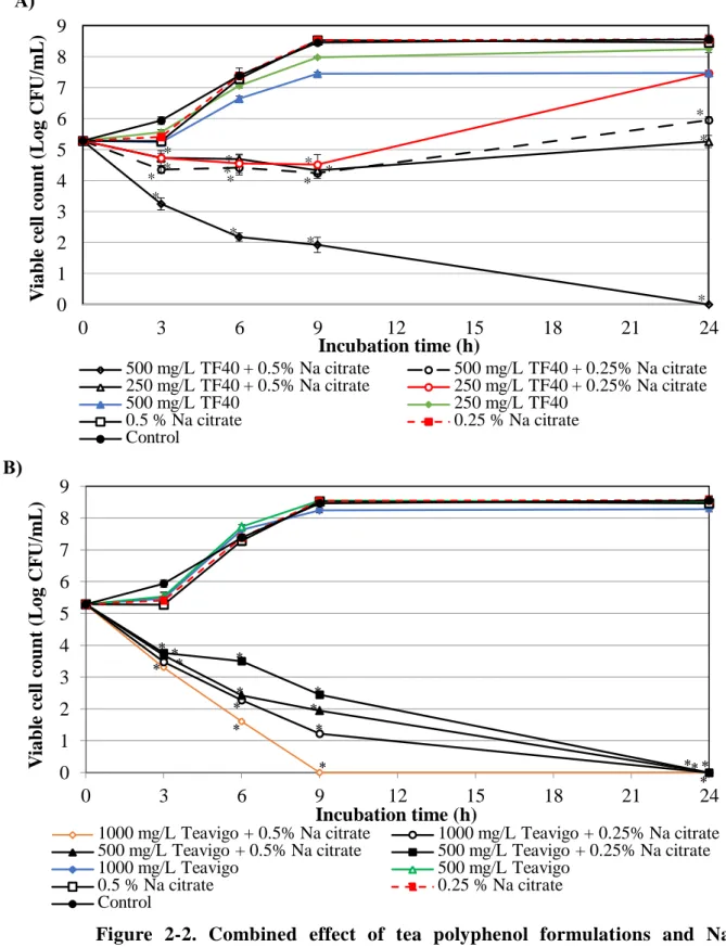
Similar to the combination with EDTA, inhibitory effects of Na citrate were observed in both the combination with TF40 and Teavigo (Fig. 2-2 A and B). A rapid decrease of the viable cell counts was observed in the combination of 1000 mg/L Teavigo and 0.5% Na citrate. The viability of E. coli O157:H7 decreased to below lower detection limit after incubation for 9 h (Fig. 2-2B). After 24 h incubation, the combination of 1000 mg/L Teavigo and 0.25% Na citrate, 500 mg/L Teavigo and Na citrate (0.5% or 0.25%), and the combination of 500 mg/L TF40 and 0.5% Na citrate a lso reduced the viable cell counts to below lower detection limit. This combined effect was not observed in the combination of 500 mg/L TF40 and 0.25% Na citrate, TF40 250 mg/L and Na citrate (0.5% or 0.25%), but their combination effects against E. coli O157:H7 were stronger than that of single use (Fig. 2-2 A and B). There is a 16 and 32- fold reduction of Na citrate concentration for inhibition of the growth of E. coli O157:H7 in the combination with Teavigo and TF40, respectively (Table 2-1 and 2-2).
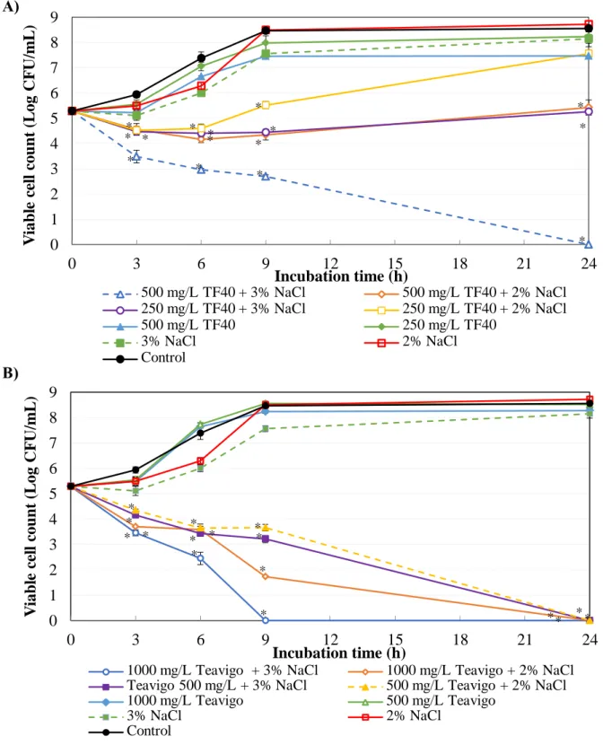
Figure 2-3 shows the time-kill curves of tea polyphenol formulations, NaCl and their combination against E. coli O157:H7. Although the combination of TF40 and NaCl did not show a significant combined effect, the combination of 500 mg/L TF40 and 3%
NaCl showed bactericidal effects after 24 h incubation (Fig. 2-3A). When the concentration of TF40 was further decreased to 250 mg/L, bactericidal effect was not observed in the combination with 2% and 3% NaCl. However, the growth of E. coli O157:H7 was inhibited by the combination of 500 mg/L TF40 and 2% NaCl, and 250 mg/L TF40 and 3% NaCl. The combination of Teavigo (1000 mg/L or 500 mg/L) and NaCl (2% or 3%) reduced viable cell counts to below lower detection limit after 24 h incubation (Fig. 2-3B).
0 1 2 3 4 5 6 7 8 9
0 3 6 9 24
Viable cell count (Log CFU/ml)
Time incubation (h)
TF40 500 mg/L + 3% NaCl TF40 500 mg/L + 2% NaCl TF40 250 mg/L + 3% NaCl TF40 250 mg/L + 2% NaCl
TF40 500 mg/L TF40 250 mg/L
3% NaCl 2% NaCl
Control
29
Figure 2-2. Combined effect of tea polyphenol formulations and Na citrate on the growth of E. coli O157:H7. The mixtures of Na citrate and TF40 (A) or Teavigo (B) were added to E. coli O157:H7 culture and incubated at 37oC. Viable cell count was determined at 0, 3, 6, 9 and 24 h incubation. Values are average ± SD for 3 separate experiments. *, P < 0.05.
B) 0 1 2 3 4 5 6 7 8 9
0 3 6 9 12 15 18 21 24
Viable cell count (Log CFU/mL)
Incubation time (h)
500 mg/L TF40 + 0.5% Na citrate 500 mg/L TF40 + 0.25% Na citrate 250 mg/L TF40 + 0.5% Na citrate 250 mg/L TF40 + 0.25% Na citrate
500 mg/L TF40 250 mg/L TF40
0.5 % Na citrate 0.25 % Na citrate
Control
*
*
*
*
*
*
*
*
* *
*
* *
*
*
0 1 2 3 4 5 6 7 8 9
0 3 6 9 12 15 18 21 24
Viable cell count (Log CFU/mL)
Incubation time (h)
1000 mg/L Teavigo + 0.5% Na citrate 1000 mg/L Teavigo + 0.25% Na citrate 500 mg/L Teavigo + 0.5% Na citrate 500 mg/L Teavigo + 0.25% Na citrate
1000 mg/L Teavigo 500 mg/L Teavigo
0.5 % Na citrate 0.25 % Na citrate
Control
* * *
*
*
*
* *
*
*
*
*
* * *
*
A)
30
Figure 2-3. Combined effect of tea polyphenol formulations and NaCl on the growth of E. coli O157:H7. The mixtures of NaCl and TF40 (A) or Teavigo (B) were added to E. coli O157:H7 culture and incubated at 37oC. Viable cell count was determined at 0, 3, 6, 9 and 24 h incubation. Values are average ± SD for 3 separate experiments. *, P < 0.05.
B) A)
0 1 2 3 4 5 6 7 8 9
0 3 6 9 12 15 18 21 24
Viable cell count (Log CFU/mL)
Incubation time (h)
500 mg/L TF40 + 3% NaCl 500 mg/L TF40 + 2% NaCl 250 mg/L TF40 + 3% NaCl 250 mg/L TF40 + 2% NaCl
500 mg/L TF40 250 mg/L TF40
3% NaCl 2% NaCl
Control
*
*
* *
*
*
* * *
*
*
*
*
*
*
0 1 2 3 4 5 6 7 8 9
0 3 6 9 12 15 18 21 24
Viable cell count (Log CFU/mL)
Incubation time (h)
1000 mg/L Teavigo + 3% NaCl 1000 mg/L Teavigo + 2% NaCl Teavigo 500 mg/L + 3% NaCl 500 mg/L Teavigo + 2% NaCl
1000 mg/L Teavigo 500 mg/L Teavigo
3% NaCl 2% NaCl
Control
* * *
*
* * *
*
* *
*
*
*
*
* *
31
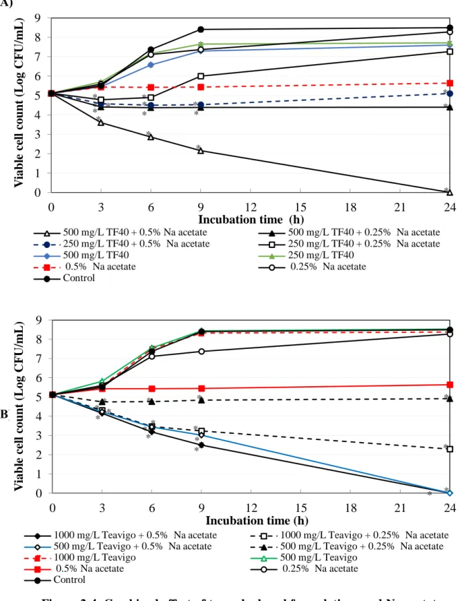
Figure 2-4. Combined effect of tea polyphenol formulations and Na acetate on the growth of E. coli O157:H7. The mixtures of Na acetate and TF40 (A) or Teavigo (B) were added to E. coli O157:H7 culture and incubated at 37oC. Viable cell count was determined at 0, 3, 6, 9 and 24 h incubation. Values are average ± SD for 3 separate experiments. *, P < 0.05.
B) A)
0 1 2 3 4 5 6 7 8 9
0 3 6 9 12 15 18 21 24
Viable cell count (Log CFU/mL)
Incubation time (h)
500 mg/L TF40 + 0.5% Na acetate 500 mg/L TF40 + 0.25% Na acetate 250 mg/L TF40 + 0.5% Na acetate 250 mg/L TF40 + 0.25% Na acetate
500 mg/L TF40 250 mg/L TF40
0.5% Na acetate 0.25% Na acetate
Control
* *
*
*
* *
*
*
* * * *
*
*
*
0 1 2 3 4 5 6 7 8 9
0 3 6 9 12 15 18 21 24
Viable cell count (Log CFU/mL)
Incubation time (h)
1000 mg/L Teavigo + 0.5% Na acetate 1000 mg/L Teavigo + 0.25% Na acetate 500 mg/L Teavigo + 0.5% Na acetate 500 mg/L Teavigo + 0.25% Na acetate
1000 mg/L Teavigo 500 mg/L Teavigo
0.5% Na acetate 0.25% Na acetate
Control
*
*
*
*
*
*
* *
* *
*
*
*
*
* *
32
Figure 2-4 shows the combined effect of the tea polyphenol formulations and Na acetate against E. coli O157:H7. The growth of E. coli O157:H7 was observed in the combination of 250 mg/L TF40 and 0.25% Na acetate, while the combination of 250 mg/L TF40 and 0.5% Na acetate, and 500 mg/L TF40 and 0.25% Na acetate slightly inhibited the growth of E. coli O157:H7 (Fig. 2-4A). In the combination of 500 mg/L TF40 and 0.5% Na acetate, the viable cell counts were decreased to below lower detection limit after 24 h incubation (Fig. 2-4A). However, the combined antibacterial effects of TF40 and Na acetate were not significantly strong compared with those of the single use (Table 2-2).
The viable cell counts slightly decreased in combination of 1000 mg/L Teavigo and 0.25%
Na acetate. Teavigo inhibited the growth of E. coli O157:H7 and significantly reduced the viable counts to below detection limit at both 1000 mg/L and 500 mg/L in the combination with 0.5% Na acetate after 24 h incubation (Fig. 2-4B).
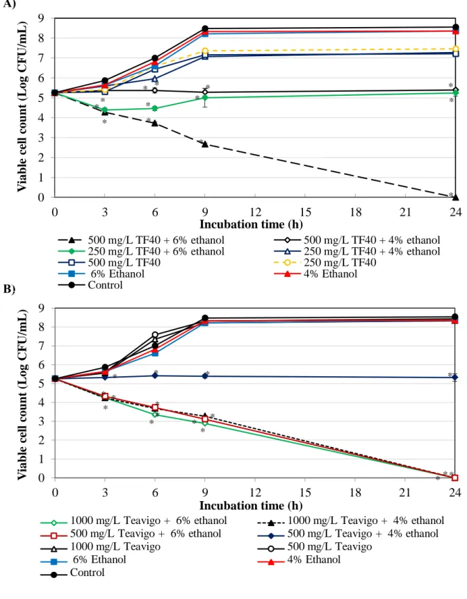
Combined effect of the polyphenol formulations and ethanol on the growth of E.
coli O157:H7 is shown in Figure 2-5. The growth of E. coli O157:H7 was observed in the combination of 250 mg/L TF40 and 4% ethanol. The combinations of 500 mg/L TF40 and 4% ethanol, and 250 mg/L TF40 and 6% ethanol slightly inhibited the growth of E. coli O157:H7. After 24 h incubation, the viability of E. coli O157:H7 was decreased to below lower detection limit in the combination of 500 mg/L TF40 and 6% ethanol (Fig. 2-5A).
The combinations of 1000 mg/L Teavigo and ethanol at both 4% and 6%, and 500 mg/L Teavigo and 6% ethanol also reduced the viable cell count to below lower detection limit after 24 h incubation (Fig. 2-5B). The growth of E. coli O157:H7 was inhibited by the combination of 500 mg/L Teavigo and 4% ethanol.
33
Figure 2-5. Combined effect of tea polyphenol formulations and ethanol on the growth of E. coli O157:H7. The mixtures of ethanol and TF40 (A) or Teavigo (B) were added to E. coli O157:H7 culture and incubated at 37oC. Viable cell count was determined at 0, 3, 6, 9 and 24 h incubation. Values are average ± SD for 3 separate experiments. *, P < 0.05.
B) A)
0 1 2 3 4 5 6 7 8 9
0 3 6 9 12 15 18 21 24
Viable cell count (Log CFU/mL)
Incubation time (h)
500 mg/L TF40 + 6% ethanol 500 mg/L TF40 + 4% ethanol 250 mg/L TF40 + 6% ethanol 250 mg/L TF40 + 4% ethanol
500 mg/L TF40 250 mg/L TF40
6% Ethanol 4% Ethanol
Control
*
* *
*
*
* *
*
*
*
* *
*
0 1 2 3 4 5 6 7 8 9
0 3 6 9 12 15 18 21 24
Viable cell count (Log CFU/mL)
Incubation time (h)
1000 mg/L Teavigo + 6% ethanol 1000 mg/L Teavigo + 4% ethanol 500 mg/L Teavigo + 6% ethanol 500 mg/L Teavigo + 4% ethanol
1000 mg/L Teavigo 500 mg/L Teavigo
6% Ethanol 4% Ethanol
Control
* *
*
* *
*
*
*
* *
* *
* * * *