Japan Advanced Institute of Science and Technology JAIST Repository https://dspace.jaist.ac.jp/ Title ドナー‐アクセプター型チオフェン共重合体の結晶構 造解析 Author(s) 茂村, 雄也 Citation Issue Date 2006-03
Type Thesis or Dissertation Text version none
URL http://hdl.handle.net/10119/3231 Rights
Crystal Structure Analysis of Thiophene Copolymers
of Electron Donor-Acceptor Type
Yuya Shigemura
School of Materials Science,
Japan Advanced Institute of Science and Technology
February 14, 2006
Introduction
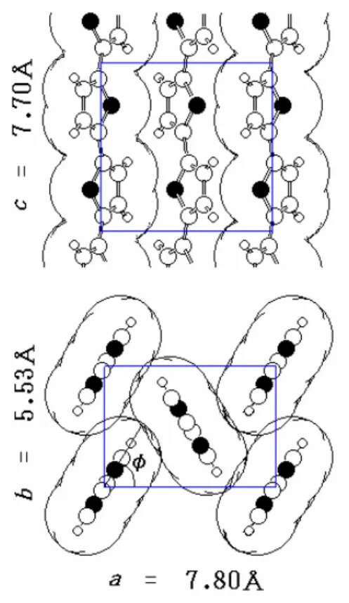
The molecular chain of polythiophene (PTh) takes all-trans planar conformation, and they are packed in the pgg manner (Figure 1). The setting angle (
φ
) between the molecular side and the ac plane is about 60° , namely, the packing is not of face-to-face stacking type. Positional disorders exist along the chain axis.In this study, the crystal structure of copolymers that consist of electron-donating thiophene and the electron-accepting arylene group with nitro groups, P(p-NPh-co-Th), P(2NPh-co-Th), P(DNTh-co-Th), and P(DNTh-co-BiTh) (schema 1), were analyzed by X-ray diffraction technique.
Experimental
The samples were offered from Prof. Yamamoto (Chemical Resource Laboratory, Tokyo Institute of Technology). X-ray measurements were carried out with CuKα radiation. The diffraction curves were obtained by a powder diffractometer equipped with a scintillation counter in the range of 2
θ
= 2-50°. Analysis was performed with linked-atom Rietveld method with standard bond lengths and bond angles.Results and Discussion
As for the XRD curve of P(p-NPh-co-Th), the peaks were observed at 2
θ
= 12° (d1 = 7.4 Å) and 24° (d2 = 3.7 Å). Positional disorder along the chainaxis was suggested by this feature. The structure to reproduce such an XRD curve was only the pgg type structure (Figure2). The setting angle is probably near to
φ
= 15°. The distinction betweenφ
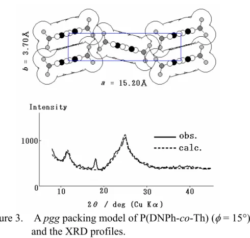
= 0-15° was difficult. As for the XRD curve of P(DNPh-co-Th), the peaks were observed at 2θ
= 11.6° (d1 = 7.6Å) and 24.7° (d2 = 3.6Å). The sharp peak observed at 2θ
=18° was attributed to the remaining catalyst particles. The structure is similarly disturbed. The simulation suggested the pgg type structure (Figure 3). The setting angle was in the range of
φ
= 0-15°.As for the XRD curve of P(DNPh-co-BiTh), the peaks were observed at 2
θ
= 11.3° (d1 = 7.8Å) and 24.7° (d2 = 3.6Å). The structure is disturbed too.The simulation suggested the pgg type structure (Figure 4). The setting angle was in the range of
φ
= 0-10°.Because P(2NPh-co-Th) was amorphous, it was not able to evaluate the structure.
Conclusion
P(p-NPh-co-Th), P(DNPh-co-Th), and P(DNPh-co-BiTh) took disordered structure of the pgg type. The setting angles were much smaller than that in PTh. Basically the packing is close to the cmm type. Electron donor-acceptor interactions may play an important role.
Figure 1. The pgg crystal structure of PTh.
Figure 2. A pgg packing model of P(p-NPh-co-Th) (
φ
= 15°), and the XRD profiles.Figure 3. A pgg packing model of P(DNPh-co-Th) (
φ
= 15°), and the XRD profiles.Figure 4. A pgg packing model of P(DNTh-co-BiTh) (