IRUCAA@TDC : Parameters for diffusion weighted magnetic resonance imaging for temporomandibular joint
9
0
0
全文
(2) 5. Bull Tokyo Dent Coll (2006) 47(1): 5–12. Original Article. Parameters for Diffusion Weighted Magnetic Resonance Imaging for Temporomandibular Joint Takamichi Otonari, Mamoru Wakoh, Tsukasa Sano, Mika Yamamoto, Mai Ohkubo and Takuya Harada Department of Oral and Maxillofacial Radiology, Tokyo Dental College, 1-2-2 Masago, Mihama-ku, Chiba 261-8502, Japan. Received 27 March, 2006/Accepted for publication 2 May, 2006. Abstract The purpose of this study was to determine optimum diffusion parameters for diffusion weighted imaging (DWI) techniques, including echo planer imaging (EPI), single-shot fast spin echo (SSFSE), and steady-state free precession (SSFP) in Magnetic Resonance Imaging (MRI) of the Temporomandibular Joint (TMJ). A polyethylene tube with distilled water was individually positioned at the external acoustic meatus foramen in each of three volunteers with normal healthy TMJs. Images were obtained using three types of DWI at differing diffusion parameters, b-factors, and diffusion moment. Signal intensity and imaging ability for various anatomical structures, including the distilled water, were evaluated from each image. The details of the anatomical structures of the TMJ were unidentifiable in the images produced with EPI and SSFSE, but were identifiable on the SSFP images. A diffusion moment value from 100mT/m*msec to 150 mT/m*msec for SSFP, in particular, restrained the signal intensity of the water, thereby protecting the comparably high image quality of the TMJ structure. In conclusion, only SSFP is capable of allowing interpretation of emerging pathologic conditions in the TMJ region, when used with a diffusion moment set at between from approximately 100mT/m*msec to 150 mT/m*msec. Key words:. Magnetic resonance imaging (MRI)—Diffusion weighted imaging (DWI)— Temporomandibular joint (TMJ)— b-factor —Diffusion moment. Introduction. condyle. Furthermore, use of T2-weighted MRI which allows elucidation of pathologic conditions by means of fluid, enables visualization of the abnormal accumulation of TMJ fluid known as TMJ effusion. TMJ effusion, which typically appears as a bright signal on T2-weighted MR images6,18,19), has been recognized as a possible sign related to pain in patients with TMJ disorders9,25,28). Therefore,. Magnetic Resonance Imaging (MRI) is a very useful imaging modality for diagnosing disorders of the Temporomandibular Joint (TMJ). This modality allows non-invasive imaging of the position and shape of the articular disc, alteration in bone marrow and change in morphology of the mandibular 5.
(3) 6. Otonari T et al.. at our hospital, T2-weighted MR images are routinely obtained from patients, with suspected TMJ disorders characterized by symptoms of inflammation. However, the clinical significance of TMJ effusion remains to be clarified. Some authors question the relationship between TMJ effusion and pain1,5,15,17). Recently, an advanced MRI technique focusing on fluid conditions, referred to as Diffusion Weighted Imaging (DWI), has come into practical use in the medical field according to Le Bihan et al.10) and Moseley et al.14). DWI reflects the diffusion coefficient of water molecules in body tissue with the application of strong Motion Probing Gradient (MPG). T2-weighted MRI reflects all water molecules in body tissue as a bright signal, whereas DWI selectively reflects water molecules in which Brownian movement is restricted as a bright signal. DWI makes it possible to detect initial inflammatory changes unidentified by T2weighted MR images14). This technique traditionally required a long time to obtain images and image quality had a low signal-to-noise ratio. However, developments in both hardware and software have overcome these faults. DWI has been applied to many cases of brain parenchyma disease, and has consequently contributed to the diagnosis of acute cerebral infarct3,4,11,13,14). The diagnosis of an acute cerebral infarct results from the fact that intracellular edemas caused by acute brain ischaemic diseases depress the diffusion of water molecules, and this abnormal phenomenon in the body tissue is then described with a bright signal on the MR image. This advanced MRI technique was also very effective in discriminating cystic lesions in the brain as a result of differing viscous behavior of contents between cystic lesions22). Recently, this technique has also been used for diseases other than those in the cerebral region, for example, in vertebral diseases2) such as fracture and neoplastic lesion. Furthermore, DWI has also been discussed as a way forward in the differentiation of benign and malignant tumors originating in musculoskeletal tissue7,26). DWI studies involving the knee joint12) and salivary gland21) have been also reported. However, to. the best of our knowledge, there have been no reports on the use of DWI in the diagnosis of the temporomandibular joint diseases. DWI offers a possible method for the representation of clinical state, especially in terms of stages of inflammatory change in disorders of the TMJ. However, the limited imaging area of the TMJ region, and adjacent anatomical structures surrounding the TMJ as typified by air and bone, may provide obstacles to obtaining adequate MR images for diagnostic purposes. Therefore, the purpose of this preliminary study was to investigate adequate imaging conditions for imaging diagnosis in the TMJ region, by comparing several types of DWI technique.. Materials and Methods MRI was performed with a 1.5 Tesla MR imager (Magnetom Symphony Maestro Class, Siemens, Muenchen, Germany). The TMJ surface coils used as receivers were 8 cm in diameter. Six normal healthy TMJs in three volunteers were subjected to MRI. None of these six TMJs showed clinical signs or any anatomical abnormalities on MRI. All volunteers gave their consent to take part in the study. The study was approved by the Ethics Committee of our institute and was carried out in accordance with the Helsinki Declaration. As described above, DWI represents the diffusion coefficient of water molecules characterized by Brownian movement on MR images as a so-called “bright signal.” To compare water signal intensity on TMJ images among several types of DWI technique, consisting of echo planer imaging (EPI), singleshot fast spin echo method (SSFSE), and steady-state free precession method (SSFP), we prepared a polyethylene tube containing distilled water (Fig. 1). The tube was 6 cm in diameter and 4 cm in bore diameter, and was individually positioned at the external acoustic meatus foramen in each of the three volunteers. First, we carried out Proton Density Weighted Imaging (PDWI) according to the usual imaging procedure at our department,.
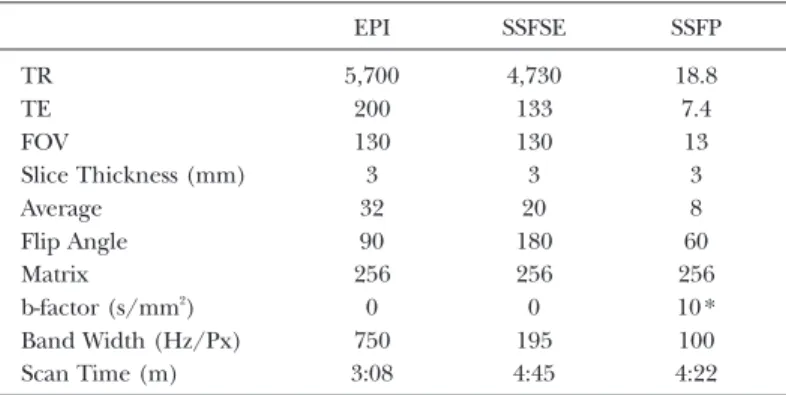
(4) 7. DWI Technique of MRI for TMJ Region. as this method is routinely applied to patients with TMJ disorders. Consecutively, a series of images was obtained using each DWI technique. To obtain a DWI, a setup motion probing gradient (MPG) strength, which affects the degree (pitch) of diffusion weight, is required. This degree is actually expressed as a b-factor for both EPI and SSFSE, and as a diffusion moment for SSFP. In an environment. A. B. Fig. 1 Polyethylene tube (seen on A) inserted into external acoustic meatus foramen (shown on B). marked by slightly affected MPG, water molecules are generated a bright signal, regardless of level of Brownian movement. On the other hand, a strong MPG degrades the ratio of signal-to-noise on MR images. To minimize the effects of diffusion weight in imaging, therefore, the numeric values of the b-factor and diffusion moment used were 0 and 10, respectively, which provided conditions with the lowest MPG in the MRI system used. Other imaging factors are shown in Table 1. A total of four images were printed out on a film to evaluate diagnostic viability. Four oral and maxillofacial radiologists independently evaluated whether the shape of the mandibular condyle could be identified as an anatomical landmark on each DWI image, referring to the PDWI. Viewing conditions were standardized with the usual ambient lighting and masking of extraneous light from the view box. Based on the results of the evaluation, another additional experiment was carried out. A total of six SSFP images was additionally obtained with diffusion moment set at 10, and varying from 50 to 250 in 50 increments. The number of TMJs and volunteers was the same as in the initial study. Thereafter, using viewer software adapted for an MR imager, a region of interest (ROI) 0.02 cm2 in size was set at the water area in the tube, the bone marrow area of the mandibular con-. Table 1 Parameters for each diffusion weighted imaging (DWI) technique. TR TE FOV Slice Thickness (mm) Average Flip Angle Matrix b-factor (s/mm2) Band Width (Hz/Px) Scan Time (m). EPI. SSFSE. SSFP. 5,700 200 130 3 32 90 256 0 750 3:08. 4,730 133 130 3 20 180 256 0 195 4:45. 18.8 7.4 13 3 8 60 256 10 * 100 4:22. * diffusion moment (* mT/m*msec) Decision factor of diffusion weight for EPI and SSFSE was b-factor, and that for SSFP as diffusion moment..
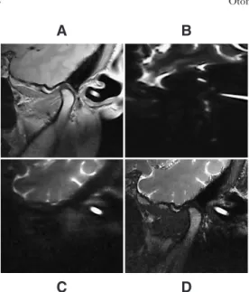
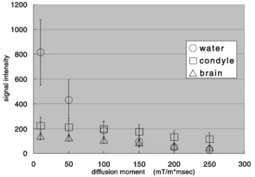
(5) 8. Otonari T et al.. A. B. and 30, 115 and 49 for maximum diffusion moment (250 mT/m*msec), respectively. As the diffusion moment value increased, the signal intensities for each area and signal-tonoise ratio commonly decreased. This tendency was particularly marked in the water area. The signal intensity in the water area was similar to that in bone marrow when the diffusion moment was set at 100 mT/m*msec, whereas the former was less than the latter, and was similar to the signal intensity of brain parenchyma set at 150 mT/m*msec.. Discussion. C. D. Fig. 2 Proton density weighted image and three types of diffusion-weighted image of TMJ from 35 years male A: Proton density weighted image routinely taken showing TMJ structures clearly. B and C: Neither echo planer imaging (EPI) image (B) nor single-shot fast spin echo (SSFSE) image (C) detecting mandibular condyle, at all. D: Only Steady-state free precession (SSFP) image presented shape of mandibular condyle.. dyle, and the brain parenchyma area on each image, and signal intensity on each ROI was measured.. Results Figure 2 shows the DWI and PDWI obtained. Nobody could identify the shape of the mandibular condyle on the images obtained with EPI and SSFSE, whereas all observers could identify the SSFP-generated image. Figures 3 and 4 show the six SSFP images obtained with varied diffusion moments and signal intensities at the water area, the bone marrow area of the mandibular condyle, and the brain parenchyma area on each image, respectively. Signal intensities measured at the water, bone marrow, and brain parenchyma areas for minimum diffusion moment (10 mT/m*msec) were 814, 223 and 141,. Diffusion-weighted MRI is a new concept that was introduced as intravoxel incoherene motion. In tissue, the apparent diffusion coefficient (ADC), which is the reference index for evaluating variation in signal intensity influenced by increase of MPG, reflects local water mobility in vivo. In recent years, DWI has been investigated extensively in the brain, and has been shown to be especially sensitive in the early detection of cerebral ischemia24). Acute brain infarct using DWI technique generates a high intensity signal, reflecting the decrease in ADC in such lesions. This may result from brain cellulous edema and narrow intercellular space causing a decrease in the number of water molecules in which Brownian movement is possible3,20,23). More specifically, failure of the sodium potassium pump causes cytotoxic cell swelling that results in a decrease in interstitial space, and, therefore, the dephasing of water spins is reduced. When MPG is applied, signal intensity in healthy cells with high diffusion power, that is a high ADC, reduces, but that in abnormal cells in ischemic regions remains high. In many clinical cases, EPI does not provide an adequate image for diagnosis, except in lesions of the brain parenchyma. This is a result of the strong magnetic susceptibility artifact produced by the presence of complex structures consisting of air, bone, soft tissue, and others, which differs from the relatively homogeneous structures found in the brain.
(6) 9. DWI Technique of MRI for TMJ Region. A. B. C. D. E. F. Fig. 3 Steady-state free precession (SSFP) images obtained with different diffusion moments A: 10 mT/m*msec B: 50 mT/m*msec C: 100 mT/m*msec D: 150 mT/m*msec E: 200 mT/m*msec F: 250 mT/m*msec When diffusion moment value increased, signal intensities for each area (water in auditory canal, bone marrow of condyle and brain parenchyma) decreased. Signal intensity in water area, in particular, dramatically decreased.. Fig. 4 Relationship between signal intensity at each area (water in auditory canal, bone marrow of condyle and brain parenchyma) and diffusion moment. region. On the other hand, SSFSE and SSFP provide images with little distortion due to the slight magnetic susceptibility artifact. In EPI and SSFSE, MPG is decided by the value of the b-factor (s/mm2), which reflects the. degree (pitch) of diffusion weight. However, MPG in SSFP is decided by diffusion moment (mT/m*msec), which multiplies MPG strength by MPG application time. In our study, DWIs produced by EPI and.
(7) 10. Otonari T et al.. SSFSE did not allow identification of the entire TMJ structure or the mandibular condyle, respectively, when b-factor was set at the minimal 0. The strong magnetic susceptibility artifact produced by the presence of complex structures in the TMJ region resulted in a degradation of image quality. We believe that, even if the value of the b-factor was increased in EPI and SSFSE, image quality would not be improved. It has been suggested that EPI and SSFSE sequences are not suitable for the clinical diagnosis of TMJ, especially with regard to inflammatory change at the primary stage of onset of disorder. The results of this study also indicate that EPI and SSFSE should be applied to regions with relatively homogeneous structures such as the brain region, as many previous reports regarding brain parenchyma disease27). In this study, only SSFP allowed identification of the mandibular condyle. Therefore, DWI produced by using SSFP sequences were additionally performed with incrementation of the value of diffusion moment. A comparison of the three regions—distilled water, brain parenchyma, and bone marrow of the mandibular condyle—with the signal intensity set at 10 mT/m*msec revealed that the minimal value of diffusion moment was highest in the distilled water area, followed by the bone marrow of the mandibular condyle, and the brain parenchyma. At 250 mT/m*msec which is the maximal value of diffusion moment, the highest value was found in the bone marrow of the mandibular condyle, followed by the brain parenchyma and distilled water. These results suggest that signal intensity of water molecules with a relatively high diffusion coefficient was restrained in strong MPG. The strong MPG produced by a diffusion moment of 250 mT/m*msec indisputably degraded the ratio of signal-to-noise in the diffusion-weighted MR images, whereas the signal intensity in water did not reach zero. Signal intensity in the distilled water area taken at 100 mT/m*msec was inferior to that in the bone marrow of the mandibular condyle, and was superior to that in the brain parenchyma. However, the signal intensity. of water at 150 mT/m*msec was inferior to those in both the bone marrow of the mandibular condyle and the brain parenchyma. In conclusion, setting the value of diffusion moment at between 100 mT/m*msec to 150 mT/m*msec for SSFP restrains the signal intensity of water, and thereby protects the comparably higher image quality of TMJ structures. In particular, a diffusion moment of 100 mT/m*msec for SSFP appears to offer a good basis for taking DWI images of the TMJ region. Andera et al.2) reported that SSFP was very effective in identifying the cause of vertebral fractures, although the anatomical structure surrounding the vertebrae is complicated, as in the TMJ region. Clinical image diagnosis of the TMJ region using DWI requires the maximum possible image quality (high ratio of signal-to-noise) of both anatomical structures and accurate description of water molecules with slight diffusion such as found in the initial stage of inflammatory change. The results of other studies suggest that DWI is capable of detecting bone marrow abnormalities, while not being so useful in differentiating spinal infection and malignancy16). However, another report has suggested that DWI offers a potential tool for therapy monitoring which would allow us to revise the way in which cancer patients are managed8). When these requirements have been met, DWI, especially SSFP, will provide an effective clinical tool in the interpretation of emerging pathologic conditions in the TMJ region, for instance, offering an alternative to T2weighted MRI in the evaluation of the abnormal accumulation of TMJ fluid.. References 1) Adame CG, Monje F, Offnoz M, Martingranizo R, Munoz M (1998) Effusion in magnetic resonance imaging of the temporomandibular joint: a study of 123 joints. J Oral Maxillofac Surg 56:314–318. 2) Andera BO, Axel S, Roland B, Reiner B, Andreas K, Maximilian R, Michael D (1998) Diffusion-weighted MR imaging of bone mar-.
(8) DWI Technique of MRI for TMJ Region. 3). 4). 5). 6). 7). 8). 9). 10). 11). 12). 13). 14). row: Differentiation of benign versus pathologic compression fracture. Radiology 207:349–356. Benveniste H, Hedlund LW, Johnson GA (1992) Mechanism of detection of acute cerebral ischemia in rats by diffusion-weighted magnetic resonance microscopy. Stroke 23: 746–754. Chien D, Kong KK, Gress DR, Buonanno FS, Buxton RB, Rosen BR (1992) MR diffusion imaging of cerebral infarction in humans. Am J Neuroradiol 13:1097–1102. Haley DP, Schiffman E, Lindgren BR, Anderson Q, Andreasen K (2001) The relationship between clinical and MRI findings in patients with unilateral temporomandibular joint pain. J Am Dent Assoc 132:476–481. Harms SE, Wilk RM, Wolford LM, Chiles DG, Milam SB (1985) The temporomandibular joint: magnetic resonance imaging using surface coil. Radiology 157:133–136. Hermann G, Abdelwahab IF, Miller TT, Klein MJ, Lewis MM (1992) Tumour and tumourlike conditions of the soft tissue: magnetic resonance imaging features differentiating benign from malignant masses. Br J Radiol 65:14–20. Herneth AM, Friedrich K, Weidekamm C, Schibany N, Krestan C, Czerny C, Kainberger F (2005) Diffusion weighted imaging of bone marrow pathologies. Eur J Radiol 55:74–83. Larheim TA, Westesson PL, Sano T (2001) MR grading of temporomandibular joint fluid: association with disk displacement categories, condyle marrow abnormalities and pain. Int J Oral Maxillofac Surg 30:104–112. Le Bihan D, Breton E, Lallemand D, Grenier P, Cabanis E, Laval-Jeantet M (1986) MR imaging of intravoxel incoherent motions: application to diffusion and perfusion in neurogenic disorders. Radiology 161:401–407. Matsumoto K, Lo EH, Pierce AR, Wei H, Garrido L, Kowall NW (1995) Role of vasogenic edema and tissue cavitation in ischemic evolution on diffusion-weighted imaging: comparison with multiparameter MR and immunohistochemistry. Am J Neuroradiol 16:1107–1115. Miller KL, Hargreaves BA, Gold GE, Pauly JM (2004) Steady-state diffusion-weighted imaging of in vivo knee cartilage. Magn Reson Med 51:394–398. Mintorovitch J, Moseley ME, Chileuitt L, Shimizu H, Choen P, Weinstein PR (1991) Comparison of diffusion and T2-weighted MRI for the early detection of cerebral ischemia and reperfusion in rats. Magn Reson Med 18:39–50. Moseley ME, Cohen Y, Mintorovitch J,. 15). 16). 17). 18) 19). 20) 21). 22). 23). 24). 25). 11. Chileuitt L, Shimizu H, Kucharczyk J, Wendland MF, Weinstein PR (1990) Early detection of regional cerebral ischemia in cats: comparison of diffusion and T2 weighted MRI and spectroscopy. Magn Reson Med 14: 330–346. Murakami K, Nishida K, Bessho K, Iizuka T, Tsuda Y, Konishi J (1996) MRI evidence of high signal intensity and temporomandibular arthralgia and relating pain. Does the high signal correlate to the pain?. Br J Oral Maxillofac Surg 34:220–224. Pui MH, Mitha A, Rae WI, Corr P (2005) Diffusion-weighted magnetic resonance imaging of spinal infection and malignancy. J Neuroimaging 15:164–170. Rudisch A, Innerhofer K, Bertram S, Emshoff R (2001) Magnetic resonance imaging findings of internal derangement and effusion in patients with unilateral temporomandibular joint pain. Oral Surg Oral Med Oral Pathol Oral Radiol Endod 92:566–571. Sano T, Yamamoto M, Okano T (2003) Temporomandibular joint: MR imaging. Neuroimaging Clin N Am 13:583–595. Sano T, Yamamoto M, Okano T, Gokan T, Westesson PL (2004) Common abnormalities in temporomandibular joint imaging. Curr Probl Diagn Radiol 33:16–24. Siesjo BK (1992) Pathophysiology and treatment of focal cerebral ischemia. Part 1: Pathophysiology. J Neurosurg 77:169–184. Sumi M, Takagi Y, Uetani M, Morikawa M, Hayashi K, Kabasawa H, Aikawa K, Nakamura T (2002) Diffusion-weighted echoplaner MR imaging of the salivary glands. AJNR 178: 959–965. Tsuruda JS, Chew WM, Mosely ME, Norman D (1990) Diffusion-weighted MR imaging of the brain: value of differentiating between extraaxial cysts and epidermoid tumors. AJNR 11:925–931. Verheul HB, Balazs R, Berkelbach van der Sprenkel JW, Tulleken CA, Nicolay K, Tamminga KS, van Lookeren Campagne M (1994) Comparison of diffusion-weighted MRI changes in cell volume in a rat model of brain injury. NMR Biomed 7:96–100. Warach S, Gaa J, Siewert B, Wielopolski P, Edelman RR (1995) Acute human stroke studies by whole brain echo planer diffusionweighted magnetic resonance imaging. Ann Neurol 37:231–241. Westesson PL, Brooks SL (1992) Temporomandibular joint: relationship between MR evidence of effusion and the presence of pain and disk displacement. AJR Am Roentgenol 159:559–563..
(9) 12. Otonari T et al.. 26) Woude HJ, Bloem JL, Pope Jr TL (1998) Magnetic resonance imaging of the musculoskeletal system. Clin Orthop 347:272–286. 27) Xu D, Henry RG, Mukherjee P, Carvajal L, Miller SP, Barkovich AJ, Vigneron DB (2004) Single-shot fast spin-echo diffusion tensor imaging of the brain and spine with head and phased array coils at 1.5 T and 3.0 T. Magn Reson Imaging 22:751–759. 28) Yamamoto M, Sano T, Okano T (2003) Magnetic resonance evidence of joint fluid with temporomandibular joint disorders. J Comput Assist Tomogr 27:694–698.. Reprint requests to: Dr. Takamichi Otonari Department of Oral and Maxillofacial Radiology, Tokyo Dental College, 1-2-2 Masago, Mihama-ku, Chiba 261-8502, Japan Tel: 043-270-3961 Fax: 043-270-3962 E-mail: [email protected].
(10)
図
関連したドキュメント
The construction of homogeneous statistical solutions in [VF1], [VF2] is based on Galerkin approximations of measures that are supported by divergence free periodic vector fields
Zhao, “Haar wavelet operational matrix of fractional order integration and its applications in solving the fractional order differential equations,” Applied Mathematics and
Preconditioning of radial basis function interpolation systems via accelerated iterated approximate moving least squares approximation. in Progress on Meshless
Further investigate use of different Matérn parameters Couple smoothing parameter to current residuals Do smoothing with an approximate smoothing kernel Apply similar ideas in
We investigate the global dynamics of solutions of four distinct competitive rational systems of difference equations in the plane1. We show that the basins of attractions of
If we represent π by a diagram (of either type), erase the point corresponding to i and the arc connected to the point (and number other points appropriately for the circular
“ Increase the Eco-friendly of Solid Waste Management System from waste collection, transfer waste, disposal waste to land. fills, compositing, and/or incinerations along with
引火性液体 : 区分4 眼に対する重篤な損傷性/ : 区分2B 眼刺激性 警告 眼刺激 可燃性液体
