Clinical Pharmacogenetics Implementation
Consortium Guideline for HLA Genotype and Use of Carbamazepine and Oxcarbazepine: 2017
Update
Elizabeth J. Phillips
1, Chonlaphat Sukasem
2,3, Michelle Whirl-Carrillo
4, Daniel J. M € uller
5,6, Henry M. Dunnenberger
7, Wasun Chantratita
8,9, Barry Goldspiel
10, Yuan-Tsong Chen
11,12, Bruce C. Carleton
13, Alfred L. George Jr.
14, Taisei Mushiroda
15, Teri Klein
4,
Roseann S. Gammal
16,17and Munir Pirmohamed
18The variant alleleHLA-B*15:02is strongly associated with greater risk of Stevens–Johnson syndrome (SJS) and toxic epidermal necrolysis (TEN) in patients treated with carbamazepine or oxcarbazepine. The variant alleleHLA-A*31:01is associated with greater risk of maculopapular exanthema, drug reaction with eosinophilia and systemic symptoms, and SJS/TEN in patients treated with carbamazepine. We summarize evidence from the published literature supporting these associations and provide recommendations for carbamazepine and oxcarbazepine use based onHLAgenotypes.
Human leukocyte antigen (HLA) genetic variation is implicated in the development of specific cutaneous adverse reactions to aro- matic anticonvulsants. The purpose of this guideline is to inter- pret HLA-B*15:02 and HLA-A*31:01 genotyping results to guide the use of carbamazepine and oxcarbazepine. Detailed guidelines regarding the selection of alternative therapies, when to conduct genotype testing, and cost-effectiveness analyses are beyond the scope of this document. The Clinical Pharmaco- genetics Implementation Consortium (CPIC) guidelines are
periodically updated at https://cpicpgx.org/guidelines and http://
www.pharmgkb.org.
FOCUSED LITERATURE REVIEW AND UPDATE
A systematic literature review focused on HLA-B*15:02 and HLA-A*31:01genotypes and carbamazepine- and oxcarbazepine- induced cutaneous adverse reactions was conducted (details in Supplemental Material).
This guideline is an update to the 2013 CPIC guideline for HLA-B*15:02 and carbamazepine use.1 The recommenda- tions provided in the original guideline have not changed and are included here. However, the scope of the existing recommendations has now expanded to include the use of carbamazepine and oxcarbazepine based on HLA-A*31:01 and HLA-B*15:02 genotypes, respectively. Furthermore, the accompanying Supplemental Material now includes resources to facilitate the incorporation of HLA genotype results into electronic health records with clinical decision support (https://cpicpgx.org/guidelines/guideline-for-carbamazepine-and- hla-b/).
1Vanderbilt University Medical Center, Nashville, Tennessee, USA;2Division of Pharmacogenomics and Personalized Medicine, Department of Pathology, Faculty of Medicine Ramathibodi Hospital, Mahidol University, Bangkok, Thailand;3Laboratory for Pharmacogenomics, Somdech Phra Debaratana Medical Center, Faculty of Medicine Ramathibodi Hospital, Bangkok, Thailand;4Department of Biomedical Data Science, Stanford University, Stanford, California, USA;
5Campbell Family Mental Health Research Institute, Centre for Addiction and Mental Health, Toronto, Ontario, Canada;6Department of Psychiatry and Pharmacology & Toxicology, University of Toronto, Toronto, Ontario, Canada;7Center for Molecular Medicine, NorthShore University HealthSystem, Evanston, Illinois, USA;8Virology Laboratory, Department of Pathology, Faculty of Medicine Ramathibodi Hospital, Mahidol University, Bangkok, Thailand;9Center for Medical Genomics, Ramathibodi Hospital, Mahidol University, Bangkok, Thailand;10Pharmacy Department, National Institutes of Health Clinical Center, Bethesda, Maryland, USA;11Institute of Biomedical Sciences, Academia Sinica, Taipei, Taiwan;12Department of Pediatrics, Duke University Medical Center, Durham, North Carolina, USA;13Division of Translational Therapeutics, Department of Pediatrics, Faculty of Medicine, University of British Columbia, and BC Children’s Hospital Research Institute, Vancouver, British Columbia, Canada;14Department of Pharmacology, Northwestern University Feinberg School of Medicine, Chicago, Illinois, USA;15Laboratory for Pharmacogenomics, RIKEN Center for Integrative Medical Science, Yokohama, Japan;16Department of Pharmacy Practice, MCPHS University, Boston, Massachusetts, USA;17Department of Pharmaceutical Sciences, St. Jude Children’s Research Hospital, Memphis, Tennessee, USA;18Department of Pharmacology, University of Liverpool, Liverpool, UK. Correspondence: M. Pirmohamed ([email protected], or [email protected])
Received 20 October 2017; accepted 20 December 2017; advance online publication 2 February 2018. doi:10.1002/cpt.1004
574 VOLUME 103 NUMBER 4 | APRIL 2018 |www.cpt-journal.com
CPIC UPDATE
210
GENES:HLA-BANDHLA-A Background
HLA-BandHLA-Aare part of a large cluster of genes known as the human major histocompatibility complex (MHC). The clus- ter contains three subgroups: class I, II, and III. TheHLA-Band HLA-Agenes are part of the class I complex, along withHLA-C.
These genes encode cell surface proteins that present intracellular antigens to the immune system. Intracellular antigens are usually the normal breakdown products of intracellular proteins and are recognized as “self.” However, if the antigen presented derives from a pathogen or, in some cases, a transplanted tissue, it may be recognized as “nonself” and trigger an immune response. HLA is inherited in a codominant fashion with one set of class I and II alleles being inherited from each parent where both have full phe- notypic expression.
Because HLA proteins present a wide variety of peptides for immune recognition, theHLAgenes are among the most highly polymorphic genes in the human genome.HLApolymorphisms were previously ascertained serologically, but standard molecular approaches that now use DNA sequence-based typing methods either by standard Sanger or next-generation sequencing have revealed much greater complexity of genetic variation within this locus. For example, according to the World Health Organization (WHO) Nomenclature Committee for Factors of theHLASys- tem (http://hla.alleles.org), there are more than 4,000 identified HLA-B alleles and more than 3,000 identified HLA-A alleles, many of which differ by more than one nucleotide from one another. Each allele is designated by the gene name followed by an asterisk and a four- or six-digit identifier giving information about the allele type (designated by the first two digits) and spe- cific protein subtype (second set of digits). The details of HLA nomenclature have been described in a previous CPIC guideline.2
The guideline presented here specifically discusses the class I HLA alleles HLA-B*15:02 and HLA-A*31:01 as they relate to carbamazepine- and oxcarbazepine-induced cutaneous adverse reactions, including Stevens–Johnson syndrome/toxic epidermal necrolysis (SJS/TEN), drug reaction with eosinophilia and sys- temic symptoms (DRESS), and maculopapular exanthema (MPE).
Genetic test interpretation
Clinical genotyping tests exist for identifyingHLA-BandHLA- A alleles, including HLA-B*15:02 and HLA-A*31:01.
Genotyping results are presented as “positive” if one or two cop- ies of the variant allele are present or “negative” if no copies of the variant allele are present. There is no intermediate genotype.
Genotype definitions for HLA-B*15:02 and HLA-A*31:01 are summarized in Table 1. Nucleotide and amino acid sequence alignments for HLA-B*15:02 andHLA-A*31:01and the corre- sponding reference sequences are available in Supplemental Figures S1–S4.
Available genetic test options
Commercially available genetic testing options change over time.
Additional information about pharmacogenetic testing can be found at the Genetic Testing Registry website (http://www.ncbi.
nlm.nih.gov/gtr/).
Incidental findings
AlthoughHLAalleles have been studied in the context of specific responses to HIV and other pathogens, there are currently no specific diseases or conditions that have been strongly linked to HLA-B*15:02 or HLA-A*31:01 independent of drug use.3–5 However,HLA-B*15:02has also been associated with SJS/TEN from phenytoin use, and otherHLA-Balleles have been strongly associated with adverse drug reactions. For example, HLA- B*57:01is associated with abacavir-induced hypersensitivity reac- tion, and HLA-B*58:01 is associated with allopurinol-induced severe cutaneous adverse reactions (including SJS/TEN and DRESS). CPIC guidelines are available to guide prescribing of phenytoin,6 abacavir,7 and allopurinol,8 based on HLA-B genotype.
Other considerations
HLA-B*15:02and HLA-A*31:01 have distinct ethnic and geo- graphical distributions that are important for evaluating popula- tion risk (see HLA-A and HLA-B Allele Frequency Table).
The frequency ofHLA-B*15:02is highest in East Asian (6.9%), Oceanian (5.4%), and South/Central Asian (4.6%) populations.
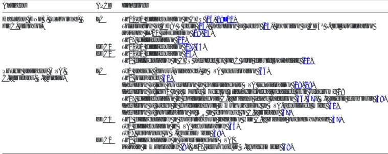
However, not all East Asian subpopulations carry this allele in such high frequencies.HLA-B*15:02frequency is much lower in Japanese (<1%) and Korean (<2.5%) populations. The allele is also quite rare in African populations (not observed), African Americans, Middle Easterners, Caucasians, and Hispanics/South Americans (<1%). In contrast, the frequency of the HLA- A*31:01 allele is higher than theHLA-B*15:02allele in Cauca- sians (3%) and Hispanic/South Americans (6%). However, it is Table 1 Assignment ofHLA-BandHLA-Agenotypes
Genotype Definition Examples of diplotypes
HLA-B*15:02negative Homozygous for an allele other thanHLA-B*15:02 *Xa/*Xa
HLA-B*15:02positive Heterozygous or homozygous variant *15:02/*Xa,*15:02/*15:02
HLA-A*31:01negative Homozygous for an allele other thanHLA-A*31:01 *Yb/*Yb
HLA-A*31:01positive Heterozygous or homozygous variant *31:01/*Yb,*31:01/*31:01
aWhere *X5anyHLA-Ballele other thanHLA-B*15:02.bWhere *Y5anyHLA-Aallele other thanHLA-A*31:01.
CPIC UPDATE
CLINICAL PHARMACOLOGY & THERAPEUTICS| VOLUME 103 NUMBER 4 | APRIL 2018 575
211
also found in high frequencies in some East Asians, specifically Japanese (8%) and South Koreans (5%), and South/Central Asians (2%). While these frequencies are helpful in determining broad population risks, they cannot replace genotypes on an indi- vidual basis.
DRUGS: CARBAMAZEPINE AND OXCARBAZEPINE Background
Carbamazepine. Carbamazepine, an aromatic anticonvulsant related to the tricyclic antidepressants, is US Food and Drug Administration (FDA)-approved for the treatment of epilepsy, trigeminal neuralgia, and bipolar disorder. Carbamazepine reduces the propagation of abnormal impulses in the brain by producing a frequency- and voltage-dependent block of sodium channels, thereby inhibiting the generation of repetitive action potentials in the epileptic focus.8,9 Carbamazepine-induced adverse effects that may have known dose- or concentration- dependency include dizziness, ataxia, and nystagmus. Other adverse effects such as aplastic anemia, hyponatremia, leukopenia, osteoporosis, liver injury, and hypersensitivity reactions such as MPE, DRESS, and SJS/TEN have a complex dose–response rela- tionship such that it is difficult to delineate a clear linear dose–
response relationship. For additional information regarding the pharmacokinetics and pharmacogenomics of carbamazepine, please refer to the PharmGKB website: http://www.pharmgkb.
org/pathway/PA165817070.10
Oxcarbazepine. Oxcarbazepine is the keto-analog of carbamaze- pine. With its similar structure, oxcarbazepine shares many thera- peutic indications and adverse effects with carbamazepine.
Furthermore, patients who have had hypersensitivity reactions to carbamazepine may also be predisposed to hypersensitivity reac- tions with oxcarbazepine; these patients should only be treated with oxcarbazepine if the potential benefit justifies the potential risk.
Linking genetic variability to variability in drug-related phenotypes
There is evidence linking the HLA-B*15:02 genotype with the risk of carbamazepine- and oxcarbazepine-induced SJS/TEN (Supplemental Table S1) and linking HLA-A*31:01 genotype with the risk of carbamazepine-induced SJS/TEN, DRESS, and MPE (Supplemental Table S2). Application of a grading system to evidence linkingHLAgenotypic variations to phenotypic vari- ability with respect to cutaneous adverse reactions indicates a high quality of evidence in the majority of cases. This body of evi- dence provides the basis for the recommendations in Table 2 andTable 3.
HLA-B*15:02. HLA-B*15:02 is specific for carbamazepine- and oxcarbazepine-induced SJS and TEN, although the data are strongest for carbamazepine. SJS is characterized by epidermal detachment affecting up to 10% of the body surface area (BSA), while TEN usually involves more than 30% of the BSA. Patients with between 10–30% of the BSA blistered are defined as having an SJS/TEN overlap syndrome. Mortality rates are typically
below 5% for SJS and can be above 30% for TEN, with sepsis being the most frequent cause of death.11Mortality from SJS/
TEN is also related to age, the drug half-life, and how early the drug is discontinued.12,13An immune-mediated etiology has been shown for these reactions, which is consistent with the anamnes- tic response often seen clinically on drug rechallenge.14In terms of the immunopathology, cytotoxic T cells, or CD81T cells (lymphocytes matured in the thymus that express the CD8 pro- tein on their surface), are involved in SJS and TEN.15,16Further discussion on the mechanism of carbamazepine-induced SJS/
TEN is presented in theSupplemental Material.
Consistent with the regional and ethnic distribution of the HLA-B*15:02 allele, studies have shown the genetic risk of carbamazepine-associated SJS/TEN to be higher in several Asian countries with increased frequency of the HLA-B*15:02 allele, including Vietnam,17 Cambodia,17 Reunion Islands,17 Thai- land,18,19 some parts of India,20 Malaysia,21and Hong Kong.22 TheHLA-B*15:02allele has not been observed in cases of SJS/
TEN in some ancestral groups, such as Japanese and Korean pop- ulations or non-Asian descendants in Europe or North Amer- ica,17,23–26where the frequency of the allele is very low. In the Han Chinese population, the sensitivity of HLA-B*15:02 as a predictive test for SJS/TEN has been estimated at 98% and spe- cificity at 97%23; the positive predictive value is estimated at 7.7% and negative predictive value at 100%.27 However, it is important to note that in one study, in a group of individuals thought to be of European origin, four of 12 individuals with SJS/TEN carried the HLA-B*15:02 allele.24 Subsequently, they were found to have some Southeast Asian ancestry. This example underscores the importance of considering the HLA-B*15:02 allele carrier status in therapeutic decision-making regardless of self-reported ethnicity.
Based on the strong evidence linking HLA-B*15:02 to carbamazepine-induced SJS/TEN, the FDA issued a Health Alert in 2007 about changes to package labeling and recommendations for genetic testing in patients treated with carbamazepine.28The FDA label for carbamazepine carries a boxed warning about the risk of SJS/TEN with the presence of the HLA-B*15:02 allele and states that patients testing positive for the allele should not be treated with carbamazepine unless the benefit clearly out- weighs the risk. The FDA label for oxcarbazepine does not carry this boxed warning, but there is mention of the association betweenHLA-B*15:02and the risk of SJS/TEN in the warnings and precautions section that advises avoiding oxcarbazepine in HLA-B*15:02 positive patients unless the benefit clearly out- weighs the risk. The positive predictive value of HLA-B*15:02 for oxcarbazepine-induced SJS/TEN is estimated to be 0.73%, which is much lower than that of carbamazepine-induced SJS/
TEN (7.7%); however, the negative predictive value for both nears 100% in Southeast Asian populations.29
HLA-A*31:01. Unlike HLA-B*15:02, the HLA-A*31:01 allele is associated with a wider range of carbamazepine hypersensitivity reactions, including MPE, DRESS, and SJS/TEN, in many differ- ent populations.30 DRESS is a severe hypersensitivity reaction characterized by generalized cutaneous eruptions with systemic
CPIC UPDATE
576 VOLUME 103 NUMBER 4 | APRIL 2018 |www.cpt-journal.com
212
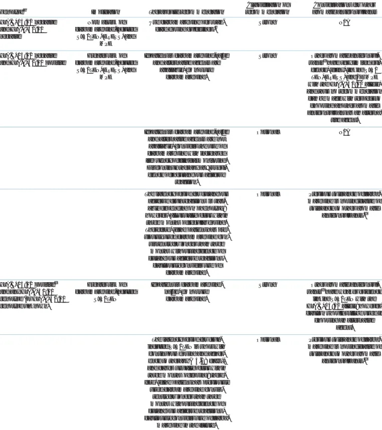
Table 2 Recommendations for carbamazepine therapy based onHLA-BandHLA-Agenotypes
Genotypea Implication Therapeutic recommendation
Classification of recommendation
Considerations for other aromatic anticonvulsants HLA-B*15:02negative
andHLA-A*31:01 negative
Normal risk of carbamazepine-induced
SJS/TEN, DRESS, and MPE
Use carbamazepine per stan- dard dosing guidelines.b
Strong N/A
HLA-B*15:02negative andHLA-A*31:01positive
Greater risk of carbamazepine-induced
SJS/TEN, DRESS, and MPE
If patient is carbamazepine-na€ıve and alternative agents are
available, do not use carbamazepine.
Strong Other aromatic anticonvul- santsdhave very limited evi-
dence, if any, linking SJS/
TEN, DRESS, and/or MPE with theHLA-A*31:01allele, and thus no recommendation
can be made with respect to choosing another aromatic anticonvulsant as an alterna-
tive agent.
If patient is carbamazepine-na€ıve and alternative agents are not available, consider the use of carbamazepine with increased frequency of clinical monitoring.
Discontinue therapy at first evi- dence of a cutaneous adverse
reaction.
Optional N/A
The latency period for cutaneous adverse drug reactions is vari- able depending on phenotype;
however, all usually occur within three months of regular dosing.
Therefore, if the patient has pre- viously used carbamazepine con- sistently for longer than three
months without incidence of cutaneous adverse reactions,
cautiously consider use of carbamazepine.
Optional Previous tolerance of carba- mazepine is not indicative of tolerance to other aromatic
anticonvulsants.d
HLA-B*15:02positivec and anyHLA-A*31:01 genotype (orHLA-A*31:01 genotype unknown)
Greater risk of carbamazepine-induced
SJS/TEN
If patient is carbamazepine- na€ıve, do not use
carbamazepine.
Strong Other aromatic anticonvul- santsdhave weaker evidence
linking SJS/TEN with the HLA-B*15:02allele; however, caution should still be used in
choosing an alternative agent.
The latency period for drug- induced SJS/TEN is short with
continuous dosing and adher- ence to therapy (4-28 days), and cases usually occur within three months of dosing; there- fore, if the patient has previously
used carbamazepine consis- tently for longer than three months without incidence of cutaneous adverse reactions, cautiously consider use of carba-
mazepine in the future.
Optional Previous tolerance of carba- mazepine is not indicative of tolerance to other aromatic
anticonvulsants.d
DRESS, drug reaction with eosinophilia and systemic symptoms; MPE, maculopapular exanthema; N/A, not applicable; SJS5Stevens-Johnson syndrome; TEN, toxic epi- dermal necrolysis.
aIf onlyHLA-B*15:02was tested, assumeHLA-A*31:01is negative and vice versa.bHLA-B*15:02has a 100% negative predictive value for carbamazepine-induced SJS/
TEN, and its use is currently recommended to guide use of carbamazepine and oxcarbazepine only. Because there is a much weaker association and less than 100% nega- tive predictive value ofHLA-B*15:02for SJS/TEN associated with other aromatic anticonvulsants, using these drugs instead of carbamazepine or oxcarbazepine in the set- ting of a negativeHLA-B*15:02test in Southeast Asians will not result in prevention of anticonvulsant-associated SJS/TEN.40 cIn addition toHLA-B*15:02, risk for carbamazepine-induced SJS/TEN has been reported in association with the most common B75 serotype alleles in Southeast Asia,HLA-B*15:08, HLA-B*15:11, andHLA- B*15:21. Although not described, the possibility of carbamazepine-induced SJS/TEN in association with less frequently carried B75 serotype alleles, such asHLA- B*15:30andHLA-B*15:31, should also be considered.dAromatic anticonvulsants include carbamazepine, oxcarbazepine, eslicarbazepine, lamotrigine, phenytoin, fosphe- nytoin, and phenobarbital.
CPIC UPDATE
CLINICAL PHARMACOLOGY & THERAPEUTICS| VOLUME 103 NUMBER 4 | APRIL 2018 577
213
manifestations that can be life-threatening, whereas MPE is a milder reaction with only the presence of rash without mucosal or organ involvement, or systemic features. Available evidence suggests an association between the presence of HLA-A*31:01 and carbamazepine-induced MPE, DRESS, and SJS/TEN, with the data strongest for DRESS and SJS/TEN in European and Japanese populations, where the allele frequency is higher; how- ever, no such evidence exists for oxcarbazepine.
In Southeast Asian populations, the strong association between HLA-B*15:02 and carbamazepine-induced SJS/TEN would over- whelm any potential association between HLA-A*31:01 and carbamazepine-induced SJS/TEN. In European, African, and Japa- nese populations where the carriage rate of HLA-B*15:02 is less than 1%, HLA-A*31:01 appears to be the primary driver of carbamazepine-induced SJS/TEN and other hypersensitivity reac- tions.HLA-A*31:01 is also a risk factor for MPE and DRESS in Han Chinese populations. The positive predictive value and number needed to test to prevent one case of all carbamazepine-induced hypersensitivity reactions (most influenced by MPE>>>DRESS) combined are most favorable for European populations, and they are estimated at 43% and 47, respectively.31Limited, if any, evidence exists to support an association betweenHLA-A*31:01and hyper- sensitivity associated with other aromatic anticonvulsants, including lamotrigine,32 oxcarbazepine, eslicarbazepine, phenytoin, fospheny- toin, and phenobarbital, and thus no recommendations can be given regarding the safety of these agents in HLA-A*31:01 positive patients. In light of evidence supporting clinical crossreactivity among aromatic anticonvulsants, however, in the instance where a severe hypersensitivity reaction has occurred with one agent, avoid- ance of the others is recommended.33
Therapeutic recommendations
The therapeutic recommendations forHLA-B*15:02and carba- mazepine remain unchanged from the original guideline,1 but in this update they are now also applicable to oxcarbazepine (Tables 2,3). These recommendations hold irrespective of the patient’s region of origin or ethnic group. For patients who are HLA-B*15:02 negative, carbamazepine or oxcarbazepine may be prescribed per standard guidelines. If a patient is carbamazepine-na€ıve or oxcarbazepine-na€ıve andHLA-B*15:02 positive, carbamazepine and oxcarbazepine should be avoided, respectively, due to the greater risk of SJS/TEN. Other aromatic anticonvulsants, including eslicarbazepine, lamotrigine, phenyt- oin, fosphenytoin, and phenobarbital, have very limited evi- dence, if any, linking SJS/TEN with the HLA-B*15:02 allele; however, caution should still be used when choosing an alternative agent. With regular dosing, carbamazepine- or oxcarbazepine-induced SJS/TEN usually develops within the first 4–28 days of therapy; therefore, patients who have been continuously taking carbamazepine or oxcarbazepine for longer than 3 months without developing cutaneous reactions are at extremely low risk (but not zero) of carbamazepine- or oxcarbazepine-induced adverse events in the future, regardless of HLA-B*15:02status.34,35
For patients who are HLA-A*31:01 negative, carbamazepine may be prescribed per standard guidelines (Table 2). If a carbamazepine-na€ıve patient also received testing for HLA- B*15:02and is positive for this allele, carbamazepine should be avoided regardless of the HLA-A*31:01 genotype result. If a patient is carbamazepine-na€ıve andHLA-A*31:01positive, and if alternative agents are available, carbamazepine should be avoided Table 3 Recommendations for oxcarbazepine therapy based onHLA-Bgenotype
Genotype Implication Therapeutic recommendation
Classification of recommendation
Considerations for other aromatic anticonvulsants HLA-B*15:02negative Normal risk of oxcarbazepine-
induced SJS/TEN
Use oxcarbazepine per standard dosing guidelines.
Strong N/A
HLA-B*15:02positive Greater risk of oxcarbazepine- induced SJS/TEN
If patient is oxcarbazepine- na€ıve, do not use
oxcarbazepine.
Strong Other aromatic anticonvul- santsahave weaker evidence
linking SJS/TEN with the HLA-B*15:02allele; however, caution should still be used in
choosing an alternative agent.
The latency period for drug- induced SJS/TEN is short with continuous dosing and adherence to therapy (4-28
days), and cases usually occur within three months of
dosing; therefore, if the patient has previously used oxcarbazepine consistently for longer than three months
without incidence of cutane- ous adverse reactions, cau-
tiously consider use of oxcarbazepine in the future.
Optional Previous tolerance of oxcarba- zepine is not indicative of tol-
erance to other aromatic anticonvulsants.a
N/A, not applicable; SJS, Stevens-Johnson syndrome; TEN, toxic epidermal necrolysis.
aAromatic anticonvulsants include carbamazepine, oxcarbazepine, eslicarbazepine, lamotrigine, phenytoin, fosphenytoin, and phenobarbital.
CPIC UPDATE
578 VOLUME 103 NUMBER 4 | APRIL 2018 |www.cpt-journal.com
214
due to the greater risk of SJS/TEN, DRESS, and MPE. Other aromatic anticonvulsants, including oxcarbazepine, have very lim- ited evidence, if any, linking SJS/TEN, DRESS, and/or MPE with theHLA-A*31:01allele, and thus no recommendation can be made with respect to choosing another aromatic anticonvul- sant as an alternative agent. If alternative agents are not available, consider the use of carbamazepine with increased frequency of clinical monitoring. Discontinue therapy at the first evidence of a cutaneous adverse reaction. As previously mentioned, since the latency period for cutaneous adverse drug reactions is known, if the patient isHLA-A*31:01positive and has previously used carbamaz- epine for longer than 3 months without incidence of a cutaneous adverse reaction, cautiously consider use of carbamazepine.
Pediatrics. Data describing the relationship between HLA- B*15:02 and HLA-A*31:01 genotype and carbamazepine- or oxcarbazepine-induced cutaneous adverse reactions in pediatric patients are scarce (Supplemental Tables S1, S2). In the absence of data suggesting a different relationship between these HLA alleles and drug-induced hypersensitivity in pediatric patients, the recommendations may be used to guide use of carbamazepine and oxcarbazepine in both adult and pediatric patients.
Recommendations for incidental findings
Aromatic anticonvulsants that are structurally similar to carba- mazepine have also been associated with SJS/TEN and HLA- B*15:02. The drug-specific evidence linking HLA-B*15:02 and SJS/TEN is discussed in the Supplemental Material and may have implications for choosing alternatives to carbamazepine in those who carry theHLA-B*15:02allele.
Other considerations
HLA-B75 serotypes. HLA-B*15:02 is the most common HLA- B75serotype allele in Southeast Asia. Other less frequently carried members of theHLA-B75serotype includeHLA-B*15:08, HLA- B*15:11, andHLA-B*15:21. The HLA proteins coded by these alleles share structural similarity and peptide binding grooves, and hence peptide binding specificities, withHLA-B*15:02and have also been reported in association with carbamazepine-induced SJS/TEN.26,36–38Currently, the majority of available data focuses on the risk of carbamazepine-induced SJS/TEN conferred by the presence ofHLA-B*15:02and is the basis for the design of effi- cient single allele molecular typing assays. However, some labs may provide high-resolutionHLA-Btyping and the possibility of carbamazepine-induced SJS/TEN with HLA-B*15:08, HLA- B*15:11, HLA-B*15:21, and even less common HLA-B75 sero- type alleles such as HLA-B*15:30 and HLA-B*15:31 where carbamazepine-induced SJS/TEN has yet to be described, needs to be considered a potential risk if this information is available.
Implementation of this guideline. The guideline supplement and CPIC website (https://cpicpgx.org/guidelines/guideline-for-car- bamazepine-and-hla-b/) contains resources that can be used within electronic health records (EHRs) to assist clinicians in applying genetic information to patient care for the purpose of drug therapy optimization (see Resources to incorporate
pharmacogenetics into an electronic health record with clinical deci- sion supportin theSupplemental Material).
POTENTIAL BENEFITS AND RISKS FOR THE PATIENT A potential benefit ofHLA-B*15:02andHLA-A*31:01testing is a reduction in the incidence of serious, and sometimes fatal, cuta- neous adverse reactions to carbamazepine and oxcarbazepine by identifying those who are at significant risk and using alternative therapy. The success of HLA-B*15:02 prospective screening in reducing the rate of SJS/TEN has been demonstrated clinically in a Chinese population.39
A potential risk of HLA-B*15:02 orHLA-A*31:01 testing is ruling out the use of carbamazepine or oxcarbazepine in patients who may not ever develop a hypersensitivity reaction to the drug.
This risk is mitigated by the fact that there are often alternatives to carbamazepine or oxcarbazepine with comparable effective- ness; however, consideration must be given to the risk of cutane- ous adverse reactions with other anticonvulsants. For example, it has been demonstrated in an Asian population that an HLA- B*15:02screening policy for carbamazepine will not decrease the overall rate of SJS/TEN if other anticonvulsants associated with SJS/TEN (e.g., phenytoin) are used instead of carbamazepine.40 The risk of phenytoin-associated SJS/TEN is described in more detail in the CPIC guideline forCYP2C9andHLA-Bgenotypes and phenytoin dosing.6Furthermore, other anticonvulsants may be associated with more unfavorable adverse effect profiles com- pared to carbamazepine or oxcarbazepine.
Although genotyping is considered reliable when performed in qualified clinical laboratories, laboratory error and sample mix-up is always a distinct possibility. If an HLA-B*15:02-negative, Southeast Asian individual who does not carry another B75 sero- type of HLA develops carbamazepine-induced SJS/TEN, for instance, the HLAtyping should be repeated to rule out sample or typing error. Genotype results are associated with a patient for a lifetime; as such, a genotyping error could have a broader impact on healthcare should other HLA-B*15:02 or HLA- A*31:01associations be identified in the future.
CAVEATS: APPROPRIATE USE AND/OR POTENTIAL MISUSE OF GENETIC TESTS
If a patient has taken carbamazepine or oxcarbazepine consis- tently for more than 3 months, it is highly unlikely that a severe cutaneous adverse reaction will occur after that time. As a result, known HLA-B*15:02 or HLA-A*31:01 genotypes will be less helpful for treatment-experienced patients compared to treatment-na€ıve patients. Furthermore, because extensive ethnic admixture has occurred globally and not all carbamazepine- and oxcarbazepine-induced cutaneous adverse reactions can be attrib- uted to HLA-B*15:02or HLA-A*31:01, clinicians should care- fully monitor all patients as standard practice.
SUPPLEMENTARY MATERIALis linked to the online version of the arti- cle at http://www.cpt-journal.com
ACKNOWLEDGMENTS
We acknowledge the critical input of Dr. M. Relling and members of the Clinical Pharmacogenetics Implementation Consortium (CPIC) of the
CPIC UPDATE
CLINICAL PHARMACOLOGY & THERAPEUTICS| VOLUME 103 NUMBER 4 | APRIL 2018 579
215
Pharmacogenomics Research Network, funded by the National Institutes of Health. CPIC members are listed here: https://cpicpgx.org/members/.
DISCLAIMER
Clinical Pharmacogenetics Implementation Consortium (CPIC) guidelines reflect expert consensus based on clinical evidence and peer-reviewed literature available at the time they are written and are intended only to assist clinicians in decision-making, as well as to identify questions for further research. New evidence may have emerged since the time the guideline was submitted for publication. Guidelines are limited in scope and are not applicable to interventions or diseases not specifically identified. Guidelines do not account for all individual variation among patients and cannot be considered inclusive of all proper methods of care or exclusive of other treatments. It remains the responsibility of the healthcare provider to determine the best course of treatment for the patient. Adherence to any guidelines is voluntary, with the ultimate determination regarding its application to be solely made by the clinician and the patient. CPIC assumes no responsibility for any injury to persons or damage to property related to any use of CPIC guidelines, or for any errors or omissions.
CPICVR is a registered service mark of the US Department of Health and Human Services.
CONFLICT OF INTEREST
The authors declare no competing interests for this work.
FUNDING
This work was funded by the National Institutes of Health (NIH) for CPIC (R24GM115264) and PharmGKB (R24GM61374). E.J.P. receives funding from the NIH: 1P50GM115305-01, 1R01AI103348-01, 1P30AI110527-01A1, 5T32AI007474-20, 1R13AR71267-01, National Health & Medical Research Council of Australia, and Australian Centre for HIV and Hepatitis Virology Research. B.C.C. receives funding from the Pharmaceutical Outcomes Programme (POPi), which has received financial support for its pharmacogenetics research from the following government-funded agencies in Canada: Canada Foundation for Innovation (CFI), Canadian Institutes of Health Research (CIHR), Genome Canada, Genome British Columbia and the Provincial Health Services Authority, the University of British Columbia, and British Columbia Children’s Hospital Research Institute. M.P. receives funding from the NIHR (NIHR Senior Investigator), MRC (MRC Centre for Drug Safety Science), the international Serious Adverse Event Consortium (iSAEC), NIHR CLAHRC North-West Coast and the Wolfson Foundation.
VC2018 American Society for Clinical Pharmacology and Therapeutics
1. Leckband, S.G.et al. Clinical Pharmacogenetics Implementation Consortium guidelines for HLA-B genotype and carbamazepine dosing.Clin. Pharmacol. Ther.94, 324–328 (2013).
2. Martin, M.A.et al. Clinical pharmacogenetics implementation consortium guidelines for HLA-B genotype and abacavir dosing.Clin.
Pharmacol. Ther.91, 734–738 (2012).
3. Das Ghosh, D.et al. Impact of genetic variations and transcriptional alterations of HLA class I genes on cervical cancer pathogenesis.Int.
J. Cancer140, 2498–2508 (2017).
4. da Silva, F.P.et al. HLA-A*31 as a marker of genetic susceptibility to sepsis.Rev. Bras. Ter. Intensiva25, 284–289 (2013).
5. Kuang, X.T.et al. Impaired Nef function is associated with early control of HIV-1 viremia.J. Virol.88, 10200–10213 (2014).
6. Caudle, K.E.et al. Clinical pharmacogenetics implementation consortium guidelines for CYP2C9 and HLA-B genotypes and phenytoin dosing.Clin. Pharmacol. Ther.96, 542–548 (2014).
7. Martin, M.A.et al. Clinical Pharmacogenetics Implementation Consortium Guidelines for HLA-B genotype and abacavir dosing:
2014 update.Clin. Pharmacol. Ther.95, 499–500 (2014).
8. Saito, Y.et al. Clinical Pharmacogenetics Implementation Consortium (CPIC) guidelines for human leukocyte antigen B (HLA-B) genotype
and allopurinol dosing: 2015 update.Clin. Pharmacol. Ther.99, 36–
37 (2016).
9. McLean, M.J. & Macdonald, R.L. Carbamazepine and 10,11- epoxycarbamazepine produce use- and voltage-dependent limitation of rapidly firing action potentials of mouse central neurons in cell culture.J. Pharmacol. Exp. Ther.238, 727–738 (1986).
10. Thorn, C.F.et al. PharmGKB summary: carbamazepine pathway.
Pharmacogenet. Tenomics21, 906–910 (2011).
11. Roujeau, J.C. & Stern, R.S. Severe adverse cutaneous reactions to drugs.N. Engl. J. Med.331, 1272–1285 (1994).
12. Garcia-Doval, I., LeCleach, L., Bocquet, H., Otero, X.L. & Roujeau, J.C. Toxic epidermal necrolysis and Stevens-Johnson syndrome: does early withdrawal of causative drugs decrease the risk of death?,Arch.
Dermatol.136, 323–327 (2000).
13. Paulmann, M. & Mockenhaupt, M. Severe drug hypersensitivity reactions: clinical pattern, diagnosis, etiology and therapeutic options.Curr. Pharm. Des.22, 6852–6861 (2016).
14. Nassif, A.et al. Drug specific cytotoxic T-cells in the skin lesions of a patient with toxic epidermal necrolysis.J. Invest. Dermatol.118, 728–733 (2002).
15. Nassif, A.et al. Toxic epidermal necrolysis: effector cells are drug-specific cytotoxic T cells.J. Allergy Clin. Immunol.114, 1209–1215 (2004).
16. Naisbitt, D.J.et al. Hypersensitivity reactions to carbamazepine:
characterization of the specificity, phenotype, and cytokine profile of drug-specific T cell clones.Mol. Pharmacol.63, 732–741 (2003).
17. Lonjou, C.et al. A marker for Stevens-Johnson syndrome. . .: ethnicity matters.Pharmacogenomics J.6, 265–268 (2006).
18. Locharernkul, C.et al. Carbamazepine and phenytoin induced Stevens-Johnson syndrome is associated with HLA-B*1502 allele in Thai population.Epilepsia49, 2087–2091 (2008).
19. Tassaneeyakul, W.et al. Association between HLA-B*1502 and carbamazepine-induced severe cutaneous adverse drug reactions in a Thai population.Epilepsia51, 926–930 (2010).
20. Mehta, T.Y.et al. Association of HLA-B*1502 allele and
carbamazepine-induced Stevens-Johnson syndrome among Indians.
Indian J. Dermatol. Venereol. Leprol.75, 579–582 (2009).
21. Chang, C.C., Too, C.L., Murad, S. & Hussein, S.H. Association of HLA-B*1502 allele with carbamazepine-induced toxic epidermal necrolysis and Stevens-Johnson syndrome in the multi-ethnic Malaysian population.Int. J. Dermatol.50, 221–224 (2011).
22. Man, C.B.et al. Association between HLA-B*1502 allele and antiepileptic drug-induced cutaneous reactions in Han Chinese.
Epilepsia48, 1015–1058 (2007).
23. Alfirevic, A., Jorgensen, A.L., Williamson, P.R., Chadwick, D.W., Park, B.K.
& Pirmohamed, M. HLA-B locus in Caucasian patients with
carbamazepine hypersensitivity.Pharmacogenomics7, 813–818 (2006).
24. Lonjou, C.et al. A European study of HLA-B in Stevens-Johnson syndrome and toxic epidermal necrolysis related to five high-risk drugs.Pharmacogenet. Genomics18, 99–107 (2008).
25. Kaniwa, N.et al. HLA-B locus in Japanese patients with anti-epileptics and allopurinol-related Stevens-Johnson syndrome and toxic epidermal necrolysis.Pharmacogenomics9, 1617–1622 (2008).
26. Kim, S.H.et al. Carbamazepine-induced severe cutaneous adverse reactions and HLA genotypes in Koreans.Epilepsy Res.97, 190–197 (2011).
27. Hung, S.I.et al. Genetic susceptibility to carbamazepine-induced cutaneous adverse drug reactions.Pharmacogenet. Genomics16, 297–306 (2006).
28. Ferrell, P.B., Jr. & McLeod, H.L. Carbamazepine, HLA-B*1502 and risk of Stevens-Johnson syndrome and toxic epidermal necrolysis: US FDA recommendations.Pharmacogenomics9, 1543–1546 (2008).
29. Chen, C.B.et al. Risk and association of HLA with oxcarbazepine- induced cutaneous adverse reactions in Asians.Neurology88, 78–
86 (2017).
30. Yip, V.L., Marson, A.G., Jorgensen, A.L., Pirmohamed, M. & Alfirevic, A. HLA genotype and carbamazepine-induced cutaneous adverse drug reactions: a systematic review.Clin. Pharmacol. Ther.92, 757–765 (2012).
31. Yip, V.L. & Pirmohamed, M. The HLA-A*31:01 allele: influence on carbamazepine treatment.Pharmgenom. Pers. Med.10, 29–38 (2017).
32. Kim, B.K.et al. HLA-A*31:01 and lamotrigine-induced severe cutaneous adverse drug reactions in a Korean population.Ann.
Allergy Asthma Immunol.118, 629–630 (2017).
CPIC UPDATE
580 VOLUME 103 NUMBER 4 | APRIL 2018 |www.cpt-journal.com
216
33. Phillips, E.J., Chung, W.H., Mockenhaupt, M., Roujeau, J.C. & Mallal, S.A. Drug hypersensitivity: pharmacogenetics and clinical syndromes.
J. Allergy Clin. Immunol.127, S60–66 (2011).
34. Tennis, P. & Stern, R.S. Risk of serious cutaneous disorders after initiation of use of phenytoin, carbamazepine, or sodium valproate: a record linkage study.Neurology49, 542–546 (1997).
35. Roujeau, J.C.et al. Medication use and the risk of Stevens-Johnson syndrome or toxic epidermal necrolysis.N. Engl. J. Med.333, 1600–
1607 (1995).
36. Kaniwa, N.et al. HLA-B*1511 is a risk factor for
carbamazepine-induced Stevens-Johnson syndrome and toxic epidermal necrolysis in Japanese patients.Epilepsia51, 2461–
2465 (2010).
37. Shi, Y.W.et al. Association between HLA and Stevens-Johnson syndrome induced by carbamazepine in Southern Han Chinese:
genetic markers besides B*1502?,Basic Clin. Pharmacol. Toxicol.
111, 58–64 (2012).
38. Jaruthamsophon, K., Tipmanee, V., Sangiemchoey, A., Sukasem, C.
& Limprasert, P. HLA-B*15:21 and carbamazepine-induced Stevens- Johnson syndrome: pooled-data and in silico analysis.Sci. Rep.7, 45553 (2017).
39. Chen, P.et al. Carbamazepine-induced toxic effects and HLA-B*1502 screening in Taiwan.N. Engl. J. Med.364, 1126–1133 (2011).
40. Chen, Z., Liew, D. & Kwan, P. Effects of a HLA-B*15:02 screening policy on antiepileptic drug use and severe skin reactions.Neurology 83, 2077–2084 (2014).
CPIC UPDATE
CLINICAL PHARMACOLOGY & THERAPEUTICS| VOLUME 103 NUMBER 4 | APRIL 2018 581
217
REVIEW
International Immunology doi:10.1093/intimm/dxz005
Advance Access publication 22 January 2019
© The Japanese Society for Immunology. 2019. All rights reserved.
For permissions, please e-mail: [email protected]
Antigen presentation and adaptive immune responses in skin
Tetsuya Honda1,*, Gyohei Egawa1,* and Kenji Kabashima1,2
1Department of Dermatology, Graduate School of Medicine, Kyoto University, 54 Shogoin-Kawahara, Sakyo, Kyoto 606-8507, Japan
2Singapore Immunology Network (SIgN) and Skin Research Institute of Singapore (SRIS), Agency for Science, Technology and Research (A*STAR), Biopolis, 8A Biomedical Grove, #06-06 Immunos, 138648, Singapore
Correspondence to: K. Kabashima; E-mail: [email protected]
*These authors contributed equally to this work.
Received 26 November 2018, editorial decision 13 January 2019; accepted 15 January 2019
Abstract
For the induction of adequate cutaneous immune responses, the antigen presentation and
recognition that occur in both the skin and skin-draining lymph nodes are essential. In each process of cutaneous immune responses, several distinct subsets of immune cells, including dendritic cells and T cells, are involved, and they elicit their respective functions in a harmonious manner.
For example, in the elicitation phase of cutaneous acquired immunity, immune cells form a specific lymphoid structure named inducible skin-associated lymphoid tissue (iSALT) to facilitate efficient antigen presentation in situ. In this short review, we will overview the mechanisms of how antigens are presented and how cutaneous adaptive immune responses are conducted in the skin, especially focusing on contact hypersensitivity, a prototypic adaptive immune response in the skin.
Keywords: contact hypersensitivity, dendritic cells, Langerhans cells, Tc1, Th1
Introduction
Skin is the forefront organ that separates our body from the outer environment and is constantly exposed to various stimuli and antigens (1, 2). To protect the host against the invasion of such foreign antigens, the skin possesses not only a physical barrier (the corneal layer and tight junc- tions) but also an immunological barrier that is composed of various immune cells residing in the skin. Among the immune cells, antigen-presenting cells (APCs), such as dendritic cells (DCs), play central roles in the induction of adaptive immunity.
When foreign antigens invade the skin they are captured by skin DCs, which subsequently migrate to skin-draining lymph nodes and undergo maturation. Therein, the migrated DCs present the antigens to naive T cells in an antigen-spe- cific manner and promote their differentiation into effector T cells (this process is referred to as ‘sensitization’), such as Th1/Tc1, Th2 and Th17 cells. Several kinds of DCs exist in the steady-state skin, and each DC subset has its own characteristics to provoke appropriate immune responses depending on the types of antigens or their routes of entry into the skin. The skin DCs also present the antigens to skin- infiltrated effector T cells in situ and activate effector T cells to produce cytokines/chemokines, which lead to antigen- specific immune responses (this process is referred to as
‘elicitation’).
In this short review, we will first discuss how various immune responses are induced by each skin DC subset in the drain- ing lymph nodes (dLNs) during sensitization. Then, we will introduce recent findings on the mechanisms of antigen pres- entation by DCs in the skin, i.e. elicitation, mainly focusing on contact hypersensitivity (CHS), a prototypic adaptive immune response in the skin.
Skin DC subsets in mice and humans
In both mice and humans, cutaneous DCs are categorized into two major subtypes in the steady state: Langerhans cells (LCs) in the epidermis, and dermal DCs (dDCs).
Although recent ontogenic analyses have revealed that LCs are a subset of tissue-resident macrophages (3, 4), they can be functionally classified as DCs because of their migratory capacity to dLNs and advanced antigen-presentation abil- ity. The dDCs are further divided into at least two subsets in both mice and humans: conventional DC1 (cDC1) and cDC2. In mice, cDC1 cells are also called XCR1+ dDCs or CD103+ dDCs and cDC2 cells are also called CD11b+ dDCs or CD301b+ dDCs. In humans, cDC1 cells are also called CD141+ or BDCA3+ dDCs and cDC2 are also called CD1c+ dDCs. For the detailed ontogeny and surface mark- ers in each dDC subset, see recent reviews by others (3–6).
22
January 2019
Downloaded from https://academic.oup.com/intimm/advance-article-abstract/doi/10.1093/intimm/dxz005/5298597 by Library,Faculty of Agriculture/Graduate School of Agriculture,Kyoto University user on 04 March 2019
218
Page 2 of 7 Antigen presentation in the skin
Each DC subset has its own functional characteristics and induces appropriate immune responses depending on the types and distribution of external antigens intruding into the skin. For example, cDC1 has a superior ability to cross-pre- sent viral and self-antigens in the skin (7, 8), whereas cDC2 induces Th2-type immune responses during sensitization with either haptens (9) or protein antigens (10). Although each DC subset has a significant functional diversity, they may play some redundant roles in a context-dependent manner.
Migration of cutaneous DCs to skin-draining lymph nodes After acquiring antigens in the skin, cutaneous DCs migrate to skin dLNs through dermal lymphatics to present the antigens to naive T cells. The essential chemokine that mediates the migration of LCs from epidermis to dermis is CXCL12 (stro- mal cell-derived factor; SDF-1) (11). In the dermis, CCL19 and CCL20 mediate the movement of LCs and dDCs to lym- phatics and to the paracortex of the skin dLNs (12).
After hapten application, dDCs reach dLNs within 24 h and peak at day 2, whereas LCs reach dLNs much more slowly than dDCs do, peaking at day 4 (13). A recent study using a novel monitoring system for cell migration, in which a photoconvertible protein KiKGR was used for cell labe- ling, further revealed the detailed kinetics of migration of each cutaneous DC subset (14). This study demonstrated that the migrated LCs in dLNs reached a plateau 4 days after photoconversion in the skin, which was consistent with the previous report. However, migration of cDC2 from skin to dLNs reached a plateau within 1 day after photo- conversion, whereas migration of cDC1 reached a plateau 3 days after photoconversion, indicating that cDC2s move faster than cDC1s to dLNs. Such different migration kinetics would probably influence the role of each DC subset during sensitization.
The role of cutaneous DC subsets in the sensitization phase of CHS
CHS is a mouse model of human contact dermati- tis, such as that induced in allergies to metals or plants and is regarded as a prototype of Th1/Tc1-type immune responses in the skin (15). Small chemicals called haptens bind to self-proteins and become antigens in CHS. Since haptens are usually small molecules (<500 Daltons), they easily pass through the corneal layer and tight junctions and can theoretically be captured by all subsets of DCs in the skin (Fig. 1). Langerin is expressed on LCs and cDC1s in mice. Extensive studies have been performed to identify the skin DC subset that establishes sensitiza- tion in CHS using various DC-depletion systems, such as mice expressing murine Langerin linked to diphtheria toxin receptor (murine Langerin–DTR mice) (16, 17), mice expressing human Langerin linked to diphtheria toxin subu- nit A (human Langerin–DTA mice) (18) and bone marrow chimera mice expressing murine Langerin–DTR (19). The results show that all subsets of DCs can mediate sensitiza- tion in CHS depending on the experimental conditions (15).
Among cutaneous DCs, cDC1 are considered to be the central APCs for the induction of Th1/Tc1 in CHS (17) despite their relatively small number in the skin. LCs also
seem to have an ability to induce Tc1/Th1 in CHS, since depletion of LCs leads to impaired Th1/Tc1 responses (16, 20), especially during sensitization with low-dose haptens (21). In addition, human LCs recognize urushiol, an antigen in poison ivy, through CD1a and induce Th17-mediated skin inflammation (22).
However, LCs may also play regulatory roles in the sensiti- zation phase of CHS depending on the context. Using human Langerin–DTA mice (18) or human Langerin–DTR mice (23), depletion of LCs leads to exacerbated CHS responses, indi- cating that LCs play regulatory roles in certain situations. As a regulatory mechanism, IL-10-mediated inhibition of CD4+ T-cell proliferation has been proposed (24). It has also been reported that LCs play a regulatory role in sensitization with innocuous haptens by tolerizing CD8+ T cells and activating regulatory T cells (Tregs) (25).
Thus, LCs seem to be endowed with functional plasticity according to their surrounding immunological environments.
The cDC2 subset can induce Th1/Tc1 responses in CHS, since simultaneous depletion of both LCs and cDC1 abro- gates, but does not completely diminish, CHS responses (17, 19). The cDC2 subset is also reported to mediate sensi- tization in CHS induced by fluorescein isothiocyanate (FITC) plus dibutyl phthalate in a thymic stromal lymphopoietin (TSLP)-dependent manner (10, 26). The current main find- ings about the role of each DC subset in the sensitization phase of CHS are summarized in Table 1.
The role of cutaneous DC subsets in epicutaneous sensitization with protein antigens
In a physiological situation, the penetration of protein antigens into the skin is blocked by the tight-junction barrier (Fig. 1).
In the steady state, LCs position their dendrites upwards, but the dendrites never cross the tight-junction barrier. However, LCs activated by various cytokines or external stimuli elon- gate their dendrites across this tight-junction barrier and capture protein antigens (27). Therefore, LCs are assumed to be the key APCs for epicutaneous sensitization with pro- tein antigens. In repetitive epicutaneous sensitization with a protein antigen, ovalbumin (OVA), LCs mediate the produc- tion of OVA-specific IgE in a TSLP-dependent manner (28).
Similarly, LCs induce IgG1 production in a mouse model of staphylococcal scalded skin syndrome (29), suggesting that LCs are important for antigen-specific antibody responses to epicutaneously applied protein antigens.
However, similar to the studies in CHS, regulatory functions of LCs, such as induction of Tregs (30) and CD4+ T-cell anergy (31), have been reported in epicutaneous sensitization with protein antigens. The ability of LCs to induce Tregs has been reported in various conditions, such as ultraviolet irradiation (32), exposure to ionizing irradiation (33) and self-antigen expression on LCs (34) or keratinocytes (35), suggesting that LCs have an ability to induce peripheral tolerance to both endogenous and exogenous protein antigens.
On the other hand, LCs are important to defend against cutaneous microbes, such as Candida albicans and Staphylococcus aureus. In a mouse model of epicutaneous C. albicans infection, Dectin-1 on LCs recognizes the bud- ding yeast form of C. albicans and induces Th17 responses
Downloaded from https://academic.oup.com/intimm/advance-article-abstract/doi/10.1093/intimm/dxz005/5298597 by Library,Faculty of Agriculture/Graduate School of Agriculture,Kyoto University user on 04 March 2019
219
Antigen presentation in the skin Page 3 of 7
(36, 37). On the other hand, cDC1 induces Th1 responses to the filamentous form of C. albicans in a Dectin-1-independent manner (37). Although cDC1 and cDC2 subsets are not involved in the Th17 differentiation in the C. albicans infection model, they mediate Tc17 responses to the skin commensal S. epidermidis (38). In mice that lack the protease ADAM17 in keratinocytes, in which the overgrowth of cutaneous S. aureus
occurs spontaneously, LCs induce the proliferation of Th17 and IL-17-producing γδ T cells in the skin (39).
Thus, each DC subset can exert roles according to the type of infecting pathogen in mice, whereas in epicutaneous OVA sensitization, LCs are mainly involved in the sensitization by exerting both pro-inflammatory and regulatory functions in a context-dependent manner (Table 1).
Hapten Protein Ag/Pathogen on skin
Corneal layer Tight junction
cDC1
cDC2
LC
Tfh(Th2) Th17Treg
Th1/Tc1
Th1/Tc1 Th2 Th1/Tc1 Th17Treg
Th2 Th1
Epidermis
Fig. 1. Possible roles of each cutaneous DC subset in the sensitization with haptens or protein antigens. Each DC subset exerts characteristic but sometimes redundant roles in sensitization with haptens and/or protein antigens. See also Table 1.
Table 1. Possible functions of skin DC subsets during epicutaneous sensitization with haptens and protein antigens
Antigens APCs Functions
Haptens (DNFB, oxazolone,
FITC, urushiol) LC Th1/Tc1 differentiation in CHS (16, 19–21)
Tolerization of CD8+ T cells (25), induction of Tregs (25), inhibition of CD4+ T-cell proliferation through IL-10 production (18, 24)
Th17 differentiation (22) cDC1 Th1/Tc1 differentiation (17, 65) cDC2 Th1/Tc1 differentiation (26)
Th2 differentiation in CHS induced by FITC plus dibutyl phthalate (10) Protein antigens (OVA,
C. albicans, S. aureus) LC Tc1 anergy (cross-tolerance) in OVA sensitization (66) Th1 tolerance (31)
Induction of IgE production in epicutaneous OVA sensitization (28, 29)
Induction of IgG1 in a mouse model of Staphylococcal scalded skin syndrome 29
Th17 differentiation in epicutaneous C. albicans yeast infection (36, 37), S. aureus dysbiosis (39) Induction of Tregs in epicutaneous immunotherapy in OVA-sensitized mice (30)
Induction of proliferation of TRM in response to C. albicans (67)
cDC1 Th1 differentiation in epicutaneous infection with C. albicans pseudohyphae (37) Tc1 differentiation in OVA sensitization (66)
Tc17 response to S. epidermidis (38) cDC2 Th2 differentiation in subcutaneous OVA/
papain immunization (9), Tc17 response to S. epidermidis (38)
DNFB, dinitrofluorobenzene.
Downloaded from https://academic.oup.com/intimm/advance-article-abstract/doi/10.1093/intimm/dxz005/5298597 by Library,Faculty of Agriculture/Graduate School of Agriculture,Kyoto University user on 04 March 2019