RESEARCH ARTICLE
Novel Babesia bovis exported proteins that modify properties of infected red blood cells
Hassan HakimiID1,2*, Thomas J. Templeton1, Miako SakaguchiID3, Junya Yamagishi4,5, Shinya Miyazaki1, Kazuhide YahataID1, Takayuki UchihashiID6, Shin-ichiro KawazuID2, Osamu KanekoID1, Masahito AsadaID1,2*
1 Department of Protozoology, Institute of Tropical Medicine (NEKKEN), Nagasaki University, Nagasaki, Japan, 2 National Research Center for Protozoan Diseases, Obihiro University of Agriculture and Veterinary Medicine, Obihiro, Hokkaido, Japan, 3 Central Laboratory, Institute of Tropical Medicine (NEKKEN), Nagasaki University, Nagasaki, Japan, 4 Division of Collaboration and Education, Research Center for Zoonosis Control, Hokkaido University, Sapporo, Japan, 5 International Collaboration Unit, Research Center for Zoonosis Control, Hokkaido University, Sapporo, Japan, 6 Department of Physics, Nagoya University, Aichi, Japan
*[email protected](HH);[email protected](MA)
Abstract
Babesia bovis causes a pathogenic form of babesiosis in cattle. Following invasion of red blood cells (RBCs) the parasite extensively modifies host cell structural and mechanical properties via the export of numerous proteins. Despite their crucial role in virulence and pathogenesis, such proteins have not been comprehensively characterized in B. bovis.
Here we describe the surface biotinylation of infected RBCs (iRBCs), followed by proteomic analysis. We describe a multigene family (mtm) that encodes predicted multi-transmem- brane integral membrane proteins which are exported and expressed on the surface of iRBCs. One mtm gene was downregulated in blasticidin-S (BS) resistant parasites, suggest- ing an association with BS uptake. Induced knockdown of a novel exported protein encoded by BBOV_III004280, named VESA export-associated protein (BbVEAP), resulted in a decreased growth rate, reduced RBC surface ridge numbers, mis-localized VESA1, and abrogated cytoadhesion to endothelial cells, suggesting that BbVEAP is a novel virulence factor for B. bovis.
Author summary
Babesia bovisis an apicomplexan intraerythrocytic protozoan parasite which causes the most pathogenic form of babesiosis in cattle. Like other apicomplexan parasites,B.bovis- induced modification of host cells is crucial for its survival. However, our knowledge of Babesiasurface exposed proteins is limited to variant erythrocyte surface antigen1 (VESA1), which is responsible for fatal cerebral babesiosis. Here we identified two novel exported proteins inB.bovisusing red blood cell (RBC) surface biotinylation and mass spectrometry. One of the proteins was determined to be essential for parasite development and pathogenicity. Induced knockdown of this protein resulted in a decreased growth rate, reduced RBC surface protrusions created by the parasite, mis-localized VESA1, and a1111111111
a1111111111 a1111111111 a1111111111 a1111111111
OPEN ACCESS
Citation: Hakimi H, Templeton TJ, Sakaguchi M, Yamagishi J, Miyazaki S, Yahata K, et al. (2020) Novel Babesia bovis exported proteins that modify properties of infected red blood cells. PLoS Pathog 16(10): e1008917.https://doi.org/10.1371/journal.
ppat.1008917
Editor: David R. Allred, University of Florida, UNITED STATES
Received: February 12, 2020 Accepted: August 20, 2020 Published: October 5, 2020
Peer Review History: PLOS recognizes the benefits of transparency in the peer review process; therefore, we enable the publication of all of the content of peer review and author responses alongside final, published articles. The editorial history of this article is available here:
https://doi.org/10.1371/journal.ppat.1008917 Copyright:©2020 Hakimi et al. This is an open access article distributed under the terms of the Creative Commons Attribution License, which permits unrestricted use, distribution, and reproduction in any medium, provided the original author and source are credited.
Data Availability Statement: All relevant data are within the manuscript and its Supporting Information files.
abrogated cytoadhesion to endothelial cells. VESA1 is a ligand for cytoadhesion of iRBCs to capillary endothelial cells which leads to blockage of capillaries and causes cerebral symptoms. The second identified protein is encoded by a large multigene family. The gene was downregulated in blasticidin-S resistant parasites, suggesting that the protein mediates entry of blasticidin-S and likely other solutes across the iRBC membrane. This is the first description of a putative channel or transporter molecule on the surface ofBabe- sia-iRBC. Our results provide new insights into the host cell modifications byB.bovisand their pathogenicity.
Introduction
Babesiosis is an emerging tick-borne disease affecting animals and humans which is caused by intraerythrocytic protozoans of the genusBabesia. The parasite infects a wide range of ver- tebrates and causes great economic losses in livestock. The burden of bovine babesiosis in tropical and subtropical regions is attributed toBabesia bovisandBabesia bigemina. While pathogenesis ofB.bigeminais mainly related to intravascular hemolysis, sequestration of B.bovis-infected red blood cells (iRBCs) in internal organs and brain produces severe clinical symptoms which occasionally result in fatality [1]. Constraints against disease control include the low efficacy of available live vaccines forB.bovis, limited treatment options, and the emer- gence of drug and acaricide resistance of tick vectors [1–3]. The combination of new drugs and vaccine intervention are required to better control the disease.
Following RBC invasion of merozoites or sporozoites injected by ticks,B.bovisparasites modify the host cell via the export of numerous proteins to the RBC cytoplasm and surface, to facilitate metabolite exchange, increase RBC rigidity, and to mediate cytoadherence in deep tissues [4–6]. Modification of iRBCs results in the production of unique surface protrusions, ridges, which are the focal points for adhesion to endothelial cells [4,7,8]. Cytoadherence causes sequestration of iRBCs in the microvasculature of internal organs, thus avoiding spleen clearance. The binding of iRBCs to unknown receptor(s) on brain microvascular endothelial cells can cause cessation of blood flow and produce cerebral symptoms [8].
Although the mechanisms of protein export to the RBC cytoplasm and surface are not known, several exported proteins have been reported inB.bovis[9–15]. The majority of known exported proteins are the products of multigene families such as variant erythrocyte surface antigen 1 (VESA1), small open reading frame proteins (SmORFs), and spherical body protein 2 (SBP2). VESA1 proteins are heterodimeric proteins encoded by the largest multigene family,ves1, inB.bovis[16]. They cluster on the surface of ridges, undergo antigenic variation, and are responsible for host immune evasion and cytoadhesion [4,11]. SmORFs are produced from the second largest gene family,smorfs, and are exported to the RBC during parasite development [15,17]. While the function of SmORFs is unknown, their gene distribution and proximity toves1genes throughout theB.bovisgenome indicates a role in VESA1 biology [17]. SBP2 is also encoded by a multigene family,sbp2. Unlikeves1andsmorfs, which are unique toB.bovis,sbp2are conserved across the genusBabesia[17–19]. SBP2 are localized to spherical bodies, organelles analogous to dense granules in other apicomplexan parasites, and are released into the RBC cytoplasm upon invasion [9]. To date SBP1, SBP2, SBP3, and SBP4 have been characterized inB.bovis[9,10,12,13] but their functions during invasion and development in the RBC are unknown. Recently, Gohil et al. (2013) identified three novel exported proteins ofB.bovisamong 214 putative exported protein annotated for the presence of a signal peptide cleavage site but negative for transmembrane domains (TM) or glycosylphosphatidylinositol (GPI) anchor attachment motif [14]. Pelle´ et al. (2015) further
Funding: This study was supported partly by grants from Japan Society for the Promotion of Science (https://www.jsps.go.jp/english/) to H.H.
(15K18783, 19K15983), M.A. (16K08021, 19K06384), S.K. (18K19258, 19H03120) and O.K.
(16F16105). This work was supported by NRCPD OUAVM Joint Research Grant of NRCPD, Obihiro University of Agriculture and Veterinary Medicine (https://www.obihiro.ac.jp/facility/protozoa/ento) to M.A. (28-11, 29-2, 30-1). H.H. is a recipient of the JSPS Postdoctoral Fellowship for foreign researchers from the Japan Society for the Promotion of Science. T.J.T. was supported by a visiting professorship to the Institute of Tropical Medicine, Nagasaki University (http://www.tm.
nagasaki-u.ac.jp/nekken/en/). The funders had no role in study design, data collection and analysis, decision to publish, or preparation of the manuscript.
Competing interests: The authors have declared that no competing interests exist.
refined this list to include 59 proteins harboring a PEXEL-like motif (PLM) preceded by a sig- nal peptide sequence [15]. PEXEL (Plasmodiumexport element) is a unique conserved penta- meric motif at the N-terminal of approximately 300 proteins inPlasmodium[20,21] which is cleaved within the endoplasmic reticulum by a protease and determines protein export [22,23]. A similar signal-mediated pathway was identified inToxoplasma gondii, but as a sort- ing signal to dense granules before release into the parasitophorous vacuole (PV) space [24].
Despite their crucial role inB.bovisvirulence and pathogenesis, iRBC surface exposed pro- teins have not been comprehensively characterized. In this study we performed biotinylation of iRBC surface proteins, allowing their extraction, purification, and proteomic analysis. We confirmed export of several candidate proteins and performed initial protein characterizations, including a family of multi-transmembrane integral membrane proteins and a novel virulence factor associated with iRBC cytoadhesion to endothelial cells.
Results
RBC surface proteomics
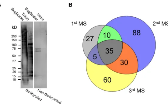
Biotinylation coupled to liquid chromatography-tandem mass spectrometry (LC-MS/MS) was used to obtain the first comprehensive surface proteome ofB.bovis-iRBCs. We selected B.bovisfor adhesion to bovine brain endothelial cells (BBECs) by panning uncloned parasites and the resulting cytoadherent parasite line was used for biotinylation (S1A Fig). The surface proteins of enriched iRBCs fromin vitroculturedB.boviswere biotinylated, extracted, puri- fied, and analyzed by LC-MS/MS in three biological replicates. Surface protein biotinylation was confirmed by live fluorescence microscopy. Biotinylation did not affect the integrity of iRBC membranes and the release of merozoites (S1B Fig). The extraction and purification of biotinylated proteins was confirmed by Western blot analysis (Fig 1A). A complete list of B.bovisproteins exported from the Scaffold software is provided in the supplemental
Fig 1. Biotinylation and liquid chromatography-tandem mass spectrometry (LC-MS/MS) ofB.bovis-iRBCs. (A) Western blot analysis of sequentially extracted proteins from biotinylated and control (non-biotinylated) samples. The image is representative of three independent experiments done with an approximately two-month interval. The membrane was probed with horseradish peroxidase (HRP)-conjugated streptavidin. (B) Venn diagram showing the number ofB.bovisproteins identified from biotinylated samples by three independent LC-MS/MS analyses.
https://doi.org/10.1371/journal.ppat.1008917.g001
information (S1 Table). In the first attempt, we lysed the iRBCs by a hypotonic solution fol- lowed by detergent protein extraction using the BugBuster reagent, which was shown to be efficient forP.falciparum-iRBCs surface proteins such as PfEMP1 [25]. LC-MS/MS analysis showed bovine hemoglobin contamination which might interfere with identification of B.bovispeptides. Therefore, in the second and third attempts, protein extraction was done fol- lowing biotinylation and saponin treatment. This modification improved the detection of total B.bovispeptides and proteins. Mass spectra obtained from LC-MS/MS were searched against B.bovisandBos taurusdatabases. Because parasite-encoded surface exposed proteins are low in abundance, we allowed protein identification by a single peptide. The number of unique peptides identified in the first, second, and third attempts from biotinylated samples were 805, 1600, and 948, respectively, in which 24–26% of identified peptides were fromB.bovisand yielded 77, 163, and 130 proteins, respectively (Table 1). Out of the 255 uniqueB.bovisprotein hits, 80 proteins detected from biotinylated samples by at least two MS analyses (Fig 1B) were further annotated for predicted export based upon the presence of a signal sequence, a trans- membrane (TM) domain, or GPI anchor. This refinement resulted in a total of 38 putative secretory proteins of which 20 had no predicted function in the database (S2 Table). The well- known surface exposedB.bovisprotein, VESA1, was identified in all three MS. VESA1 lacks an N-terminal signal sequence but has a C-terminal TM domain [11]. One SmORF and one SBP2, the products of two multigene families inB.bovis, SBP1 and SBP3 were also detected.
These proteins have an N-terminal signal sequence and were shown to be exported into the iRBC cytoplasm [9,10,12,13,15]. Of the 38 identified proteins 32 possessed a PEXEL-like export motif (PLM; RxL or RxxL) proposed by Pelle´ et al. [15].
Validation of localization for a subset of candidates
To evaluate the localization of putative exported proteins, we transformed parasites with plasmid constructs expressing target molecules fused with 2 myc epitopes (S2A Fig). We selected 10 pro- teins from the candidate list based on the three criteria for secretion and their abundance in the MS analysis (S3 Table) and further evaluated together with two positive control proteins, SBP3 and a variant of SmORF. Indirect immunofluorescence antibody test (IFAT) revealed that sig- nals of three proteins were detected in iRBCs among selected 10 candidates. Two proteins showed signals inside the parasite and the edge of the iRBCs like SBP3 and SmORF, namely, BBOV_III000060 (Bb60-mtm) and BBOV_III011920 (Bb11920-mtm) (Fig 2A). These proteins possess ten TM domains each and are paralogs encoded by a multigene family (termedmulti- transmembrane(mtm)family). Each possess an RxL motif at amino acid positions 24–27. The
Table 1. Summary of identified proteins.
1stMS 2ndMS 3rdMS
Biotinylated Non-biotinylated Biotinylated Non-biotinylated Biotinylated Non-biotinylated
Peptides� % Peptides� % Peptides % Peptides % Peptides % Peptides %
Bos taurus 612 76.0 179 89.9 1184 74.0 261 89.7 701 74.3 227 83.2
Babesia bovis 193 24.0 20 10.1 416 26.0 30 10.3 247 26.2 46 16.8
Total 805 199 1600 291 948 273
Proteins % Proteins % Proteins % Proteins % Proteins % Proteins %
Bos taurus 255 76.8 114 82.0 389 70.5 119 79.9 259 66.6 97 68.8
Babesia bovis 77 23.2 25 18.0 163 29.5 30 20.1 130 33.4 44 31.2
Total 332 139 552 149 389 141
�The peptide numbers are from BugBuster protein extract
https://doi.org/10.1371/journal.ppat.1008917.t001
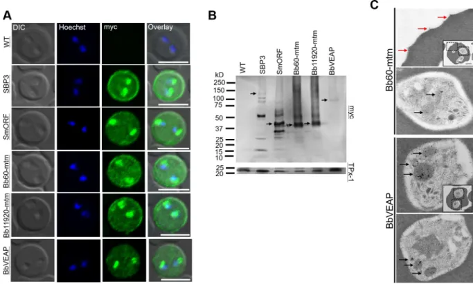
third protein, BBOV_III004280 (BbVEAP in theFig 2A) showed signals inside iRBCs and para- sites, but not from the edge of the RBC (Fig 2A). The deduced amino acid sequence of BBO- V_III004280 possesses a signal peptide with an RxL motif at amino acid position 185–188 and is conserved among all piroplasms exceptB.microti. We designated BBOV_III004280 as VESA export-associated protein (VEAP) gene because knockdown of this gene suppressed VESA export as described below. The remaining seven candidates did not show signals in iRBCs by IFAT, suggesting that they were not exported to iRBCs (S2B Fig). Thus, we decided to further characterize three novel proteins that were exported to iRBC. Target protein expression in trans- genic parasites was confirmed by Western blotting (Fig 2B). SBP3 and SmORF showed several bands indicating possible processing during export or degradation during protein extract prepa- ration [15]. The products of themtmgenes Bb60-mtm and Bb11920-mtm showed bands at the expected size of 49 kDa plus band smears. BbVEAP showed a single band corresponding to the anticipated size of 100 kDa. Immunoelectron microcopy (IEM) revealed that Bb60-mtm is local- ized in the spherical bodies of merozoites and on the iRBC surface close to ridge structures (Fig 2C,S3 Fig). BbVEAP was detected in merozoite spherical bodies (Fig 2C,S3 Fig).
A multigene family encoding Bbmtm is expanded inB.bovis
Of the three novel proteins that were exported into iRBC, Bb60-mtm and Bb11920-mtm are paralogous proteins with similar architectures of ten TM domains each and molecular weights
Fig 2. Expression and localization analysis of candidate proteins determined by indirect immunofluorescence antibody test (IFAT), Western blotting, and immunoelectron microscopy (IEM). (A) IFAT of parental wild type (WT) and transgenicB.bovislines expressing myc-tagged target proteins. The parasites were reacted with anti-myc antibody (α-myc, green) and nuclei were stained with Hoechst 33342 (Hoechst, blue). Scale bar = 5μm. (B) Western blot analysis of transgenicB.bovisexpressing myc-tagged proteins and WT parasite (WT). The expected full-length bands of the proteins are indicated with black arrows. (C) Immunoelectron microscopic analysis of transgenicB.bovis expressing Bb60-mtm or BbVEAP tagged with myc epitopes. Anti-myc antibody shows concentration of Bb60-mtm and BbVEAP in spherical bodies (black arrows) and Bb60-mtm expression on the iRBC surface (red arrows). Scale bar = 1μm.
https://doi.org/10.1371/journal.ppat.1008917.g002
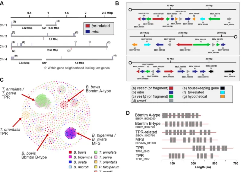
of approximately 49 kDa. Via BLAST searches of PiroplasmaDB and GenBank, we found 44 total copies in theB.bovisgenome and named this newly identified expanded gene family mtm(Fig 3A,S4 Table). The genes are typically telomeric and located in gene neighborhoods containingves1andsmorfgenes, two gene families encoding important exported proteins in B.bovis(Fig 3B). InP.falciparum, exported proteins are also typically localized to clusters within telomeric regions [26]. The familymtmexists in anotherBabesiaspecies from sheep (Xinjiang), which is closely related toB.bovis[27], and to date the family is unique to these two species (Table 2). To examine in other Piroplasmida the possible expansion of gene fami- lies that encode multi-transmembrane proteins, we used TMHMM-2.0 to screen whole genome and proteome datasets (Table 2,Fig 3C and 3D). Specifically, a major facilitator super- family (mfs)-like gene is greatly expanded inB.ovataandB.bigemina; whiletpr(T.parva repeat) is significantly expanded inTheileriaspp. [28,29], though several homologs oftprexist inBabesiaspp. Homology clustering shows thattprinT.parvaandT.annulatamake one clus- ter whileT.orientalis tprforms a separate cluster (Fig 3C). Homology clustering ofmtm
Fig 3. Expansion and distribution of genes encoding multi-transmembrane proteins in piroplasms. (A) Distribution ofmtmandtpr-related genes in theB.bovis genome. (B) Arrangement of multigene families (ves1,mtm,smorf, andtpr-related genes) in the nuclear genome ofB.bovis. (C) Homology clustering based on sequence similarities of genes with more than eight TM domains in piroplasms,Plasmodium falciparum, andT.gondii. (D) Schematics of selected gene products with multiple TM domains in piroplasms. Box indicates predicted TM domain.
https://doi.org/10.1371/journal.ppat.1008917.g003
revealed the existence of two clusters forB.bovis: A-type and B-type, and Bb60-mtm and Bb11920-mtm belong to B-type (Fig 3C,S4 Table). In summary, although orthologous rela- tionships and expansions of genes encoding homologous multi-transmembrane proteins are not generally conserved across theBabesiaandTheileriagenera, what is conserved is a theme of lineage-specific expansions of gene families that encode multi-transmembrane proteins which are candidates for export to the iRBC.
Bbmtm is associated with BS uptake
The presence of expanded gene families encoding predicted exported multi-transmembrane proteins across piroplasmida suggests that intraerythrocytic development of these parasites requiresde novochannel or transporter activity across the iRBC membrane. New permeability pathways have been described for theP.falciparum-iRBC membrane [30,31], and the activity of a plasmodial surface anion channel (PSAC) was shown to be determined by the protein products ofP.falciparum clag3.1andclag3.2genes [32] within the expandedclaggene family. Orthologs ofclaggenes are absent in piroplasmida, butBabesiaparasites increase iRBC permeability to sev- eral organic solutes including sorbitol, suggesting the existence of channels or transporters [33].
Selection ofP.falciparumunder high BS concentration resulted in silencing of bothclag3genes, suggesting that resistance to BS happens through epigenetic downregulation ofclaggenes [34].
To gain insights intomtmfunction, we produced two BS-resistant lines ofB.bovisby exposing the parasites to increasing concentrations of BS. These parasites showed increased half maximal inhibitory concentration (IC50) of BS compared to wild type (WT) (9.4 and 10.7 vs 2.3μg/mL, Fig 4A), and delayed lysis in sorbitol lysis assays compared to WT (Fig 4B). RNA-seq analysis revealed that transcripts of several genes including onemtm(Bb60for BS-resistant line 1 and BBOV_III000010 (Bb10) encoding an A-type Bbmtm for BS-resistant line 2) among manymtm family members was less abundant than in WT, suggesting that thesemtmsare potentially linked to BS uptake activity of iRBCs (Fig 4C,S5 Table). The sensitivity to BS was partially reversed in parasites cultured in the absence of BS for approximately two months (5 and 2.9μg/mL, respec- tively), suggesting epigenetic regulation of this drug resistance. RNA-seq and qRT-PCR con- firmed the recovery of downregulatedBb60andBb10in the corresponding revertant parasite lines (Fig 4D,S4 Fig). Episomal overexpression of Bb60-mtm and Bb10-mtm in BS-resistant line 1 and 2 showed fluorescence signals inside the parasite and the edge of the iRBCs as expected (Fig 4E) and made these parasites more sensitive to BS (Fig 4F; IC50of 7.8 and 2.3μg/mL for BS- res1-Bb60 and BS-res2-Bb10, respectively), supporting our hypothesis on their role in BS uptake.
However, episomally overexpressing Bb60-mtm with 10 or 100 nM WR99210 (Bb60-10 or Bb60-100 lines, respectively) in WT BS-sensitive line did not change IC50to BS (2.8 or 2.5μg/
mL, respectively), perhaps indicating saturation of the channel activity (S5 Fig).
Characterization of BbVEAP
Our three attempts to disrupt the BbVEAP gene locus, the third protein in our list which is exported into iRBC, using CRISPR/Cas9 system were unsuccessful, suggesting possible
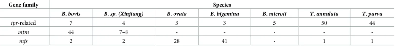
Table 2. Distribution oftpr-related,mtmandmfsgenes in Piroplasmida.
Gene family Species
B.bovis B.sp.(Xinjiang) B.ovata B.bigemina B.microti T.annulata T.parva
tpr-related 7 4 3 3 5 50 44
mtm 44 7–8 - - - - -
mfs 2 2 28 41 - 1 1
https://doi.org/10.1371/journal.ppat.1008917.t002
Fig 4. Blasticidin S-resistance inB.bovisis linked with downregulation ofBb60. (A) Growth inhibition curves of parasite lines in the presence of different concentrations of BS (μg/mL). All data are expressed as mean±SEM of triplicate cultures. (B) Osmotic lysis ofB.boviswild type (WT) and BS-resistant line 1 in the presence of sorbitol. iRBCs were enriched and the lysis experiment was performed at 37˚C. The graph is representative from two biological replicates done within a two-week interval. (C) The scatter diagram showing the differential expression of genes inB.bovisWT and BS-resistant lines. The horizontal and vertical axes represent the log 2-fold expression of WT and BS-resistant parasites, respectively. The upregulated and downregulated genes are shown in red and blue colors, respectively. (D) Scatter diagram showing the differential expression of genes inB.bovis WT and BS-sensitive revertant lines. The expression of downregulatedmtmsin BS-resistant lines,Bb60andBb10, recovered in revertant lines (green dots). (E) Indirect immunofluorescence antibody test of transgenicB.bovisBS-resistant lines episomally expressing myc-tagged Bb60-mtm or Bb10-mtm stained with anti-myc (green). The parasite nuclei were stained with Hoechst 33342 (Hoechst, blue). Scale bar = 5μm. (F) Growth inhibition curves of a panel of parasite lines in the presence of different concentrations of BS (μg/mL). Bb60-mtm and Bb10-mtm are episomally overexpressed under 100 nM WR99210 in BS-resistant lines 1 and 2, respectively. All data are expressed as mean±SEM.
https://doi.org/10.1371/journal.ppat.1008917.g004
essentiality for the parasite. To functionally characterize BbVEAP we inserted a glucosamine (GlcN)-inducibleglmSriboswitch together with 2 myc epitopes at the 3’ end of the BbVEAP open reading frame (ORF) [35]. Integration of theglmSsequence into the endogenous locus was confirmed by PCR (Fig 5A) and the expression of the myc-tagged protein was confirmed by Western blot analysis with the predicted band size (Fig 5A and 5B). In the absence of GlcN, glmS-tagged parasites demonstrated a roughly 80% reduction in basal BbVEAP protein expres- sion by Western blot analysis (Fig 5B and 5C). This reduction did not affect the growth of par- asites compared to the control (Fig 5D). The reduction of BbVEAP protein expression without addition of GlcN could be due to leakiness of theglmSsystem, decrease of mRNA stability or translation due toglmSsequence at its 3’ end, or endogenously produced GlcN byB.bovisas is reported forTrypanosoma cruzi[36], an observation for future verification. Addition of GlcN resulted in a dose dependent parasite growth reduction of BbVEAP-myc-glmSlines compared to the control BbVEAP-myc parasite (S6 Fig). Significant reduction of BbVEAP protein expression (82–92% reduction) with 2.5 mM GlcN, the maximum concentration without effect on the control parasite, was confirmed by Western blot analysis (Fig 5B and 5C). This knock- down resulted in a significant decrease in the growth rate accompanied by a significant increase of ring stage (immature) parasites and a decrease of binary form (mature) parasites, suggesting a defect in parasite development (Fig 5D and 5E).
Because BbVEAP is deposited in spherical bodies, we examined whether the knockdown of BbVEAP affected structure of these organelles by transmission electron microcopy (TEM).
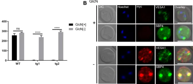
While no clear changes were seen in spherical body structure, we noticed fewer RBC surface ridges (Fig 5F). Scanning electron microscopy (SEM) images further revealed that the ridges were less protrusive with GlcN treatment in addition to a reduction in number (S7 Fig). To exclude the contribution of parasite stage on ridge numbers, we quantified the ridges in the mature stage of the parasite (binary form) using TEM images. GlcN treatment significantly reduced the number of ridges on the surface of iRBCs (Fig 5G). Because ridges are the focal point for adhesion ofB.bovis-iRBCs to endothelial cells [8], we examined whether knockdown of BbVEAP affected the cytoadhesion of iRBCs to BBECs. For this purpose, we used a cloned parasite from the cytoadherentB.bovisline (S1A Fig) to generated additional transgenic para- site lines in which a myc-glmSsequence was inserted within the 3’ end of the BbVEAP ORF the same way as the BbVEAP-myc-glmSline. The parasites were treated with GlcN for 3 days and cytoadhesion assays were conducted. BbVEAP expression was dramatically reduced in two BbVEAP-knockdown clones with GlcN treatment as determined by Western blot analysis (S8A Fig). While the addition of GlcN had no effect on the binding ability of WT parasites, cytoadhesion was abrogated in two BbVEAP-knockdown clones (Fig 6A). Although the expressed amount of VESA1 appears to be unchanged by Western blotting (S8A Fig), its distri- bution was affected; parasites without GlcN treatment showed a punctate pattern in the iRBC resembling the expression of VESA1 on ridges while in parasites with GlcN treatment signal was only detectable within the parasite cytoplasm (Fig 6B). Unlike VESA1, the expression and localization of SBP4, another exported protein into iRBC, was not affected when treated with GlcN (Fig 6B,S8A Fig).
Discussion
In this study we used iRBC surface biotinylation coupled with mass spectrometry to character- ize the surface proteome ofB.bovis. This approach was successfully used to describe thePlas- modiumparasite surface and iRBC surface parasite-encoded proteome [37,38]. We prioritized 38 proteins for potential export into iRBC, based upon their abundance in the above assay and subsequent annotation for export motifs. This list includes known exported proteins, such as
Fig 5. Induced knockdown of BbVEAP using theglmSriboswitch system decreases growth rate and ridge numbers. (A) Schematic of CRISPR/
Cas9 plasmid to insert myc-glmSsequences within theBbVEAPgene locus and agarose gel electrophoresis of diagnostic PCR to confirm integration of myc-glmSsequence at the 3’ end of theBbVEAPORF.rap-3’NR,rhoptry associated protein3’ noncoding region;hdhfrorf,human dihydrofolate reductaseORF;ef-1aIG,elongation factor-1αintergenic region;tpx-3’NR,thioredoxine peroxidase-13’ noncoding region; U6-3’NR,B.bovis U6 spliceosomal RNA3’ noncoding region; gRNA, guide RNA; and HR, homologous region.glmS-C1 andglmS-C2 indicate transgenic lines
independently generated and following "-1" and "-2" indicate 2 independent clones. (B) Western blot analysis of 2 independently generated BbVEAP- myc-glmSclones and a control BbVEAP-myc parasite line expressing myc-tagged BbVEAP without theglmSelement from the endogenous gene locus in the presence or absence of glucosamine (GlcN). Anti-TPx-1 antibody was used to detect TPx-1 protein as a loading control. The image is representative of three independent experiments done within an approximately one-week interval. (C) Densitometry of BbVEAP protein levels in all conditions measured relative to the control parasite (GlcN-untreated BbVEAP-myc line) incubated in the presence or absence of GlcN. (D) Growth of BbVEAP-myc-glmSand control BbVEAP-myc lines in the presence or absence of GlcN. Initial parasitemia was 0.1% and parasitemia was
VESA1, SBPs, and SmORF, thus validating the study methodology. Of the novel exported pro- tein candidates, 2 Bbmtm proteins belong to a multigene family; and a protein we have termed BbVEAP which is largely conserved within piroplasma. It is noted that our method resulted in the biotinylation and identification of merozoite surface and abundant proteins (S2B Fig), pos- sibly due to biotin entry into or lysis of RBCs and parasites.
The Bbmtm genes are expanded to 44 gene copies in theB.bovisgenome, the same number as the expansion ofsmorfgenes. Likesmorfs,mtmsare typically located within gene neighbor- hoods, often telomeric, containingves1multigene family members. VESA1, SmORFs, and Bbmtms are exported proteins and close association of their gene loci in the genome of B.bovismay suggest a common epigenetic control of expression. Although themtmgene fam- ily is unique toB.bovis, proteins with a similar multi-transmembrane structure such as MFS and TPR are expanded in other piroplasms, indicating a conservation of lineage-specific expansion of multi-transmembrane proteins. UnlikePlasmodiumandT.gondii, which main- tain a PV membrane (PVM) through their developmental cycle in the host cell, the PVM rup- tures within minutes of invasion byBabesia, like a related piroplasm,Theileria[39,40].
Therefore, these parasites are in direct contact with the host cell cytoplasm and this may allow the parasite to export proteins possessing multiple TM domains to the host cell. To our knowl- edge this is distinct fromPlasmodium, for which no known multi-transmembrane proteins are exported across the PVM. ThePlasmodiumCLAG/RhopH1 proteins that is localized on the iRBC membrane possess two transmembrane domains, although lacking a ‘classical’
monitored for 3 days with daily culture medium replacement. The data are shown as mean±S.D. for three independent experiments performed with a one-week interval. (��,P<0.01;���,P<0.001;����,P<0.0001; determined by multiplettest). (E) Proportion of ring, binary, and multiple stages in different parasite lines in the presence or absence of GlcN on day 3 post GlcN introduction. The data are shown as mean±S.D. (�,P<0.05;��, P<0.01; ns, not significant,P�0.05; determined by multiplettest). (F) Transmission electron microscopy images of BbVEAP-myc-glmSin the presence (+) or absence (-) of GlcN. Black arrows indicate spherical bodies and red arrow shows ridges. Scale bar = 0.5μm. (G) Quantification of ridge numbers on the surface of iRBCs of BbVEAP-myc-glmSparasites in the presence or absence of GlcN at day 3 post GlcN introduction. Ridge numbers per 10μm of iRBC circumference were quantified only in mature stage iRBCs (binary form) (����,P<0.0001; determined by Mann- WhitneyUtest).
https://doi.org/10.1371/journal.ppat.1008917.g005
Fig 6. BbVEAP knockdown abrogates binding of iRBCs to endothelial cells. (A) Cytoadhesion assay of BbVEAP-myc-glmSand WT parasites in the presence (+) or absence (-) of GlcN. All data are expressed as mean±SEM of triplicate assay (����,P<0.0001; determined by paired Student’sttest). (B) Indirect immunofluorescence microscopy test of BbVEAP-myc-glmSparasite in the presence (+) or absence (-) of GlcN (α- myc, red;α-VESA1 andα-SBP4, green). The parasite nuclei were stained with Hoechst 33342 (Hoechst, blue). Scale bar = 5μm.
https://doi.org/10.1371/journal.ppat.1008917.g006
transporter or channel multi-TM structure, and are involved in the permeability pathway across the RBC membrane, also termed PSAC; but they are introduced via merozoite rhoptry secretion during the RBC invasion [32,41,42].
The function of Bbmtm, MFS, or TPR in piroplasms is not known but their structure sug- gests a transporter or channel activity. It was shown thatB.divergens, a zoonoticBabesiaspe- cies that infects cattle and immunocompromised humans, increases RBC permeability to various organic solutes, indicating the existence of parasite produced channels or transporters on the surface of iRBC [33]. Similarly, we have seen increased permeability of RBC to sorbitol when they were infected withB.bovis, which was reduced in BS-resistant lines. We found that only eitherBb60orBb10was downregulated in each of two BS-resistant lines among allmtm gene family members and revertants recovered the transcription level of respectivemtm, indi- cating a link between expression of thesemtmsand BS resistance. These observations support the idea that Bbmtm acts as a channel or transporter, and if this is the case then Bb60-mtm or Bb10-mtm can transport BS across the RBC membrane. AlthoughBb60andBb10were down- regulated in BS-resistant lines, the expression of othermtmswas unchanged in comparison to control parasites, suggesting stable epigenetic regulation, or a substrate specificity for each mtm. Transcripts of several other genes were also reduced in the BS-resistant parasite lines, which might contribute to this resistance, and needs future evaluation.
In this study we identified BbVEAP and found that this protein is involved in ridge forma- tion, VESA1 expression on iRBC, and binding of iRBCs to endothelial cells, which establishes BbVEAP as a novel virulence factor ofB.bovis. Although distribution of VESA1 in the iRBC was affected by BbVEAP knockdown, the mechanism behind this observation is unclear.
Because we could not co-precipitate these two proteins by immunoprecipitation experiments (S8B Fig), they may not directly interact. Recently it was shown that upregulation of SBP2 truncated copy 11 inB.bovisreduced binding of iRBCs to endothelial cells and its virulence in cattle [43,44]. However, it is unclear how SBP2 truncated copy 11 affects the export or surface expression of VESA1 or other putative cytoadhesion ligands. A knobless line ofP.falciparum has been described, and though the surface expression of PfEMP1, the functional homolog of VESA1 inP.falciparum, is reduced in these parasites they remained able to bind to endothelial cells [45,46]. RidgelessB.bovishas not been reported and VESA1 is the only known ligand for cytoadhesion ofB.bovisiRBCs [4]. Depletion of BbVEAP abrogated cytoadhesion indicating its role in the export or distribution of VESA1 and/or other putative ligands to the iRBC sur- face. Additionally, BbVEAP has a single ortholog in all piroplasmida that includes parasite spe- cies lacking ridge-like structures on iRBCs, suggesting a piroplasm-conserved function other than cytoadhesion. Considering the essentiality of this gene and conservation across piroplas- mida, including the generaTheileria, targeting this protein could be promising for the devel- opment of pan piroplasmida therapeutics.
Spherical bodies are large secretory organelles within the apical end ofBabesiaspp. [47].
The products of two multigene family inB.bovis, SBP2 and SmORFs, are localized in spherical bodies [9,15]. In this study we found two novel proteins released from spherical bodies to iRBC, Bbmtm and BbVEAP. It was proposed that the PLM in SBP2 and SmORFs works as a retention signal in spherical bodies [15]. Bbmtm and BbVEAP also have PLM, suggesting the importance of PLM for the protein export to iRBC via spherical bodies inBabesiaparasites.
Surface exposed proteins are targets of immunity and thus are vaccine targets, such as PfEMP1-VAR2CSA which is expressed on the surface ofP.falciparumiRBCs and is responsi- ble for pathogenesis of pregnancy-associated malaria [48]. VAR2CSA-based vaccines are cur- rently in clinical trials [49]. ThePlasmodiumnew permeability pathway, PSAC, is produced by Plasmodiumand is inserted into the iRBC membrane for nutrient acquisition. Since nutrient acquisition is essential for malaria parasite survival, PSAC is considered a promising target for
the development of antimalarials [50]. Targeting channels or transporters on theBabesia- iRBC membrane may lead to the development of pan anti-babesiosis drugs. Further character- ization of our proteome data set could pave the way for identification of novel vaccine and drug targets and further development of new control strategies for bovine babesiosis caused by B.bovis.
Methods
Parasite culture and transfection
B.bovisTexas strain was obtained from Washington State University and kept in continuous culture using a microaerophilic stationary-phase culture system composed of purified bovine RBCs at 10% hematocrit and GIT medium (Wako Pure Chemical Industries, Japan).
The transfection ofB.boviswas done as described [51,52]. Briefly, 100μL ofB.bovis-iRBCs were mixed with 10μg of plasmid constructs in 100μL of Amaxa Nucleofector human T-cell solution. Transfection was done using a Nucleofector device, program v-024 (Amaxa Biosys- tems, Germany). Ten nanomolar WR99210 was added one day after the transfection to select a transgenic parasite population.
Enrichment ofB.bovis-iRBCs
Enrichment ofB.bovis-iRBCs was done using a Histodenz solution. The solution was prepared by dissolving 27.6 g of Histodenz (Sigma-Aldrich) in 100 mL of Tris-buffered solution (5 mM Tris-HCl, 3 mM KCl, and 0.3 mM Na2-EDTA, pH 7.5). The iRBCs were layered on the surface of 80% Histodenz solution in GIT medium and centrifuged for 30 min at 2500 x g with a swinging bucket rotor. The iRBCs at the bottom of the tube were used for biotinylation assays.
Biotinylation and protein extraction
Biotinylation of iRBC surface proteins was done as described [25,37]. In the first attempt, pro- teins were serially extracted using a hypotonic solution (20X diluted PBS) and BugBuster pro- tein extraction reagent (Novagen) containing the endonuclease benzonase. MS showed high bovine hemoglobin contamination. Thus, in the second and third attempts biotinylated iRBCs were initially treated with 0.2% (w/v) saponin on ice for 15 min to remove hemoglobin. Pro- tein extraction was done serially first by resuspending the parasite pellets in BugBuster protein extraction reagent containing the endonuclease benzonase. Following centrifugation and removing extracted proteins, the remaining parasite pellet was lysed with a solution containing 150 mM NaCl, 5 mM EDTA, 50 mM Tris pH8.0, 1.0% Triton-X 100 (w/v), and protease inhib- itor cocktail (Complete Mini, Roche) at 4˚C for 1 h. The protein extract was incubated with Dynabeads MyOne Streptavidin C1 (Invitrogen) with rotation at 4˚C for 1 h. The beads were washed 5 times with wash buffer containing 0.1% SDS, 400 mM urea, 150 mM NaCl, and 50 mM Tris-HCl (pH 8.0) to remove unbound proteins. Bound proteins were eluted from the beads by boiling for 5 min in 1x Sample Buffer (25 mM Tris (pH 6.8), 2.5% w/v SDS, 2.5% v/v glycerol, 0.08% w/v bromophenol blue, and 5% beta-mercaptoethanol). Proteins were assessed for biotinylation via Western blotting using horseradish peroxidase (HRP)-conjugated strepta- vidin (1:40,000, Invitrogen).
Liquid chromatography-tandem mass spectrometry (LC-MS/MS)
The purified biotinylated proteins were subjected to LC-MS/MS as described [53]. The sam- ples were briefly electrophoresed on SDS-polyacrylamide gel electrophoresis. The gel contain- ing proteins were excised, fixed with acetic acid/methanol solution, and subjected to LC–MS/
MS analysis at the W. M. Keck Biomedical Mass Spectrometry Laboratory, University of Vir- ginia, USA. The data analysis was performed by database searching using the Sequest search algorithm against theBos taurusandB.bovisreference strain in UniProt and SwissProt. Filter- ing and extraction of data was performed using Scaffold version 4.8.9 (Proteome Software Inc.). Protein identifications were accepted if they could be established at greater than 90%
probability and contained at least 1 identified peptide. Quantitative value (normalized total spectra) was used to show an estimate of protein abundance between biotinylated and non- biotinylated samples. PiroplasmaDB-34 [54] was used for annotation of the target proteins.
Cytoadhesion assay
Cytoadhesion assays were done as described [55]. Briefly, bovine brain endothelial cells (BBECs; Cell Applications Inc., USA) were seeded in 6 well plates containing cover glasses (Matsunami Glass, Japan).B.bovis-iRBCs with 2–5% parasitemia and 1% hematocrit were added to the BBEC culture. The cells were incubated for 90 min with gentle agitation every 15 min. Nonadherent iRBCs were washed away with Hanks-balanced salt solution. Cells on the cover glasses were fixed with methanol and stained with Giemsa’s solution and the number of bound iRBCs were counted for 500 BBECs.
Plasmid construction
The schematic of the plasmid expressing myc-tagged target proteins is shown inS2 Fig. The primers used for plasmid construction are listed inS6 Table.B.bovis elongation factor-1α intergenic region-B (ef-1αIG-B), ORF of the gene of interest (GOI), andthioredoxine peroxi- dase-1(tpx-1) 3’ noncoding region (NR) were PCR-amplified fromB.bovisgenomic DNA. A DNA fragment containingB.bovis actin5’ NR (act5’NR),human dihydrofolate reductase (hdhfr), andB.bovis rhoptry associated protein3’ NR (rap3’NR) was amplified from aB.bovis green fluorescent protein (GFP)-expressing plasmid [51].ef-1αIG-B was cloned into the EcoRI site of pBluescript SK using In-Fusion HD Cloning Kit (Takara Bio Inc., Japan). Subse- quently, the ORF of GOI tagged with 2 myc epitopes,tpx-13’NR, andact5’NR-hdhfr-rap 3’NR were cloned into SmaI to make the final plasmid for episomal expression of myc-tagged proteins.
The CRISPR/Cas9 system was employed to delete BBOV_III004280 or insert myc andglmS sequences into the 3’ end of the original locus [56]. Briefly, homologous regions (HRs) were PCR-amplified fromB.bovisgenomic DNA and inserted into the BamHI site of the BbU6- Cas9-hDHFR plasmid. Single guide RNA (sgRNA) was inserted into the AarI site of BbU6- Cas9-hDHFR using T4 DNA ligase (New England Biolabs, USA). Parasites were transfected as described [51,52] and the obtained transgenic parasites were cloned by limiting dilution before analysis. Diagnostic PCR was done using glms-F-IF and 4280-3NR-integR (S6 Table) to con- firm insertion of myc-glmS.
Development of BS-resistantB.bovisand comparative transcriptomics B.bovisWT parasites were initially cultured with 1μg/mL blasticidin-S solution (BS; Thermo Fisher Scientific, USA), and the BS concentration was increased stepwise. Two independent resistant lines that propagate under 4μg/mL BS were produced. To produce revertant para- sites, BS-resistant lines were cultured without BS for two months. Comparative transcrip- tomics betweenB.bovisWT and its BS-resistant derivatives or revertant parasites were performed by RNA-seq. RNA was extracted from parasites using TRIzol reagent (Invitrogen).
Libraries were constructed using a TruSeq Stranded mRNA Library Preparation Kit (Illumina, USA), according to the manufacturer’s protocol and the products were subjected to
Novaseq6000 (Illumina) with the 150-bp paired-end protocol. Acquired reads were mapped againstB.bovisT2Bo reference genome obtained from PiroplasmaDB-37 using HISAT2 [57].
Read data normalization and differential expression were obtained using Cufflinks with the default parameters [58]. Complementary DNA was synthesized using SuperScript III Reverse Transcriptase (Invitrogen) with random primers.
Bioinformatics analysis
SignalP-5.0 and -3.0 were used to predict putative signal peptides [59]. TMHMM-2.0 was used to predict TM domains [60]. GPI anchors were predicted using PredGPI [61]. To identify mul- tigene families encoding multiple TM domains, whole genome sets of protein sequences for B.bovis,B.bigemina,B.ovata,B.microti,Theileria annulata,T.parva,T.orientalis,P.falcipa- rum, andT.gondiiwere evaluated using TMHMM-2.0 and proteins were selected having equal to or more than eight TM domains and at least one TM domain average per 100 amino acids. Mutual homology among the selected proteins were identified by BLASTP [62] and pro- teins with more than a 200 bit-score and by eye were regarded as homologs. The overall rela- tionships were visualized with Gephi [63] using a Fruchterman–Reingold layout.
SDS-PAGE and Western blotting
Parasite proteins were extracted using 1.0% Triton-X 100 (w/v) in PBS and protease inhibitor cocktail (Complete Mini, Roche) at 4˚C for 1 h. The protein fractions were separated by elec- trophoresis and transferred to polyvinylidene difluoride membranes (Millipore, USA). The membranes were probed with mouse anti-myc monoclonal antibody (1:500; 9B11, Cell Signal- ing Technology, USA), rabbit anti-TPx-1 polyclonal antibody (1:250 [52]), rabbit anti-VESA1 polyclonal antibody (1:100), or rabbit anti-SBP4 polyclonal antibody (1:1000 [13]) at 4˚C over- night. Washing was done in PBS supplemented with 0.05% Tween-20 (TPBS). Secondary probe of membranes was done with HRP-conjugated goat anti-mouse or rabbit IgG (1:8,000;
Promega, USA). Protein bands were visualized using ECL Select Western Blotting Detection Reagent (GE healthcare) and detected by a chemiluminescence detection system (LAS-4000 mini; Fujifilm, Japan).
Immunoprecipitations were performed as described [64]. Parasite pellets were prepared by saponin treatment of BbVEAP-myc tagged parasites and cross-linked with 1 mM DSP (Sigma). Proteins were extracted with 1% Triton X-100 (w/v) in PBS (containing 1 mM EDTA, 10% glycerol, and protease inhibitors) at 4˚C for 1 h. The extracted proteins were incu- bated with anti-myc mouse mAb (9B11, Cell Signaling) at room temperature for 4 h with gen- tle rotation. The solution was then mixed with protein G Sepharose 4 fast flow (GE
Healthcare) and incubated with rotation at 4˚C overnight. The mixture was centrifuged and the beads were washed with 0.5% Triton X-100 (w/v) in PBS (containing 1 mM EDTA, 10%
glycerol, and protease inhibitors). To elute proteins, the beads were mixed with 2.4μg/μL of c- myc peptide (Thermo Fisher Scientific) and incubated at 4˚C for 12 h. The beads were centri- fuged and the supernatant was collected as an immunoprecipitated (IP) fraction.
Indirect immunofluorescence antibody test (IFAT)
IFAT was done on thin blood smears from cultured parasites that had been air-dried and fixed in a 1:1 acetone:methanol mixture at−20˚C for 5 min [65]. Smears were immunostained with mouse anti-myc monoclonal antibody (9B11) at 1:500 dilution in TPBS and incubated at 37˚C for 60 min. Double immunostaining of smears was done with rabbit anti-VESA1α(antisera against peptide YNQVVHYIRALFYQLYFLRK; Medical & Biological Laboratories Co., ltd, Japan) at 1:50 dilution, or rabbit anti-SBP4 at 1:1000 dilution. The smears were incubated with