Acta Med. Nagasaki 45 : 45-47
Kissing Aneurysm of the Distal Anterior Cerebral Artery: Preoperative CT Angiography and Surgical Management: A Case Report
Makio KAMINOGO 1), Masaharu TANI 1), Jun-Ichi KAWAKUBO 1), Hideki ISHIMARU 2), Hideyuki HAYASHI 2), Shobu SHIBATA 1)
1) Department of Neurosurgery, Nagasaki University School of Medicine 2) Department of Radiology, Nagasaki University School of Medicine
We describe a patient with mirror-image aneurysms in the bilateral distal anterior cerebral artery (ACA). The larger aneurysm was clearly disclosed with digital subtraction angiography (DSA), but the smaller one could not be definitely
identified. The bilateral aneurysms were confirmed with computed tomographic (CT) angiography, which showed the right ACA aneurysm to be hidden behind the left ACA aneu- rysm, likely buried in the cingulate gyrus. During surgery, the left ACA aneurysm was clipped first. The right ACA aneurysm was exposed by a small subpial resection of the cingulate gyrus, and the right ACA aneurysm, which strongly adhered to the surrounding tissue, was safely dissected. Multiple aneu- rysms associated with a distal ACA aneurysm are not rare. We conclude that further examination with CT angiography is im- portant when kissing aneurysms are suggested by DSA.
Introduction
The prevalence of distal anterior cerebral artery (ACA) aneurysm is relatively low, and our personal experience in this area is rather limited'-'). Aneurysms of the dis- tal ACA have some unique characteristics. They tend to bleed, irrespective of their size'-". The incidence of multiple aneurysms is higher in cases of distal ACA aneurysms than in cases of other location, and addi- tional aneurysms are not infrequently detected in the contralateral or same distal ACA1-41. The aneurysm in mirror position to the distal ACA aneurysm is not al- ways identified preoperatively'). We treated a patient in which computed tomographic (CT) angiography was helpful in the diagnosis and operative management of kissing aneurysms of the distal ACA.
Address Correspondence : Makio Kaminogo, M.D.
Department of Neurosurgery, Nagasaki University School of Medicinel-7-1 Sakamoto, Nagasaki 852-8501, Japan
TEL: +81-95-849-7375, FAX: +81-95-849-7378 E-mail: kaminogo@net. nagasaki-u. ac. jp
Case report
Magnetic resonance (MR) angiography in a 42-year- old woman suggested unruptured distal ACA aneurysms.
The woman had undergone MR angiography for brain screening called "Brain Check-up"". She had no history
of severe headache or other neurological symptoms.
Digital subtraction angiography (DSA) clearly disclosed a distal ACA aneurysm at the origin of the right anterior internal frontal artery and suggested an aneurysm in mirror position on the left ACA (Fig. 1). Multiplanar re- formatted (MPR) CT images clearly depicted two aneu- rysms and indicated that the larger right ACA aneurysm was located just behind the smaller left ACA aneurysm (Fig. 2). Surgery was performed via interhemispheric approach. The smaller aneurysm of the left ACA was explored, and its neck was dissected (Fig. 3, left) and clipped. The right ACA aneurysm was not yet apparent, but, based on the preoperative CT angiography, we thought that the right ACA aneurysm was buried in the cingulate gyrus, behind the left ACA aneurysm. A small subpial resection of the right cingulate gyrus was performed to safely expose the right aneurysm and right ACA (Fig. 3, right). After this exposure, the right an- eurysm was clearly dissected from the left aneurysm and clipped. The postoperative course in this case was uneventful, and postoperative angiography confirmed complete obliteration of both aneurysms.
Discussion
Distal ACA aneurysms are sometimes accompanied by additional aneurysms on the same or the opposite ar- tery. Laitinen found aneurysms bilaterally in 3 of 14
(21 %) patients with distal ACA aneurysm". Sindou re-
ported bilateral aneurysms in 2 out of 19 (11 %) patients
with distal ACA aneurysms9). Other reported incidences
are 10 to 39 %4. 10). But because the prevalence of distal
Figure 1. Right carotid angiogram, right oblique view (left), anteroposterior view (center), and left oblique view (right), clearly disclose the aneurysm (arrow) of the right distal anterior cerebral artery (ACA). An aneurysm in mirror position on the left distal ACA (arrowhead) is also suggested, but is not definitely di- agnosed by angiography.
Figure 2A. The slice position for multiplanar reformatted (MPR) images.
Figure 2B. MPR images (a to f: left anterior to right posterior) clearly show aneurysms in bilateral distal anterior cerebral artery (ACA). The spatial relation between these aneurysms is also demonstrated.
L-An = left aneurysm, R-An = right aneurysm,* = left anterior
cerebral artery,' = right anterior cerebral artery
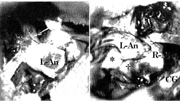
Figure 3. Intraoperative photograph. The aneurysm of the left ACA is exposed (left). After clipping the neck of the left aneurysm, subpial resection of the cingulate gyrus is performed, and the right ACA an- eurysm, strongly adhering to the left ACA aneurysm, is exposed (right).
L- An = left aneurysm, R- An = right aneurysm,* = left anterior cerebral artery,' = right anterior cerebral artery, CG = resected cingulate gyrus.
ACA aneurysms is low and is reported at 2.3% - 5.6 p/01,2,4,5) , most neurosurgeons have rather limited experi- ence in this area.
Yasargil has indicated that the distal ACA aneurysm presents particular surgical problems, including a nar- row working space in the interhemispheric fissure and callosal cistern, strong adhesion between the cingulate gyri, a broad-based aneurysmal neck, a hard-to-identify parent artery, and fixation of aneurysmal dome on the pial layer 4). Ohno et al emphasized the technical diffi- culty in surgical treatment of the distal ACA aneu- rysm due to the broad neck configuration and coexis- tence of other aneurysms'). In the present case, MPR images obtained by helical CT clearly demonstrated the kissing aneurysms and their spatial relation. When we first clipped the left ACA aneurysm following dis- section of the neck, we considered that retraction of the left aneurysm and left ACA without identification of the right lesion might injure the right aneurysm and induce its premature rupture. Thus, we performed subpial resection of the right cingulate gyrus around the left ACA aneurysm Strong adhesion of the right ACA aneurysm to the left ACA aneurysm and left ACA was verified. The right ACA aneurysm was care-
fully and successfully dissected from the surrounding structures.
Careful angiographic examination is required in cases of distal ACA aneurysm to detect other aneurysms,
and CT angiography is useful in diagnosing the kissing aneurysms and evaluating their positional relation.
The small subpial resection in lieu of retraction of a superficial aneurysm can be used to safely dissect the second aneurysm.
References
1) de Sousa A, Dantas R, de Cardoso G, Costa B. Distal anterior cere- bral artery aneurysms. Surg Neurol 52: 128-136, 1999
2) Hernesniemi J, Tapaninaho A, Vapalahti M, Niskanen M, Kari A, Luukkonen M. Saccular aneurysms of the distal anterior cerebral
artery and its branches. Neurosurgery 31: 994-999, 1992
3) Ohno K, Monma S, Suzuki R, Masaoka H, Matsushima Y, Hirakawa K. Saccular aneurysms of the distal anterior cerebral artery.
Neurosurgery 27: 907-913, 1990
4) Yasargil MG. Distal anterior cerebral artery aneurysms (pericallosal
artery aneurysms). In: Yasargil MG ed. Microneurosurgery. Vol II, Stuttgart: Georg Thieme Verlag, pp. 224-231, 1984
5) Yoshimoto T, Uchida K, Suzuki J. Surgical treatment of distal an- terior cerebral artery aneurysms. J Neurosurg 50: 40-44, 1979 6) Mori T, Fujimoto M, Shimada K, Shin H, Sakakibara T, Yamaki T.
Kissing aneurysms of distal anterior cerebral arteries demonstrated
by magnetic resonance angiography. Surg Neurol 43: 497-499, 1995 7) Yoshimoto T, Mizoi K. Importance of management of unruptured
cerebral aneurysms. Surg Neurol 47: 522-526, 1997
8) Laitinen L, Snellman A. Aneurysms of the pericallosal artery. A study of 14 cases verified angiographically and treated mainly by direct surgical attack. J Neurosurg 17: 447-458, 1960
9) Sindou M, Pelissou-Guyotat I, Mertens P, Keravel Y, Athayde AA.
Pericallosal aneurysms. Surg Neurol 30: 434-440, 1988
10) Wisoff JH, Flamm ES. Aneurysms of the distal anterior cerebral artery and associated vascular anomalies. Neurosurgery 20: 735-741, 1987