Introduction
Congenital anomalies of the internal carotid artery (ICA) classified as agenesis, aplasia, and hypoplasia by Lie24) are rare. However, the associated incidence of intracranial aneurysms is quite high. Its incidence has been reported as 25-67%1,19,23,33,39).
The current literature on these complicated aneurysms does not discuss aneurysmal size or treatment, and there have been only a few reports on large or giant aneurysms associated with these anomalies of the ICA. We present a case of a giant, irregularly shaped aneurysm of the anterior communicating artery associated with hypoplasia of the unilateral ICA with secondary occlusion, and review the literature.
Case Presentation
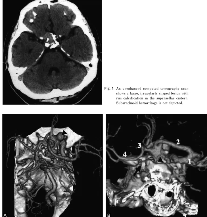
A 62-year-old man presented with complaints of a continual severe headache and was admitted to our hospital. There was no evidence of subarachnoid hemorrhage based on computed tomography (CT). However, CT revealed a large, irregularly shaped lesion with rim calcification in the suprasellar cistern (Fig. 1). Three-dimensional CT angiography (3D-CTA) showed an irregularly shaped aneurysm of the anterior communicating artery. The intracranial portion of the left ICA was not delineated (Fig. 2A, B). A right ICA arteriogram revealed the irregularly shaped aneurysm of the anterior communicating artery (10×12×28 mm), in which both the left anterior cerebral artery (ACA) and left middle cerebral artery (MCA) were filled by the right ICA via the anterior communicating artery (Fig. 3A). A left common carotid angiogram revealed occlusion of the cervical portion
1) Department of Neurosurgery, Fujigaoka Hospital, Showa University 2) Department of Neurosurgery, Higashi-Totsuka Memorial Hospital
●Abstract●
Objective: There have been few reports on the association of large or giant aneurysms with congenital absence or hypoplasia of the internal carotid artery (ICA). We present a case of a giant anterior communicating artery aneurysm associated with hypoplasia of the unilateral ICA with secondary occlusion.
Case presentation: A 62-year-old man was diagnosed with a giant anterior communicating artery aneurysm (10×12×28 mm) associated with hypoplasia of the unilateral ICA. A left common carotid angiogram revealed occlusion of the cervical portion of the left ICA. A right internal carotid arteriogram showed the aneurysm. Both the left anterior cerebral artery and left middle cerebral artery were filled from the right ICA via the anterior communicating artery. Due to the complicated morphology and calcified wall of the aneurysm, we performed endovascular treatment using a combination of 18- and 10-series microcatheters to make separate coil frames in the aneurysmal sac. Twenty-seven Guglielmi detachable coils with a total length of 419cm were placed in the aneurysm, which resulted in satisfactory occlusion. The patient was discharged without complications, and there was no evidence of aneurysmal recurrence for three years.
Conclusion: Large or giant aneurysms associated with congenital absence or hypoplasia of the ICA are rare. In their treatment them, it is important to avoid a decrease in collateral blood flow. The endovascular procedure is a reasonable therapeutic option.
●Key Words●
agenesis, embolization, giant aneurysm, hypoplasia, internal carotid artery
(Received September 22, 2008:Accepted February 9, 2009)
<Corresponding address: Itokawa H, 1-30, Fujigaoka Aoba-ku Yokohama city E-mail: [email protected]>
of the left ICA with a stump at the bifurcation (Fig. 3B).
Left vertebral angiography showed normal anatomy, with filling of both the posterior cerebral artery territory and the posterior fossa. CT at the level of the skull base showed
hypoplasia of the left carotid canal (Fig. 4).
Resting cerebral blood flow (CBF) and vascular reactivity to acetazolamide were calculated by single photon emission CT. No global or focal reduction of the CBF was detected in Fig. 1 An unenhanced computed tomography scan
shows a large, irregularly shaped lesion with rim calcification in the suprasellar cistern.
Subarachnoid hemorrhage is not depicted.
A B
Fig. 2 Three-dimensional computed tomography angiography, A:superior view shows an irregularly shaped aneurysm of the anterior communicating artery (arrowhead) B:Posteroanterior view. The intracranial portion of the left internal carotid artery is not delineated (arrow). This view delineates a well-defined neck separating the aneurysm (arrowhead) from the both anterior cerebral arteries (1: right internal carotid artery, 2: right anterior cerebral artery, 3: left anterior cerebral artery, 4: left middle cerebral artery).
the resting state; however, vascular reactivity was slightly decreased in the left cerebral hemisphere.
Because the aneurysm had complicated morphology and a
calcified wall, aneurysmal clipping was considered difficult, and we decided to perform endovascular treatment. Due to the difficulty in making a frame with a single coil for this irregularly shaped aneurysm, we used a combination of 18- and 10-series microcatheters to make separate frames in the intrasellar and suprasellar portions of the aneurysm. The first 18-microcatheter (Excelsior 1018, Boston Scientific, Natick, MA, USA) was positioned within the intrasellar portion, and the second 10-microcatheter (pre-shaped Prowler-10, J-type, Cordis, Miami, FL) was introduced within the suprasellar portion (Fig. 5A, B). A Guglielmi detachable coil (GDC-18, 9-mm 2D helix×30-cm, Boston Scientific, Natick, MA) was initially deployed from the Excelsior 1018 catheter, which was stable across the aneurysmal neck without compromising the flow in the parent artery (Fig. 5C). Then, the second coil (GDC-10, 3D 10-mm
×30-cm) was deposited via the Prowler-10 catheter, and a stable coil frame was formed in the suprasellar portion of the aneurysm (Fig. 5D). The residual aneurysmal sac was filled with small coils from the distal sac of the aneurysm in piecemeal fashion, aiming at the neck through both catheters.
Twenty-seven coils with a total length of 419 cm were placed in the aneurysm, resulting in satisfactory occlusion (Fig. 5E).
A B
Fig. 3
A: Right internal carotid angiogram (anteroposterior view) showing that the left anterior and middle cerebral arteries are supplied by the right internal carotid artery via the anterior communicating artery, and the presence of a large, irregularly shaped aneurysm of the anterior communicating artery.
B: Left common carotid arteriogram (lateral view) shows occlusion of the cervical portion of the left internal carotid artery with a stump.
Fig. 4 Computed tomography at the skull base shows a hypoplastic bony carotid canal on the left side (arrowheads).
Post-operative left vertebral angiography revealed the left ophthalmic artery supplied via the posterior communicating artery (Fig. 5F). The patient was discharged the following day without complications, and a 3 year-follow up angiogram of the right ICA revealed no recurrence of the aneurysm (Fig. 5G).
Discussion
Hypoplasia of the ICA generally refers to incomplete development of an ICA and a hypoplastic bony carotid canal can be detected by thin slice bone CT. In our case, we
recognized a left ICA occlusion with a large carotid stump in the neck. The ipsilateral ACA and MCA were supplied by the contralateral ICA through the anterior communicating artery (Fig. 2). However, CT of the skull base showed the left carotid canal to be hypoplastic (Fig. 3). Based on these findings, we diagnosed hypoplasia of the unilateral ICA with secondary occlusion. Possible reasons for occlusion of the hypoplastic ICA may include atherosclerosis, dissection, embolic phenomena or vasculitis12,23).
There are many reports of intracranial aneurysms associated with congenital absence or hypoplasia of the I Fig. 5
A: The right carotid arteriogram (oblique view) shows a large, irregularly shaped aneurysm of the anterior communicating artery.
B: The tip of the Excelsior 1018 microcatheter (arrow) is seen in the intrasellar portion of the aneurysmal sac, and that of the Prowler-10 microcatheter (arrowhead) in the suprasellar portion.
C: A Guglielmi detachable coil (GDC-18 2D 9-mm ×30-cm) is inserted via the Excelsior 1018 catheter, and is positioned near the neck without compromising the parent vessel or anterior communicating artery by the coil loops.
D:An additional GDC-10 coil (GDC-10 3D 10-mm ×30-cm) is inserted via the Prowler-10 catheter.
E:Post-embolization angiogram shows complete occlusion of the aneurysm with patency of the anterior communicating artery.
F: Post-embolization left vertebral angiography, lateral view. The left ophthalmic artery is supplied via the posterior communicating artery.
G:At the three-year follow up, a right internal carotid artery angiogram reveals no recurrence of the aneurysm.
A B C D
E F G
CA1-5,8-14,16-19,22-42). In the majority of these reports, the size and shape of the associated aneurysms have not been provided in detail. Therefore, we estimated the aneurysmal morphology by referring to published figures. Most of the associated aneurysms were 10 mm or less in size, and there were no distinctive characteristics of the aneurysmal shape compared with the usual aneurysm population. Including our case, nine of 49 (18.4%) reported aneurysms were at least 10 mm in the greatest dimension (Table 1)5,13,17,26,33,36,38). Only 2 aneurysms (2/49, 4.1%) among them were giant aneurysms, 25 mm or more in the greatest diameter. The greatest diameter of aneurysm in our case was 28 mm , the other basilar aneurysm measuring 66 mm17). These 9 aneurysms occurred in various locations of the cerebrovasculature and did not have any distinctive topographical features. Therefore, etiological factors may include not only the hemodynamic forces, but also congenital factors related to developmental failure of the ICA.
Treatment of large aneurysms associated with ICA agenesis or hypoplasia is difficult. Clipping in 3 cases and coiling in one case have been reported. However, only 2 cases among them were successful. Another case of a large aneurysm was managed conservatively due to technical complications. Anegawa et al. reported a large aneurysm associated with bilateral ICA ageneses, which was treated by clipping5). Twelve days after the operation, the patient died due to secondary cerebral edema, and the authors emphasized the importance of avoiding a decrease in the collateral flow during operation.
The most important point in treating aneurysms associated with an absent or hypoplastic ICA like this case is to maintain
collateral blood flow. We selected an interventional treatment for the preservation of collateral flow. It is preferable to use lower profile instruments to avoid flow compromise during intervention. Although techniques using a balloon or a stent can be applied for wide-necked aneurysms, the risks of these methods include vessel dissection and thromboembolic complications, and temporary vessel occlusion may also increase the risk of ischemic events7). Conversely, the advantage of using two microcatheters is the smaller profile of the instruments compared to that of a balloon or stent6,15,20,21). In our case, we used a combination of 10- and 18-series microcatheters for packing with in the complicated shape of the aneurysmal sac not only to achieve sufficient embolization, but to avoid compromising collateral blood flow during embolization.
References
1) Afifi AK, Godersky JC, Menezes A, et al: Cerebral hemiatrophy, hypoplasia of internal carotid artery, and intracranial aneurysm. A rare association occurring in an infant. Arch Neurol 44:232-235, 1987.
2) Aizawa R, Saiki I, Sakurai H, et al: Cerebral mycotic aneurysm with agenesis of both internal carotid arteries, report of a case. No To Shinkei 25:1737-1750, 1973(in Japanese).
3) Amacher AL, Drake CG, Ferguson GG: Posterior circulation aneurysms in young people. Neurosurgery 8:315-320, 1981.
4) Amano T, Inamura T, Matsukado K, et al: Ruptured saccular aneurysm of a dolichoectatic internal carotid artery in a patient with agenesis of the contralateral internal carotid artery: case report. Neuro Med Chir (Tokyo) 44:20-23, 2004.
5) Anegawa S, Torigoe R, Aikawa Y, et al: A case of bilateral absence of internal carotid with cerebral aneurysm. No To
Sugiura et al. (1997) 67 / F rt. agenesis BA trunk 21 mass effect coiling
Present case 62 / M lt. hypoplasia Acom 10×12×28 headache coiling
Abbreviations: Acom; anterior communicating artery, A1 and A2; the segments of anterior cerebral artery, BA; basilar artery, F; female, 4m*; 4-month, IC; internal cerebral artery, ICH; intracerebral hemorrhage, M; male, MCA; middle cerebral artery, Oph; ophthalmic artery, PCA; posterior cerebral artery, SAH; subarachnoid hemorrhage
Shinkei 39:517-525, 1987.
6) Baldi S, Mounayer C, Piotin M, et al: Balloon-assisted coil placement in wide-neck bifurcation aneurysms by use of a new, compliant balloon microcatheter. AJNR 24:1222-1225, 2003.
7) Baxter BW, Rosso D, Lownie SP: Double micro-catheter technique for detachable coil treatment of large, wide-necked intracranial aneurysms. AJNR 19:1176-1178, 1998.
8) Bernini FP, Cioffi FA, Muras I, et al: Agenesis of the right internal carotid artery associated with an aneurysm of the anterior communicating artery. Case report. Acta Neurochir (Wien) 54:257-263, 1980.
9) Burmester K, Stender A: 2 cases of unilateral aplasia of the internal carotid artery in simultaneous aneurysm formation in the anterior area of the circle of Willis. (On the problem of the combination of saccular aneurysms of the cerebral arteries with other abnormalities). Acta Neurochir (Wien) 9:367-378, 1961.
10) Carella A, Federico F, Germinario L, et al: Angiographic finding of agenesis of one internal carotid artery associated with two aneurysms of the omolateral posterior cerebral artery. Acta Neurol (Napoli) 30:281-285, 1975.
11) Cohen MM, Kristiansen K: Association of aneurysm with anomalies of the arteries at the base of the brain. Zentralbl Gesamte Neurol Psychiatr 143:11, 1957.
12) Czarnecki EJ, Silbergleit R, Mehta BA, et al: Absence of the supraclinoid internal carotid artery in association with intracranial aneurysms. Neuroradiology 40:11-14, 1998.
13) Handa J, Matsuda M, Koyama T, et al: Internal carotid aneurysm associated with multiple anomalies of cerebral arteries. Neuroradiology 2:230-233, 1971.
14) Hawkins TD, Scott WC: Bilateral rete carotidis in man. Clin Radiol 18:163-165, 1967.
15) Horowitz MB, Gupta R, Jovin T: The dual catheter technique for coiling of wide-necked cerebral aneurysms. An under- reported method. Interventional Neuroradiology 11:155-160, 2005.
16) Ide M, Jimbo M, Yamamoto M, et al: Posterior cerebral artery aneurysm associated with unilateral internal carotid artery agenesis-Case report. Neurol Med Chir (Tokyo) 35:825-829, 1995.
17) Katakura R, Yoshimoto T, Suzuki J: A case of a giant aneurysm of the basilar artery. Acta Neurochir (Wien) 49:87-93, 1979.
18) Kataoka K, Taneda M: Cerebral aneurysm with bilateral carotid occlusion. Report of two cases. Neurol Med Chir (Tokyo) 22:744-750, 1982.
19) Kunishio K, Yamamoto Y, Sunami N, et al: Agenesis of the left internal carotid artery, common carotid artery, and main trunk of the external carotid artery associated with multiple cerebral aneurysms. Surg Neurol 27:177-181, 1987.
20) Kwon OK, Kim SH, Kwon BJ, et al: Endovascular treatment
of wide-necked aneurysms by using two microcatheters:
techniques and outcomes in 25 patients. AJNR 26:894-900, 2005.
21) Kwon SC, Shin YS, Kim HS, et al: A double catheter technique for elongated middle cerebral artery bifurcation aneurysm. Interventional Neuroradiology 12:41-44, 2006.
22) Lagarde C, Vigouroux R, Perrouty P: Terminal agenesis of internal carotid, aneurysm of anterior communicating;
radiologic documents. J Radiol Electrol Arch Electr Medicale 38:939-941, 1957.
23) Lee JH, Oh CW, Lee SH, et al: Aplasia of the internal carotid artery. Acta Neurochir (Wien) 145:117-125, 2003.
24) Lie TA: Congenital anomalies of the carotid arteries.
Excerpta Medica, Amsterdam, 35-51, 1968.
25) Lhermitte F, Gautier JC, Poirier J, et al: Hypoplasia of the internal carotid artery. Neurology 18:439-446, 1968.
26) Moyes PD: Basilar aneurysm associated with agenesis of the left internal carotid artery. Case report. J Neurosurg 30:608 -611, 1969.
27) Naito T, Mikami Y, Nobuoka J, et al: Agenesis of internal carotid artery associated with aneurysm of anterior communicating artery. No Shinkei Geka 5:593-596, 1977 (in Japanese).
28) Nakai H, Kawata Y, Aizawa S, et al: Unilateral agenesis of the internal carotid artery in a patient with ruptured aneurysm of the anterior communicating artery; a case report. No Shinkei Geka 20:893-898, 1992 (in Japanese).
29) Nishimura S, Shima T, Okada Y, et al: Agenesis of the unilateral internal carotid artery, presenting subarachnoid hemorrhage. No To Shinkei 31:137-145, 1979 (in Japanese).
30) OkunoT, Nishiguchi T, Hayashi S, et al: A case of carotid superior cerebellar artery anastomosis associated with bilateral hypoplasia of the internal carotid artery represented as the rupture of posterior cerebral artery-posterior communicating artery aneurysm. No Shinkei Geka 16:1211- 1217, 1988 (in Japanese).
31) Rosen IW, Mills DF, Nadel HI, et al: Angiographic demonstration of congenital absence of both internal carotid arteries. Case report. J Neurosurg 42:478-482, 1975.
32) Sakurai Y, Kowada M, Fukazawa H: Agenesis of the internal carotid artery with an intracranial aneurysm: Report of a case. No To Shinkei 24:1661-1666, 1972 (in Japanese).
33) Servo A: Agenesis of the left internal carotid artery associated with an aneurysm on the right carotid syphon. Case report. J Neurosurg 46:677-680, 1977.
34) Shigemori M, Kojo N, Miyagi J, et al: Agenesis of the left internal carotid artery associated with an aneurysm of the anterior communicating artery. Neurol Med Chir (Tokyo) 20:73-79, 1980.
35) Sugita K: Microneurosurgical Atlas. Berlin, Springer-Verlag, 98-99, 1985.
36) Sugiura Y, Miyamoto T, Takehara S, et al: Basilar artery
absence of the internal carotid artery associated with cerebral Thieme Verlag, 58-59, 308, 1984.