Japan Advanced Institute of Science and Technology
Title 液晶性多糖からのミクロポーラス細胞工学基板の作成
Author(s) Sornkamnerd, Saranyoo Citation
Issue Date 2018‑03
Type Thesis or Dissertation Text version ETD
URL http://hdl.handle.net/10119/15330 Rights
Description Supervisor:金子 達雄, マテリアルサイエンス研究科
, 博士
Preparation of Microporous Cell-engineering Scaffolds from Liquid Crystalline Polysaccharide
Kaneko Laboratory, s1540009, Saranyoo Sornkamnerd Background
Scaffold is a significant material of cell-engineering treatment. It possesses important functions of cells supporting materials that allowed for cells growth and new tissue formation. In order to become a cells supporting material, the scaffold need basic requirements such as biocompatibility, biodegradability, high porosity, and shape orientation. The microporous materials are the general formation of scaffolds. It has high water adsorption capacity and abundant interconnecting pore. The high water content that resembles the native tissue allowed for cells attachment and penetration. Sacran (Figure 1), polysaccharide, is extracted from Aphanothece sacrum cyanobacteria. The polymer contains various kinds of sugar residues such as Glc, Gal, Man, Xyl, Rha, Fuc, Ara, GalN, and Mur. It also consists of many functional groups such
as hydroxyl, carboxylic, sulfate and amide. The amide sugar, acting like glycosaminoglycan, is the main content found in the extra cellular matrix. By this reason, sacran was selected for scaffold preparation. Moreover, liquid crystal behavior (LC) was observed in sacran solution. In the field of polymer orientation study, experiment conducted on LC has been considered to be a challenging
R, R’, R’’=
Fucose Rhamnose Xylose
O
HO OH
OH
OH O
OH OH
OH HO
O
HO OH
OH HO
Figure 1. Chemical structure of sacran, a LC polysaccharide.
practice. Thus, sacran is one of the most suitable materials for making scaffolds with orientation controllability. Here a new microporous scaffold using LC polysaccharide with controlled orientation is presented. This scaffold was prepared by simple methods of solvent casting and freeze-drying. The characteristic in pore size, porosity, water adsorption capacity and mechanical properties were clarified. Moreover, the cell orientation capacity was confirmed.
Aim:
(i) To prepare microporous materials scaffolds using sacran polymer.
(ii) To study the biocompatibility of the scaffolds.
(iii) To prepare sacran hydrogels with micro-patterned on the surface.
(iv) To study the orientation property of sacran anisotropic porous and micro-patterned hydrogels.
(v) To evaluate the orientation of cell on sacran materials.
Results and Discussions:
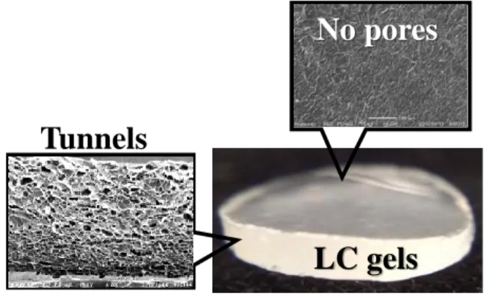
Chapter II, the surface selective microporous hydrogels with porous structure on side surface and flat on the top (Figure 2) were prepared by a combination of solvent casting and freeze drying techniques. Sacran water solution was casted at 60 °C to produce in-plane orientation thin films.
The thin films were physical cross-linked at temperature 60, 80, 100, 120 and 140 °C without cross-linking agent. Then swollen hydrogels with in-plane orientation were created by water immersion of that cross-linked films. Finally, the swollen hydrogels were subjected to freeze dry
Tunnels
LC gels No pores
Figure 2. Surface selective porous hydrogels with tunnels on side surface while did not showed porous morphology on the top surface.
process. The final products revealed an in-plane porous structure like a tunnel with pore size and porosity of 10-35 μm and 42-80 %, respectively. This is due to the sublimation of water on side surface parallel to the in-plane orientation of sacran polymer chains. In addition, they showed proper mechanical properties in a broad application. At high cross-linking temperature, the anisotropic porous materials showed low porosity, fine-size pores, and minimal water adsorption.
Conversely, the mechanical properties value such as moduli, cross-linking degree and toughness were very high. For low temperature cross-linking, the opposite set of values were observed. The water adsorption was between 9 to 186 times to that of dry material, and the elastic modulus was 3 to 585 kPa. The results reveals that the properties of the materials depends on temperature cross- linking. The surface selective microporous hydrogels were successfully prepared and precisely controlled for their properties.
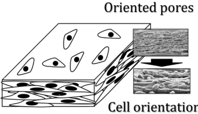
There are various applications of porous materials, and the tissue engineering scaffold is considered to be one of the most significant. In chapter III, the biocompatibility and cell orientation capacity were studied using mouse fibroblast cell L929 as a model in the cell culture experiment. The surface selective microporous hydrogels showed favorable cell compatibility property. The morphology of cells attachment was analyzed. The cells orientation on side surfaces is parallel to the in-plane orientation of polymer chains. The scaffolds can be altered to mimic the
Cell orientation Oriented pores
Figure 3. Fibroblast L929 cells attached on surface selective porous scaffold. Randomly orientation is presented on the top surface whereas perfectly orientation is revealed side surface. Additionally, the cell density on the top surface was lower than that of side surface.
native tissue that represents uni-direction of the muscle orientation (Figure 3). Moreover, the water contact angle and protein adsorption were studied on the materials which were annealed at 100, 120 and 140 °C. The water contact angle was revealed to be 95 to 37°, and the protein adsorption were 36 to 96 μg per 1 mg. In the results, water contact angle, protein adsorption and cell orientation are related to cross-linking temperature, similar to the above-mentioned properties.
However, the cell attached on top of the surface were randomly oriented. Another method was employed to control the cell orientation on the top surface of the scaffolds.
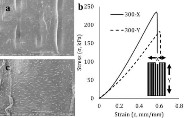
In chapter IV the scaffolds were casted on polystyrene, with micro-patterned on its surface. The pattern was set in a bar-shape mold with a diameter of 400 μm. The bars were arranged in parallel. The space between bars was fixed at 200, 250 and 300 μm. Sacran scaffolds with surface orientation were prepared with the same procedure to that of surface selective porous scaffolds except for the mentioned patterned substrate surface. The pattern of the scaffold revealed orientation perpendicular to that of bar molds. During the drying process, LC domains were slightly arranged to form an in-plane orientation like a layer. Looking at the side of bar molds, the top point of each bar has the sacran layer accumulated. The point is called nucleation point of orientation. Then the ends of polymer chains are aligned between bars. Polarization optical
0 50 100 150 200 250
0 0.2 0.4 0.6 0.8
Stress (σ, kPa)
Strain (ɛ, mm/mm) 300-X 300-Y
X
Y
a
c
b
Figure 4. Sacran film with micro-patterned on the top surface (a) showed anisotropic mechanical property (b) and one direction of fibroblast L929 cells orientation (c).
microscope technique was used to confirm the orientation of LC domains, and the results showed a clear and complete visible orientation. After that, the mouse fibroblast cell L929 was used in cell culture experiment. The distribution of cell orientation degree mimics the polymer orientation on the top surface. Finally, the orientation of cell was efficiently controlled on sacran LC polymer (Figure 4).
Conclusions
The microporous scaffolds with cell-orientation capacity was successfully prepared using sacran LC polymer. They revealed favorable results of pore properties, water adsorption capacity and mechanical properties. Furthermore biocompatibility and cell alignment were also confirmed. The angle of cell attached on materials was highly oriented, mimicking the native tissue behavior.
According to the development of technology for human’s bioengineering, the field of tissue engineering scaffolds is growing and progressing continuously. Today, the scaffolds are mainly the work of laboratory and research. However, it has the potential to be utilized, especially to save many lives on this planet, in the future.
Keywords: sacran, scaffold, liquid crystalline gels, cell-orientation, cell-engineering