Pediatric Cardiology and Cardiac Surgery 32(5): 387
‒396 (2016)
Review
【ホットトピックス】
胎児心臓診断における新しい指標
河津 由紀子1, 2)
1)市立豊中病院小児科
2)大阪府立母子保健総合医療センター小児循環器科
New Indicators in Fetal Echocardiology Yukiko Kawazu1, 2)
1) Department of Pediatrics, Toyonaka Municipal Hospital, Osaka, Japan
2) Department of Pediatric Cardiology, Osaka Medical Center for Maternal and Child Health, Osaka, Japan
In Japan, there has recently been a marked increase in fetal heart diagnosis concomitant with an overall increase in the diagnosis of prenatal diseases. With advances in imaging technology, congenital heart disease, which previously was difficult to diagnose in the fetus, can now be successfully diagnosed prenatally. Furthermore, it is now possible to evaluate cardiac function and predict prognosis after birth. In this article, I identify new indicators in fetal echocardiography and provide comprehensive yet concise commentaries as follows: (i) cardiac function assessment by tissue Doppler imaging (TDI), myocardial performance (Tei) index, and cardiovascular profile score (CVPS); (ii) anatomical diagnosis by I-shaped sign, post LA space index (PLAS index), and U-sign;
and (iii) prediction of the status and prognosis after birth by assessment of pulmonary venous flow pattern and pulsatility index (PI) during maternal hyperoxygenation testing in hypoplastic left heart syndrome, foramen ovale, and the ductus arteriosus in transposition of great arteries.
Keywords: prenatal diagnosis, congenital heart disease, fetal echocardiography, new indicator
近年,出生前診断が広がりを見せるなかで,胎児心臓診断が本邦においても急速に増加している現状 である.画像技術の進歩も相まって,従来は胎児診断が困難とされていた先天性心疾患が診断される ようになり,また胎児期から先天性心疾患の心機能を評価することや出生後の状態・予後を予測する ことも可能となってきた.それら胎児心臓診断における新しい指標として,①心機能評価に対する もの:組織ドップラー法(
tissue Doppler imaging: TDI
),Tei index
(myocardial performance index:
MPI
),cardiovascular profile score
(CVPS
)②心形態診断:ʻI-shaped
ʼsign, Post LA space index
(PLAS
index
),U-sign
③生後状態の評価・予後判定:左心低形成症候群における肺静脈血流パターンと母体酸素負荷テストでの
pulsatility index
(PI
),完全大血管転位における卵円孔と動脈管,をピックアップ してそれぞれについての解説を行う.はじめに
胎児心臓病学は
1980
年頃にイギリスのLindsey
Allan
らによって開始された分野である.以降,様々な出生前診断法の開発と需要も伴って世界的に胎児心 臓超音波検査(以下,胎児心エコー)による胎児心臓
診断が広まった.本邦においても
1994
年に日本胎児 心臓病研究会(現・学会)が創設され,2000
年前後 より胎児心エコーは全国的に急速に普及した.そして2006
年には先進医療として認定され,2010
年には正 式な専門医療行為として厚生労働省に認定,保険償還 され,更に広がりを見せている現状である.胎児心臓 著者連絡先:〒560‒0055大阪府豊中市柴原町4‒14‒1 市立豊中病院小児科 河津由紀子doi: 10.9794/jspccs.32.387
病学が開始された当初はエコー画像も不明瞭であり形 態診断自体も困難な状態であったが,その後,画像も 飛躍的に改善することにより,現在では形態診断ばか りでなく心機能評価や出生後の状態および予後の予測 も可能となってきた.一方で,胎児心臓病学は対象が 胎児であるため,出生後の循環とは異なることや在胎 週数の進行によって状態が変化するという特殊性があ ることから,その臨床研究におけるエビデンスレベル の評価が難しい分野でもある.
今回,胎児心臓診断における新しい指標について,
まず,心機能評価に対するもの,心形態診断に対する もの,生後の状態・予後判定に有用なもの,の
3
つに 分類した.その上で,近年になって論文発表され,か つ引用文献とされている指標を,比較的エビデンスレ ベルの高いものとして今回ピックアップして解説する.胎児心機能評価
胎児心機能を評価する方法として,
2014
年に発行 さ れ たAmerican Heart Association
(AHA
) のSci- entific Statement
1)に記載されているものであり,か つすでに循環器内科領域では汎用されているが,胎児 心臓診断としては比較的新しいものを提示する.1
)組織ドップラー法(tissue Doppler imaging:
TDI
)TDI
とは,心室壁の動きが血流に比べて遅いこと から,血流由来のドップラー信号を除去して壁運動由 来のドップラー信号だけを取り出すことによって,心 筋局所の運動を定量的に評価する方法である.成人領 域では,心不全における拡張能の評価と局所壁運動の 評価に汎用されている指標である.パルス組織ドップ ラー法とカラー組織ドップラー法の二種類がある.パルス組織ドップラー法(
pulse TDI
)は,サンプ ルボリュームを一カ所設定し,房室弁(成人では主に 僧帽弁)の弁輪速度の計測をして心機能評価(拡張 能)に用いられることの多い,汎用性の高い方法であ る2).一方,カラー組織ドップラー法(
color TDI
)は,断層心エコー(もしくは
M
モード心エコー)にカラー 組織ドップラーを重ねて表示して,速度成分を色の明 るさ(プローベに向かう心筋の動きを赤で,遠ざかる 動きを青で)で表示する方法である.心筋上に関心領 域(region of interest: ROI
)を設定すると,その局所 壁運動情報について縦軸を速度,横軸を時間としてプ ロファイル表示できる2).成人では心室中隔の基部と側壁の基部に
ROI
を設定して駆出期ピークのずれを 計測して同期不全の評価に用いられる.胎児心臓診断において,
TDI
は心筋の運動や時間 間隔の解析に有用とする報告が多く見られる.2005
年に
Harada
らは,従来困難であった胎児の右室機能を評価するための方法として,
TDI
を正常60
胎児に おいて在胎週数ごとに計測し評価した3).2006
年に はNii
らが,胎児完全房室ブロックへの進行を予測す る方法として胎児房室(AV
)間隔をTDI
で在胎週数 ごとに計測して報告した4).胎児TDI
の有用性につ いて,2008
年の国際産婦人科超音波医学会(ISUOG
)consensus statement
では,ʻoptional recommendation
ʼ としている5)が,最近もTDI
を評価した論文が散 見 さ れ る.2015
年 にAxt-Fliedner
ら は,pulse TDI
を利用して左心低形成症候群(HLHS
)14
胎児の右 室機能を正常28
胎児と比較して評価している6).2016
年にはWillruth
らが,正常160
胎児において,color TDI
でROI
を左右心室壁の弁輪部に設定してICT
(isovolumic contraction time
),IRT
(isovolumic relaxation time
),ET
(ejection time
)を計測した値 を正常値として報告している7).このように多くの報告があることからも,胎児心機 能を
TDI
で評価することは今後臨床面での使用が期 待される.2
)Tei index
(myocardial performance index:
MPI
)Tei index
は,パルスドップラーで得られた波形から,心室流入の終了から再開始までの時間(
a
)と心室 駆出血流持続時間(駆出時間)(b
)より,(a − b
)/b
と して算出される.(ICT
+IRT
)/ET
と等しい(Fig. 1
).収縮能低下でも拡張能低下でもいずれも
Tei index
の 増大として反映され,総合的な心機能を表す指標とし て利用されている.また,時間分解能に優れたパルス ドップラーを利用するため,心拍数の早い小児にお いても有用とされている8).産科領域において,胎児Tei index
は近年,双胎間輸血症候群(TTTS
),子宮 内発育遅延(FGR
)の評価のために利用されること が多い9, 10).胎児心臓診断においても
Tei index
は,これまで 多くの論文で胎児の心機能評価として引用されている.
Ebstein
奇形,動脈管閉鎖など,心機能が変化するような心臓病では異常値を示す11, 12).
Inamura
ら は2005
年にEbstein
奇形を含めた三尖弁異形成疾患 群において,LV Tei index
と生命予後とが関係すると 報告している11).2013
年にGhawi
らは,それまでの13
年間で報告された胎児Tei index
の正常値をまと めた上で,胎児自験例の計測から正常値は,左室Tei index 0.464 + / − 0.08
,右室Tei index 0.466 + / − 0.09
と報告しており,成人における左室0.38 + / − 0.05
, 右室0.28 + / − 0.04
に比し高値となっている13).3
)cardiovascular profile score
(CVPS
)CVPS
は,産科領域において胎児心不全の予後評価 に使用されてきた指標である.具体的には,胎児水 腫,静脈ドップラー派形,心拡大,心機能,動脈ドッ プラー派形という5
つのカテゴリーを各々2
点とし て計10
点満点として点数を評価し,低値であるほど 心不全を疑う指標である14)(Table 1
).胎児水腫,先 天性心疾患(CHD
),発育不全を伴う胎児の予後に関 係することが報告されている15‒18).心機能低下やそ のリスクのある胎児において,計測して経過を見るの に有用であろう.2008
年にMakikallio
らは子宮内発育遅延の胎児75
例においてその出産後の生命予後や脳性麻痺合併のFig.
1
Tei index
Tei index is calculated as (a−b)/b, where a is the interval between cessation and onset of the mitral or tricuspid inflow and b is the left or right ventricu- lar ejection time.
Table 1
Cardiovascular profile score (CVPS)
Cardiovascular profile score is 10 if there are no abnormal signs and reflects 2 points for each of 5 categories: hydrops, venous Doppler, heart size, cardiac function, and arterial Doppler. AEDV indicates absent end-siastolic velocity; dP/dt, charge in pres- sure over time of tricuspid regurgitant jet; DV, ductus venosus; FS, ventricular fractional shortening; LV, left ventricle; MR, mitral valve regurgitation; MV, mitral valve; REDV, reversed end-diastolic velocity; RV, right ventricle; TR, trisucpid valve regur- gitation; TV, tricuspid valve; UA, umbilical artery; and UV, umbilical vein. Reproduced with permission from the reference 16).
検討から,
CVPS
がそれらの症例では低値であったと 報告している17).Wieczorek
らは,CHD
を持つ胎児131
例において後方視的検討を行い,CVPS8
未満が 周産期死亡のリスクになることを報告している15).2013
年にはStatile
らが,高心拍出病変(奇形腫やガ レン大静脈瘤など)を合併した胎児35
例について検 討し,死亡した10
例のうち80
%がCVPS8
未満であっ たと報告している18).心形態診断
胎児心臓診断において,臨床的に最も必要とされ期 待されているのは,心臓の解剖学的な形態異常をより 正確に診断することである.この
20
年間で画像の向 上と技術や経験の蓄積によって,特に四腔断面像で描 出されるような心内の形態異常については容易に産科 スクリーニングで指摘されるようになってきた.一方で,心内の形態異常を認めない
CHD
である完全大血 管転位(TGA
)および総肺静脈還流異常(TAPVC
) は胎児期に診断できない時代が続いていたため,そ の胎児診断法の開発が求められていた.そして2013
年にIshii
らがTGA
の診断法としての ʻI-shaped
ʼsign
を報告し19),2014
年にKawazu
らがTAPVC
の診断 法 と し て の ʻPost-LA space index
(PLAS index
)ʼを 報告した20).また心内に異常を認めない血管の異常 として,血管輪を含めた右側大動脈弓の検出に有用 な指標としてのU-sign
も活用されているので提示し た21‒25).いずれもそれらの有用性を検証した論文も 報告されていることから胎児心臓診断における新しい 指標として提示する.1
)ʻI-shaped
ʼsign
TGA
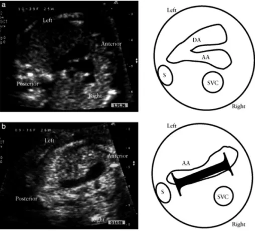
は生後早期に手術を要し,かつ一部には出 生直後に処置を要し救命できないことも起こる重症Fig.
2
I-shaped sign
Ultrasound images showing three vessels and trachea view in a fetus with a structurally normal heart (a) and in a fetus with d-transposition of the great arteries (dTGA) in the second trimester of pregnancy (b), with corresponding schematic diagrams. (a) Normal view shows aortic (AA) and ductal (DA) arches in tangential cross-section, forming a V-shape pointing to the posterior thorax on the left side of the spine (S). (b) I-shaped sign in dTGA is seen as a wide-sweeping aortic arch shaped like the letter “I” (black bar in schematic diagram) at the level of three vessels and trachea view.
Reproduced with permission from the reference 19).
CHD
である26).そのため胎児診断が生命予後の改善 に有用とされている疾患であるが,血管の異常であっ て四腔断面の異常がないため,胎児期には診断が困難 な疾患とされていた.I-shaped sign
は,TGA
においてthree vessels and trachea view
を描出した際に,大動脈が前方にある右 室から起始して長く後方に向かい動脈管弓と合流する までが「I
」の字のように描出されることを見いだし,TGA
の胎児診断に有用であると2013
年にIshii
らが 報告した指標である19)(Fig. 2
).具体的には31
例 のTGA
症例で後方視的に確認したところ,I-shaped sign
を30
例に認めたとしている.その後2015
年に はPalatnik
らが,I-shaped sign
の有用性を検討し,TGA
の胎児24
例のうち23
例(95.8
%)でI-shaped sign
陽性であったことからもこの指標がTGA
の胎児 診断において簡便かつ迅速に診断するのに有用と述べ ている27).今後もその有用性が期待される指標であ る.2
)Post LA space index
(PLAS index
)心内の形態異常を合併しない単独の総肺静脈還流 異常(
isolated TAPVC
)は,四腔断面の異常がない ため,TGA
と同様,従来は胎児診断が困難な疾患と されていた.2003
年にValsangiacomo
らはTAPVC
において左房と下行大動脈に ʻwide gap
ʼ があると述 べ28),2006
年にInamura
らはTAPVC
の四腔断面で はLA
後方に腔を二つ(共通肺静脈腔と下行大動脈)認める ʻ
double balloon sign
ʼ を報告し29),2007
年にBerg
らは ʻthe area behind heart
ʼ がCHD
診断に重要 であると報告した30).そして
2014
年にKawazu
らは,左房‒下行大動脈間距離を下行大動脈径で除した値を
PLAS index
とし,isolated TAPVC
の胎児診断に有用と報告した20).具 体的には,胎児isolated TAPVC8
例と正常胎児101
例において,PLAS index
(=左房‒下行大動脈間距離(
LD
)/
下行大動脈径(DA
))(Fig. 3
)を比較したとこ ろ,TAPVC
症例は有意に高値であり(p
<0.0001
),PLAS index
が1.27
以上であるとTAPVC
が疑わしい と報告した(感度100
%,特異度99
%).2016
年に はAkkurt
らが,在胎20
〜24
週の正常胎児165
例に おいてPLAS index
の計測を行い,その値が0.65 + /
− 0.13
と低値であったことと在胎週数による変動が なかったことを報告しており31),Isolated TAPVC
の 胎児診断に期待される指標と考えられる.ただ一方でPLAS index
低値であった偽陰性例の報告もある32)こ とから,単独で使用するのではなく他の指標も合わせ て診断することが必要かもしれない.3
)U-sign
U-sign
は胎児大動脈弓の異常を検出する指標として
2002
年にAchiron
らが提唱したものである21).3vessels trachea view
において,正常であれば大動脈 と肺動脈がcolor doppler
にて「V
」の字を形成して いるが,18,347
例中19
例には「U
」の字型を認めて,うち
18
例が右側大動脈弓(1
例は重複大動脈弓)で あったと報告している.2003
年にChaoui
らは胎児 心エコーでcolor doppler
を利用する際に右側大動脈 弓でU-sign
を認めると述べている22)(Fig. 4
).その 後も右側大動脈弓の検出に有用な指標として多く報告 されており,合併するCHD
や血管輪の診断に有用と されている23‒25).Fig.
3
Post LA space index (PLAS index)
Measurement of the left atrium‒descending aorta distance (LD) in four-chamber view on fetal echocardiography.
(a) Schematic diagram of LD and descending aorta diameter (DA). (b) LD and DA in a normal fetal heart. (c) LD and DA in a fetus with infracardiac total anomalous pulmonary venous connection. LA, left atrium; LV, left ventricle; RA, right atrium; RV, right ventricle. Reproduced with permission from the reference 20).
Fig.
4
U-sign: Three-vessel view with two antegrade flow stripes
Tiny pulmonary trunk with antegrade perfusion in mild pulmonary stenosis (left) in a fetus with Ebsteinʼs anomaly.
Right-sided aortic arch (Ao) with trachea (Trach) entrapped between the aortic arch and pulmonary trunk (Tp) (center).
Retrograde perfusion across the isthmus due to increased cerebral perfusion in a fetus with vein of Galen aneurysm (right). Reproduced with permission from the reference 22).
Fig.
5
Intrauterine pulmonary venous flow and restrictive foramen ovale in fetal hypoplastic left heart syndrome
Left: (a) Normal fetal pulmonary vein Doppler spectrum is composed of forward flow in ventricular systole (S) and diastole (D), with cessation of flow during atrial systole. (b) Reverse flow during atrial contraction in PV spectrum is commonly observed in fetal left heart obstruction. Measurement of velocity‒time integral for forward (VTIF) and reverse (VTIR) flow is demonstrated. Right: Three pulmonary vein flow patterns identified in fetuses with left heart obstruc- tion. (A) Continuous forward flow with a small a-wave reversal (VTIR/VTIF < 0.18). (B) Continuous forward flow with an increased a-wave reversal (VTIR/VTIF ≥0.18). (C) To-and-fro flow pattern with absent early diastolic forward flow.
Reproduced with permission from the reference 33).
生後状態の評価・予後判定
胎児心臓診断において,検査施行時の心機能を評 価して形態異常を診断した上で最終的に必要となる のが,児の出生後の状態および予後の推定である.
CHD
の中でも最重症とされるHLHS
の予後判定に ついて,2004
年にTaketazu
らは肺静脈の血流パター ンが有用であることを報告し33),2010
年にはSzwast
らが母体酸素負荷テストでのpulsatility index
(PI
) による肺血管の反応性の評価が有用であると報告し た34).また同じく重症CHD
であるTGA
について,1999
年にMaeno
らが胎児心エコーでの卵円孔(FO
) と動脈管(DA
)の形態がその出生直後の状態評価に 有用であることを報告した26).1
)HLHS
における肺静脈血流パターンHLHS
は出生後,血行動態的にFO
開存が不可欠 な疾患であるため,胎児心エコーにおいても従来はFO
の形態を直接評価していた.一方で,Taketazu
らは2004
年に,FO
自体でなく肺静脈血流パターン の評価の方が生後の状態をよく反映することを報告 した33).具体的には胎児HLHS 40
例において,肺 静脈血流速度信号の面積(=Velocity Time Integral
(
VTI
))を計測し,その逆行性血流と順行性血流の比(
VTIR/VTIF
)が0.18
未満(A
),0.18
以上(B
),拡張 早期順行性血流のないto-and-fro
パターン(C
)の3
パ ターンに分類した(Fig. 5
).その結果,生後B
ではA
より卵円孔が有意に小さく,C
は全例卵円孔閉鎖し ており生直後に緊急処置を要し予後不良であったと述 べている.その後,2005
年にMichelfelder
らが胎児HLHS 41
例をまとめた報告では,肺静脈血流パターンはもっと多様であるとしているが,やはり肺静脈 血流により生後緊急
BAS
(Balloon atrio-septostomy
) の 必 要 性 が 予 測 で き た と し て い る35).2008
年 にChintala
らも胎児HLHS
(FO
狭小化のない)27
例 と正常胎児66
例の肺静脈血流パターンを比較して,FO
狭小化がなくてもHLHS
においては肺静脈血流パ ターンが異なっていることを報告している36).いず れの報告もHLHS
においては肺静脈血流パターンの 確認が重要であると結論している.2
)HLHS
における母体酸素負荷テストでのpulsatil- ity index
(PI
)PI
は,パルスドップラー法による血流波形におい て,収縮期と拡張期の血流速度に平均血流速度を加味 して算出し血管抵抗を評価する方法である(=収縮期Fig.
6
Pulmonary venous Doppler flow patterns and maternal hyperoxygenation testing
Top panel illustrates the pulmonary venous Doppler flow pattern in a fetus with hypoplastic left heart syndrome and restrictive interatrial communication. Bottom two panels demonstrate maternal hyperoxygenation (MH) testing in the same fetus. The middle panel demon- strates distal branch pulmonary artery Doppler flow pattern during the room air phase of the study, whereas the bottom panel demonstrates the same Doppler flow pattern during MH. Despite the presence of a restrictive interatrial communication by previously reported criteria, there is significant vasoreactivity in response to MH. At birth, this fetus had adequate oxygen saturation with breathing room air. Reproduced with permission from the reference 34).
最高血流速度
−
拡張末期血流速度/
平均血流速度).つ まり高値で血管抵抗が高いことを示唆する.母体への酸素投与による
PI
の変化については,2002
年 にBroth
ら が 肺 低 形 成 の 胎 児29
例 に お い て,母体酸素投与に反応した(PI
が20
%以上低下し た)群15
例では生後1
例のみ死亡に対し,反応しな かった群では14
例中11
例が死亡したことより,母 体酸素投与によるPI
の変化が肺低形成の予測因子と なりうることを報告した37).心疾患児に対しては,2010
年にSzwart
らは43
例のHLHS
胎児の母体に対 し60
%酸素10
分間吸入の前後で肺動脈のPI
の低下10
%をカットオフ値として,生後緊急BAS
を要したrestrictive
群と心房中隔が開存していた群に分類でき た(感度100
%,特異度94
%)と報告した34)(Fig. 6
).2016
年にはEnzensberger
らが22
例のHLHS
胎児の 母体に対し100
%酸素10
分間吸入前後での肺静脈PI
を計測することによって,酸素負荷により胎児FO
閉 鎖もしくは狭小化の6
例中2
例でPI
低下,つまり血 管抵抗の低下により肺還流が増加し,酸素負荷が胎児HLHS
の肺血管障害の評価の一助になりうることを 示唆した38).3
)TGA
における卵円孔と動脈管TGA
,中でも生後緊急BAS
を要するようなTGA
においては,その出生後の状態を評価して予測して おくことが重要である.1999
年にMaeno
らは,胎 児TGA16
例を検討し,胎児期のFO
狭小とDA
狭 小が生後早期の予後に関連することを報告した26). 具体的には16
例中6
例に胎児期FO
の異常(fixed, flat, redundant
)があって生後5
例でFO
狭小を認めた.また
5
例に胎児期DA
狭小があり,生後は6
例 に狭小を認めた.DA
が最も細かった4
例中2
例にFO
異常もあり,その2
例は出生直後に死亡,その他 の1
例は肺高血圧が継続した.2004
年にJouannic
ら は,胎児TGA119
例においてFO
またはDA
狭小の 評価を行い,24
例でいずれかの異常を認めた(FO23, DA5
,両方)4)が,13
例で生後低酸素血症と代謝性 アシドーシスをあり緊急BAS
施行,FO
狭小とDA
狭小両方の2
例は出生直後に死亡したと報告してい る39).しかし一方で胎児エコーによる予測は特異度84
%,感度54
%,FO
狭小とDA
狭小を合わせると特 異度100
%,感度31
%と特異度は高いが感度は低い ため他の指標も必要と結論している.2011
年にPunn
らは,TGA
の新生児期緊急BAS
を胎児期に予測する ものとして,新たに「hypermobile septum
」(Fig. 7
) と「reverse diastolic DA shunt
」の二つが有用である と報告した40).具体的には,胎児TGA26
例におい て卵円孔のhypermobility
(一次孔を両心房間で揺れ る),restriction, flat, redundancy
を検証し,またDA
径とシャントパターンも検証した.26
例中14
例で緊 急BAS
を施行し,うち9
例がhypermobile
であり,8
例で拡張期DA
逆行を認めていた.Hypermobile
と 拡張期DA
逆行は緊急BAS
と有意に相関があったと している.いずれの報告も,胎児TGA
においてはFO
とDA
の観察がその出生後の予後推定に重要であ ると結論付けている.ま と め
胎児心臓診断における新しい指標として,
1.
心機能Fig.
7
Hypermobile atrial septum
Biatrial view demonstrating septum primum flap (*) oscillating between the left and right atria during various phases of the cardiac cycle. DAo, descending aorta; LA, left atrium; RA, right atrium. Reproduced with permission from the reference 40).
評価 ①
TDI
②Tei index
③CVPS
,2.
心形態診断①
I-shaped sign
②PLAS index
③U-sign
3.
生後 状態の評価・予後判定 ①HLHS
における肺静脈血 流パターン ②母体酸素負荷テストでのPI
,③TGA
における卵円孔と動脈管,について概説した.多くが すでに胎児心臓診断において活用されている指標であ るが,今後も症例を重ねることによって更に有用性が 実証されることと期待される.利益相反
本論文について,開示すべき利益相反(COI)はない.
引用文献
1) Donofrio MT, Moon-Grady AJ, Hornberger LK, et al:
Diagnosis and treatment of fetal cardiac disease: A sci- entific statement from the American Heart Association.
Circulation 2014; 129: 2183
‒2242
2) Comas M, Crispi F: Assessment of fetal cardiac function using tissue Doppler techniques. Fetal Diagn Ther 2012;
32: 30
‒38
3) Harada K, Ogawa M, Tanaka T: Right ventricular pre-ejection myocardial velocity and myocardial acceler- ation in normal fetuses assessed by Doppler tissue imag- ing. J Am Soc Echocardiogr 2005; 18: 370
‒374
4) Nii M, Hamilton RM, Fenwick L, et al: Assessment of fetal atrioventricular time intervals by tissue Doppler and pulse Doppler echocardiography: Normal values and correlation with fetal electrocardiography. Heart 2006; 92:
1831
‒1837
5) Lee W, Allan L, Carvalho JS, et al: ISUOG consensus statement: What constitutes a fetal echocardiogram?
Ultrasound Obstet Gynecol 2008; 32: 239
‒242
6) Axt-Fliedner R, Graupner O, Kawecki A, et al: Evaluation of right ventricular function in fetuses with hypoplastic left heart syndrome using tissue Doppler techniques.
Ultrasound Obstet Gynecol 2015; 45: 670
‒677
7) Willruth A, Steinhard J, Enzensberger C, et al: Fetal colour tissue Doppler imaging (cTDI): Biventricular ref- erence ranges for the time segments of the cardiac cycle in second and third trimesters of gestation. Arch Gynecol Obstet 2016; Epub ahead of print
8) Ishii M, Tsutsumi T, Himeno W, et al: Sequential evalu- ation of left ventricular myocardial performance in chil- dren after anthracycline therapy. Am J Cardiol 2000; 86:
1279
‒1281, A9
9) Raboisson MJ, Fouron JC, Lamoureux J, et al: Early inter- twin differences in myocardial performance during the twin-to-twin transfusion syndrome. Circulation 2004;
110: 3043
‒3048
10) Hernandez-Andrade E, Crispi F, Benavides-Serralde JA, et al: Contribution of the myocardial performance index and aortic isthmus blood flow index to predicting mor- tality in preterm growth-restricted fetuses. Ultrasound Obstet Gynecol 2009; 34: 430
‒436
11) Inamura N, Taketazu M, Smallhorn JF, et al: Left ven- tricular myocardial performance in the fetus with severe
tricuspid valve disease and tricuspid insufficiency. Am J Perinatol 2005; 22: 91
‒97
12) Mori Y, Rice MJ, McDonald RW, et al: Evaluation of sys- tolic and diastolic ventricular performance of the right ventricle in fetuses with ductal constriction using the Doppler Tei index. Am J Cardiol 2001; 88: 1173
‒1178 13) Ghawi H, Gendi S, Mallula K, et al: Fetal left and right
ventricle myocardial performance index: Defining normal values for the second and third trimesters
‒single tertiary center experience. Pediatr Cardiol 2013; 34: 1808
‒1815 14) Huhta JC, Paul JJ: Doppler in fetal heart failure. Clin
Obstet Gynecol 2010; 53: 915
‒929
15) Wieczorek A, Hernandez-Robles J, Ewing L, et al: Predic- tion of outcome of fetal congenital heart disease using a cardiovascular profile score. Ultrasound Obstet Gynecol 2008; 31: 284
‒288
16) Hofstaetter C, Hansmann M, Eik-Nes SH, et al: A cardio- vascular profile score in the surveillance of fetal hydrops.
J Matern Fetal Neonatal Med 2006; 19: 407
‒413
17) Makikallio K, Rasanen J, Makikallio T, et al: Human fetal cardiovascular profile score and neonatal outcome in intrauterine growth restriction. Ultrasound Obstet Gyne- col 2008; 31: 48
‒54
18) Statile CJ, Cnota JF, Gomien S, et al: Estimated cardiac output and cardiovascular profile score in fetuses with high cardiac output lesions. Ultrasound Obstet Gynecol 2013; 41: 54
‒58
19) Ishii Y, Inamura N, Kawazu Y, et al:
ʻI-shaped
ʼsign in the upper mediastinum: A novel potential marker for ante- natal diagnosis of d-transposition of the great arteries.
Ultrasound Obstet Gynecol 2013; 41: 667
‒671
20) Kawazu Y, Inamura N, Shiono N, et al:
ʻPost-LA space index
ʼas a potential novel marker for the prenatal diag- nosis of isolated total anomalous pulmonary venous con- nection. Ultrasound Obstet Gynecol 2014; 44: 682
‒687 21) Achiron R, Rotstein Z, Heggesh J, et al: Anomalies of
the fetal aortic arch: A novel sonographic approach to in-utero diagnosis. Ultrasound Obstet Gynecol 2002; 20:
553
‒557
22) Chaoui R, McEwing R: Three cross-sectional planes for fetal color Doppler echocardiography. Ultrasound Obstet Gynecol 2003; 21: 81
‒93
23) Berg C, Bender F, Soukup M, et al: Right aortic arch detected in fetal life. Ultrasound Obstet Gynecol 2006; 28:
882
‒889
24) D
ʼAntonio F, Khalil A, Zidere V, et al: Fetuses with right aortic arch: A multicenter cohort study and meta-analy- sis. Ultrasound Obstet Gynecol 2016; 47: 423
‒432 25) Hsu KC, Tsung-Che Hsieh C, Chen M, et al: Right aortic
arch with aberrant left subclavian artery
‒prenatal diagno- sis and evaluation of postnatal outcomes: Report of three cases. Taiwan J Obstet Gynecol 2011; 50: 353
‒358 26) Maeno YV, Kamenir SA, Sinclair B, et al: Prenatal features
of ductus arteriosus constriction and restrictive foramen ovale in d-transposition of the great arteries. Circulation 1999; 99: 1209
‒1214
27) Palatnik A, Gotteiner NL, Grobman WA, et al: Is the
“