PEDRAMHAMRAH, MD,EDITOR
Stevens-Johnson Syndrome/Toxic Epidermal Necrolysis e A Comprehensive Review and Guide
to Therapy. I. Systemic Disease
S
AHARK
OHANIM, MD,
1*S
OTIRIAP
ALIOURA, MD, P
HD,
2*H
AJIRAHN. S
AEED, MD,
3*E
SENK. A
KPEK, MD,
4G
UILLERMOA
MESCUA, MD,
2S
AYANB
ASU, MBBS, MS,
5P
RESTONH. B
LOMQUIST, MD,
6C
HARLESS. B
OUCHARD, MD,
7J
OHNK. D
ART, DM, FRCO
PHTH,
8X
IAOWUG
AI, P
HD,
3J
OSÉA.P. G
OMES, MD,
9D
ARRENG. G
REGORY, MD,
10G
EETHAI
YER, MD, FRCS G
LASGOW,
11D
EBORAHS. J
ACOBS, MD,
3,12A
NTHONYJ. J
OHNSON, MD,
13S
HIGERUK
INOSHITA, MD, P
HD,
14I
ASONS. M
ANTAGOS, MD,
15J
ODHBIRS. M
EHTA, MBBS,
16V
ICTORL. P
EREZ, MD,
2S
TEPHENC. P
FLUGFELDER, MD,
17V
IRENDERS. S
ANGWAN, MBBS, MS,
5K
IMBERLYC. S
IPPEL, MD,
18C
HIES
OTOZONO, MD, P
HD,
14B
HASKARS
RINIVASAN, MD, MS,
11D
ONALDT.H. T
AN, FRCS, FRCO
PHTH, FAMS,
16R
ADHIKAT
ANDON, MD, FRCO
PHTH, FRCSE
D,
19S
CHEFFERC.G. T
SENG, MD, P
HD,
20M
AYUMIU
ETA, MD, P
HD,
14 ANDJ
AMESC
HODOSH, MD, MPH
3ABSTRACT The intent of this review is to comprehen- sively appraise the state of the art with regard to Stevens Johnson syndrome (SJS) and toxic epidermal necrolysis (TEN), with particular attention to the ocular surface com- plications and their management. SJS and TEN represent two ends of a spectrum of immune-mediated, dermato- bullous disease, characterized in the acute phase by a febrile illness followed by skin and mucous membrane necrosis and detachment. The widespread keratinocyte death seen in SJS/TEN is rapid and irreversible, and even
with early and aggressive intervention, morbidity is severe and mortality not uncommon. We have divided this review into two parts. Part I summarizes the epidemiology and immunopathogenesis of SJS/TEN and discusses systemic therapy and its possible benefits. We hope this review will help the ophthalmologist better understand the mecha- nisms of disease in SJS/TEN and enhance their care of patients with this complex and often debilitating disease.
Part II (April 2016 issue) will focus on ophthalmic manifestations.
Accepted for publication October 2015.
From1Vanderbilt Eye Institute, Vanderbilt University School of Medicine, Nashville, TN;2Bascom Palmer Eye Institute, University of Miami Miller School of Medicine, Miami, FL;3Massachusetts Eye and Ear Infirmary, Har- vard Medical School, Boston, MA;4The Wilmer Eye Institute, Johns Hop- kins University School of Medicine, Baltimore, MD; 5LV Prasad Eye Institute, Hyderabad, India; 6University of Texas Southwestern Medical Center, Dallas, TX;7Loyola University, Chicago, IL;8Moorfields Eye Hospi- tal, NHS Foundation Trust, London, UK;9Federal University of São Paulo, Brazil; 10Rocky Mountain Lions Eye Institute, University of Colorado School of Medicine, Aurora, CO;11Dr G Sitalakshmi Memorial Clinic for Ocular Surface Disorders, Sankara Nethralaya, India;12Boston Foundation for Sight, Boston, MA;13United States Army Institute of Surgical Research, San Antonio, TX;14Kyoto Prefectural University of Medicine, Kyoto, Japan;
15Boston Children’s Hospital, Harvard Medical School, Boston, MA;
16Singapore National Eye Centre, Singapore Eye Research Institute, Singapore; 17Cullen Eye Institute, Baylor College of Medicine, Houston, TX;18Weill Cornell Medical College, New York, NY;19Dr. Rajendra Prasad Centre for Ophthalmic Sciences, All India Institute of Medical Sciences, Delhi, India;20Ocular Surface Center, Ocular Surface Research & Education Foundation, Miami, FL
Funded in part by an unrestricted grant to the Department of Ophthal- mology, Harvard Medical School, Mass. Eye & Ear, from Research to Pre- vent Blindness, NY, NY.
The authors have no commercial or proprietary interest in any concept or product discussed in this article.
Single-copy reprint requests to James Chodosh, MD (address below).
Corresponding author: James Chodosh, MD, MPH, Massachusetts Eye and Ear Infirmary, Harvard Medical School, 243 Charles St., Boston, MA 02114, USA. Tel: 617-573-6398. Fax: 617-573-4324. E-mail address:
*These authors contributed equally.
©2016 Elsevier Inc. All rights reserved.The Ocular SurfaceISSN: 1542- 0124. Kohanim S, Palioura S, Saeed HN, Akpek EK, Amescua G, Basu S, Blomquist PH, Bouchard CS, Dart JK, Gai X, Gomes JAP, Gregory DG, Iyer G, Jacobs DS, Johnson AJ, Kinoshita S, Mantagos IS, Mehta JS, Perez VL, Pflugfelder SC, Sangwan VS, Sippel KC, Sotozono C, Srinivasan B, Tan DTH, Tandon R, Tseng SCG, Ueta M, Chodosh J. Stevens-johnson syn- drome/toxic epidermal necrolysisea comprehensive review and guide to therapy. i. systemic disease. 2016;14(1):2-19.
Clinical Practice
KEY WORDS apoptosis, drug-induced disease, immune- mediated disease, keratinocyte death Stevens-Johnson Syndrome, toxic epidermal necrolysis
I. INTRODUCTION
T
he spectrum of disease defined by Stevens-Johnson Syndrome (SJS), the more severe toxic epidermal necrolysis (TEN), and their intermediate (SJS/TEN overlap) characterize a severe immunologic dermato- bullous condition with high mortality and significant long- term morbidity. SJS/TEN is characterized by widespread keratinocyte death and epidermal necrosis resulting in split- ting of subepidermal layers with attendant tissue loss at skin and mucosal surfaces.1 The diagnosis of SJS/TEN is made upon recognition of defining clinical signs and skin biopsy demonstrating full-thickness necrosis of the epidermis and keratinocyte apoptosis, with minimal involvement of the un- derlying dermis.2-5
The purpose of Part I of this review is to summarize the most up-to-date information on SJS/TEN, with particular attention to pathogenesis and systemic therapy. SJS/TEN is a rare disease, and there is a paucity of centralized information on best care practices. This comprehensive review critically evaluates contemporary concepts of pathophysiology and
the therapies currently in use for patients with the disorder.
However, the authors wish to emphasize that the patho- physiology of SJS/TEN is still a matter of debate, and the best systemic therapy for SJS/TEN beyond general supportive burn care remains highly controversial among burn center physicians, often even within the same burn center. The ophthalmic manifestations of SJS/TEN and their manage- ment will be covered in Part II.
To provide a comprehensive, in-depth, and authoritative review of this complex entity, we assembled a group of authors who are leaders in their respectivefields with expe- rience and publications in very specific areas addressed by the review. All authors made substantial contributions in writing and revising the manuscript in their areas of exper- tise. Each author met Harvard Medical School criteria for authorship on a scholarly paper.
II. EPIDEMIOLOGY A. Incidence
The estimated annual incidence (cases/million popula- tion/year) of SJS/TEN ranges from 0.4 to 7 cases per million population,6-8making it a rare disease.9 There are sugges- tions that the incidence in certain areas of the world may be higher. In a retrospective study of 404 hospitalized patients in South India with acute cutaneous drug reactions over a 9-year period, 19.5% were diagnosed with SJS/TEN, some- what higher than reported in other countries.10 SJS/TEN carries a significant risk of mortality, ranging from 1-5%
in SJS and 25-40% in TEN.7,11-15 Unfortunately, despite continued efforts, mortality rates remain significant.4SJS pre- dominantly affects children and adolescents, whereas TEN occurs in all ages, from premature infants to the elderly.4 The incidence of cutaneous drug reactions including TEN is 2.7 times higher in the elderly than in younger patients, and mortality from TEN is twice as high in the elderly (51% vs 25%). However, SJS/TEN is more likely recurrent in children.
In one series, 18% of 55 children developed recurrent SJS up to 7 years after the index episode, with three children experi- encing more than one recurrence.16
B. Risk Factors
1. Non-Pharmaceutical Triggers
While SJS/TEN most often represents an idiosyncratic reaction to systemic medications, there are uncommon exceptions and the disorder can be idiopathic.17,18 SJS/
TEN has been associated with vaccination19-21 and expo- sure to industrial chemicals and fumes.19,22,23 TEN has also occurred in patients consuming natural remedies and traditional Chinese herbal medications.24-27 Infection with Mycoplasma pneumoniae is a controversial cause of SJS, because Mycoplasma has also been associated with erythema multiforme and, in addition, can cause a primary mucositis.28-35 Herpes virus infections have been asso- ciated with SJS,36,37 and reactivation of herpes simplex virus has been associated with SJS recurrences, particularly in children.16,38 Two cases of TEN have been reported in which the skin manifestations occurred specifically OUTLINE
I. Introduction II. Epidemiology
A. Incidence B. Risk Factors
1. Non-Pharmaceutical Triggers 2. Offending Medications 3. Medication Cross-Reactivity III. Clinical Presentation
IV. Classification
V. Mortality from Toxic Epidermal Necrolysis VI. Differential Diagnosis
VII. Pathogenesis of SJS/TEN
A. Genetic Susceptibility to SJS/TEN B. Immunology of Acute SJS/TEN C. Mechanisms of Cell Death in SJS/TEN VIII. Acute Systemic Therapy
A. Supportive Care B. Therapeutic Agents
1. Systemic Corticosteroids
2. Human Intravenous Immune Globulin 3. Plasmapheresis
4. Granulocyte Colony Stimulating Factor 5. Cyclosporine
6. TNF-alpha Inhibitors 7. Cyclophosphamide
C. Effect of Systemic Treatments of Acute SJS/TEN on Ocular Disease
IX. Summary and Conclusions
SJS/TEN: I. SYSTEMIC DISEASE / Kohanim, Palioura, Saeed, et al
THE OCULAR SURFACE / JANUARY 2016, VOL. 14 NO. 1 / www.theocularsurface.com 3
in sun-exposed areas39,40 or after radiation therapy.41,42 Photo-induced TEN has also been reported from cloba- zam.39 Patients with brain tumors treated with radiation appear to be more susceptible to SJS/TEN when given phenytoin.43-45 TEN and other drug reactions are also more common in HIV/AIDS.4
2. Offending Medications
More than 200 offending medications have been impli- cated as triggers of SJS/TEN, with new drugs implicated almost as soon as they are on the market.4Common causes include sulfonamide antibiotics (trimethoprim/sulfamethoxazole), aromatic anticonvulsants (e.g., phenytoin, phenobarbital, and carbamazapine), beta-lactam antibiotics, nevirapine, abacavir, non-steroidal anti-inflammatory medications, allopurinol, lamotrigine, tetracyclines, quinolones, and others.1,4,46-49 There are reports of TEN from intranasal mupirocin50and from antiglaucoma, antibiotic, and over-the-counter (OTC) eye drops.51-55 One report described TEN after use of OTC oral pseudoephedrine.56 Other reports detail SJS/TEN after ingestion of medicines for the common cold.57,58
Genetic and environmental variables lead to differences in susceptibility in various populations. In a case-control study in an Asian population, carbamazepine, phenytoin, and allopurinol were the most common offending agents.59 In general, SJS/TEN develops within thefirst 8 weeks after starting a new medication.60Greater than 90% of SJS/TEN cases infirst-time users of antiseizure medications occurred in thefirst 63 days of therapy, with the risk of a serious cuta- neous reaction estimated to be in the range of 1 to 10 per 10,000 new users of this class of medication.61
3. Medication Cross-Reactivity
The potential for cross-reactivity between medications to induce recurrent SJS/TEN is a frequent concern of both patients and caregivers. There is no evidence that SJS/TEN in response to one class of medication raises the risk for SJS/TEN with a biochemically different class of medica- tions.4,62However, there is cross-reactivity between different beta-lactam antibiotics, such as penicillins and cephalospo- rins,63 so caution is advised. The antiepileptic agents, car- bamazepine, phenytoin, and phenobarbital are all aromatic compounds and show cross-reactivity in SJS/TEN. Also, antiepileptic-associated SJS/TEN is ten times more likely to occur in patients who have been previously treated with another anti-epileptic medication.4 However, a reaction to a sulfonamide antibiotic does not imply sensitivity to sulfon- amide non-antibiotic drugs (such as thiazide diuretics or COX-2 inhibitors).64,65
III. CLINICAL PRESENTATION
The pattern of clinical signs and symptoms at onset of SJS/TEN varies somewhat among affected patients, but in general, a prodrome of fever, malaise, cough, rhinorrhea, and anorexia is followed by inflammation and ulcerations of the ocular, oral, and genital mucosa. Approximately one day after the onset of mucositis,66,67 a painful generalized erythematous vesiculobullous rash develops (Figure 1).
There is a characteristic but not pathognomonic epidermal separation and sloughing with application of shear forces on the skin (positive Nikolsky sign).68Epidermal necrolysis with a sparse dermal monocytic infiltrate is the defining sign on histopathologic studies of the skin biopsy specimens
Figure 1. Acute presentations of SJS/TEN. A. Maculopapular rash on trunk. B. Raised bullae and target lesions on extremity. C. Early oral mucositis. D. Skin sloughing on trunk.
SJS/TEN: I. SYSTEMIC DISEASE / Kohanim, Palioura, Saeed, et al
from involved areas.4 Widespread necrolysis involving the skin surface occurs in most patients, with gradual onset over a period of 2-15 days. Fingernail involvement leads to nail loss and deformation.69However, even with severe skin involvement, the hairy portion of the scalp is typically spared.70 Repeated sloughing may occur in areas of re- epithelialization.66
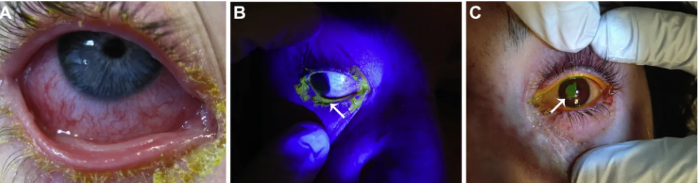
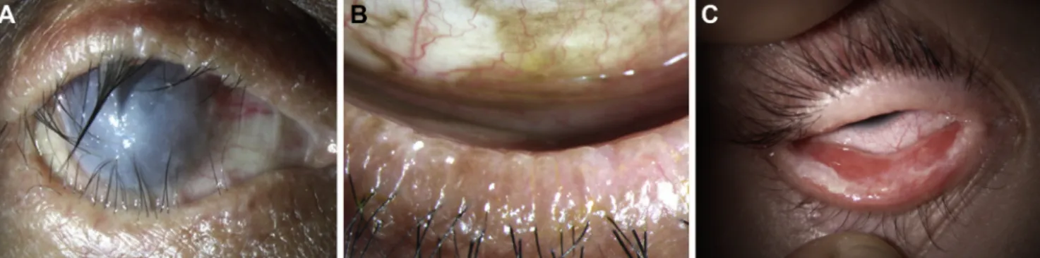
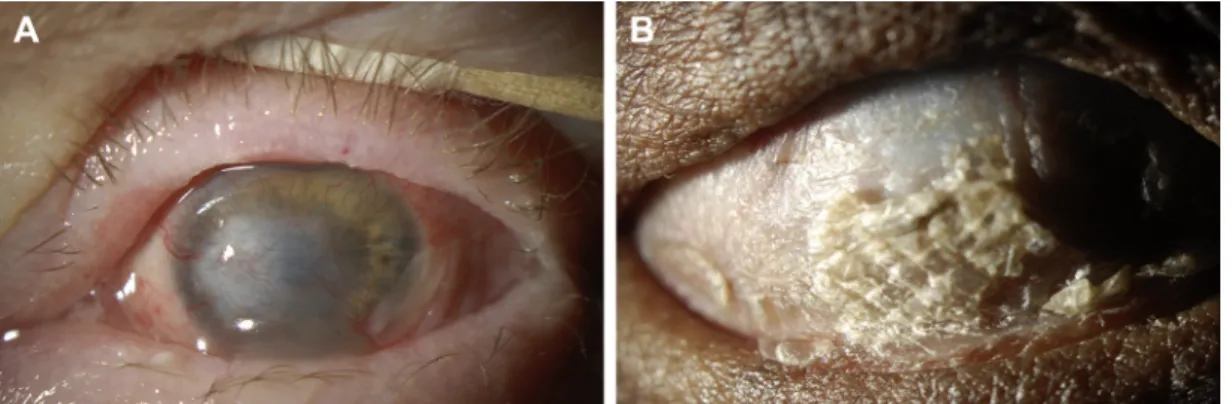
TEN is often associated with instability of major body sys- tems.71Affected patients may develop severe inflammation of internal mucosal surfaces, including the gastrointestinal and respiratory tracts.72 Major metabolic abnormalities, sepsis, multi-organ failure, pulmonary embolism, and gastrointes- tinal hemorrhage can occur.66,67,73Anemia and lymphopenia are common. Neutropenia is a particularly poor prognostic sign.74-76 Serious pulmonary disease may be present even without obvious radiographic abnormalities, leading to dys- pnea, tachypnea, and hypoxemia.77 Survivors may be left with chronic and debilitating sequelae permanently affecting their quality of life (Figure 2).
The ocular complications described later in this review are generally acknowledged as the most debilitating residual effects of SJS/TEN. However, skin scarring and pigmentation;
vaginal, urethral, and anal strictures; vulvar adenosis; penile phimosis; dental abnormalities; esophageal strictures; and dry mouth due to decreased salivary flow,4,66,67,78,79 also commonly reduce function and the quality of life following SJS/TEN. However, it is important to recognize that the acute and chronic manifestations of SJS/TEN vary among patients, and a modest subset survive their disease without apparent sequelae.
SJS/TEN can take a significant psychological toll on survivors80 and their immediate family members. A study of unsolicited internet posts by SJS/TEN survivors revealed that they had many unanswered questions long af- ter the event, and they desired to connect to other survivors to share their experiences.81Survivors had concerns about effects on fertility, fear of recurrences, and genetic inheri- tance of the disease. One study showed that survivors often choose to avoid medications altogether, and may fear becoming sick and ever needing medications.82 It is particularly poignant that patients who develop SJS/TEN due to a psychiatric therapeutic may subsequently avoid essential treatment for their mental illness. Similarly, those with chronic medical complications of SJS/TEN71 may avoid potentially beneficial medications out of anxiety about recurrence.
IV. CLASSIFICATION
Various classification schemes for SJS/TEN have been proposed, each with its own limitations. Mucosal involve- ment and the percentage of affected body surface area (BSA) are most useful in categorizing the disorder. In 1993, Bastuji-Garin et al83 divided the SJS/TEN spectrum into three major categories: SJS, defined by epidermal detach- ment of <10% of the BSA in association with widespread erythematous or purpuric macules or flat atypical targets;
SJS/TEN overlap, defined by epidermal detachment of 10%
to 30% of BSA plus widespread purpuric macules or flat atypical targets; and TEN defined by epidermal detachment of>30% of the BSA coupled with widespread purpuric mac- ules orflat atypical targets. Bastuji-Garin and coworkers also subclassified TEN as being with or without spots (purpuric macules).83
Another acute dermatobullous disorder often confused with SJS/TEN is bullous erythema multiforme. The clinical presentations and histopathology of erythema multiforme and SJS/TEN are distinctly different (Table 1).84-86 Ery- thema multiforme is characterized by epidermal detachment
of<10% BSA, coupled with localized typical target lesions
or raised atypical targets. Erythema multiforme is typi- cally caused by infection, most commonly herpes simplex virus. It most commonly presents with a minimal degree of mucosal involvement, with the skin biopsy characterized histologically by a lichenoid infiltrate, basal epidermal
Table 1. Classification for EM/SJS/TEN
Classification Bullous EM SJS SJS/TEN Overlap TEN
Detachment <10% <10% 10-30% >30%
Typical target lesions Yes No No No
Atypical target lesions Yes, raised Yes,flat Yes,flat Yes,flat
Adapted from.83
Figure 2. Schematic of body systems frequently affected in chronic SJS/TEN. In general, every body system affected in acute SJS/TEN shows chronic manifestations to variable degrees, later on. Other body systems can also be affected.71
SJS/TEN: I. SYSTEMIC DISEASE / Kohanim, Palioura, Saeed, et al
THE OCULAR SURFACE / JANUARY 2016, VOL. 14 NO. 1 / www.theocularsurface.com 5
necrosis, and moderate dermal inflammation. In contrast, SJS/TEN is typically associated with drugs, presents with prominent mucositis, and is identified histologically by full thickness epidermal necrosis with minimal underlying dermal inflammation. The current classification of SJS/
TEN used by most clinicians derives from that proposed by Bastuji-Garin et al.83,87
V. MORTALITY FROM TOXIC EPIDERMAL NECROLYSIS Various investigators have attempted to use clinical and pathological findings to predict the risk of mortality from TEN. SCORTEN (SCORe of TEN) is a mathematical model that has proven to be generally accurate in pre- dicting the risk of death from TEN.88,89 It is intended to be completed within 24 hours of admission and again on day 3 of hospitalization.89 The SCORTEN uses 7 independent risk factors to predict the risk of mortality from TEN88:
1. Age above 40 years 2. Presence of malignancy
3. Heart rate>120 beats per minute 4. Initial epidermal detachment>10%
5. Serum urea>10 mmol/L 6. Serum glucose>14 mmol/L 7. Serum bicarbonate<20 mmol/L
Using the SCORTEN system, each criterion is worth one point, with each additional point associated with a signifi- cant increase in mortality (with an increase in the odds ratio by a factor of 3.45).84For example, SCORTEN of 0-1 por- tends a 3.2% mortality, while SCORTEN of 5 or greater pre- dicts a mortality of 90% (Table 2). Other clinical parameters previously reported to be predictive of mortality include thrombocytopenia, leukopenia, delay in hospital admission, and treatment with antibiotics or corticosteroids prior to admission.74-76,90
Quinn and colleagues noted that some skin biopsy specimens from TEN patients exhibit more than minimal dermal inflammation, and showed a correlation between dermal mononuclear cell counts with disease severity and mortality.91 In their study of 37 cases, quantification of dermal mononuclear cells was almost as accurate in
predicting outcomes as SCORTEN (68% using mean cell count, vs 71% with SCORTEN).
VI. DIFFERENTIAL DIAGNOSIS
As mentioned above, erythema multiforme is considered to be an entirely different disease entity than SJS/TEN.
The former is marked by a more abbreviated course, occurs in a younger age group, and is most often associated with infection.86 Erythema multiforme frequently recurs, and skin lesions may be positive for herpes simplex virus and interferon gamma84 and show significantly lower levels of proinflammatory cytokines than in SJS/TEN.92In one study, the proportion of patients with involvement of multiple mucosal sites was significantly greater in SJS/TEN.93 SJS/
TEN is most commonly caused by medications. SJS/TEN is characterized clinically by skin lesions located predomi- nantly on the trunk, which appear as widespread,flat, atyp- ical targets or purpuric macules, and involvement of at least two mucosal sites.4
Other important entities in the differential diagnosis in patients with SJS/TEN include staphylococcal scalded skin syndrome, linear IgA bullous dermatosis, paraneoplas- tic pemphigus, acute graft-versus-host disease, drug-induced pemphigoid and pemphigus, and acute generalized exan- thematous pustulosis. These entities are distinguished by specific clinicalfindings on the skin and mucous membranes and on histopathology (Table 3).4
VII. PATHOGENESIS OF SJS/TEN
The pathogenesis of SJS/TEN is both complicated and controversial. However, available evidence points to a synthesis of genetic and innate immune mechanisms leading to keratinocyte cell death by apoptosis and secondary epidermal necrosis (Figure 3).94A 2008 review by Nickoloff examines various theories for the mechanisms underlying SJS/TEN, including altered drug metabolism, immune- mediated mechanisms, and activation of death receptors on keratinocytes.95
A. Genetic Susceptibility to SJS/TEN
There are several points to keep in mind regarding genetic susceptibility to SJS/TEN. First, the genetic risk factors are drug-specific. Second, genetic risk factors vary among populations and/or ethnic groups. Third, genetic testing for human leukocyte antigen (HLA)-B*1502 is avail- able and recommended by the U. S. Food and Drug Admin- istration for one drug, carbamazepine, in at-risk (Asian) populations, and more such recommendations are likely to follow. In patients of Han Chinese descent, HLA-B*1502 was strongly associated with carbamazepine-induced SJS/
TEN,96 and pretesting reduced the rate of SJS/TEN from carbamazepine in Hong Kong patients requiring anti- epileptic medications.97 Other HLA loci also appear to confer an increased risk of SJS/TEN.98-111HLA-B12 in one study of 44 TEN survivors was found to be more commonly detected,112 and HLA-DQB1*0601 was associated with an increased risk of SJS/TEN.113 The HLA-B*5801 allele was Table 2. SCORe of TEN (SCORTEN) level and predicted
mortality
SCORTEN Mortality
0-1 3.2%
2 12.1%
3 35.3%
4 58.3%
5 90.0%
Adapted from.88
SJS/TEN: I. SYSTEMIC DISEASE / Kohanim, Palioura, Saeed, et al
found to be present in 100% of 51 patients experiencing severe cutaneous reactions to allopurinol versus 15% of 135 tolerant patients).114
HLA-B*5701 confers an increased risk of hypersensitivity reactions to abacavir,4and the presence of all three variants HLA-B*5701, HLA-DR7, and HLA-DQ3 was 100% pre- dictive of developing a hypersensitivity reaction. Genetic associations have also been demonstrated for compound medications, such as over-the-counter cold medications.57,58 One particularly interesting association was found between SJS/TEN and potentially dominant negative single nucleotide polymorphisms in the IKZF1 gene, which codes for the tran- scription factor Ikaros.115Studies in Japanese patients also showed increased risk of eye involvement associated with HLA-A*0206 and HLA-B*44:03.57,116-118 Single nucleotide polymorphism analyses using candidate genes associated with innate immunity,118-121allergy,122,123and apoptosis124 revealed that polymorphisms in the TLR3 and EP3 genes may also be strongly associated with SJS in the Japanese pop- ulation. A putative imbalance between TLR3 and EP3 was postulated to play a role in the ocular surface disease severity in SJS/TEN. Another recent study showed a genetic polymor- phism in the IFN-gamma gene in Mexican patients with SJS.125Taken together, these reports suggest a complex role for immunogenetics in SJS/TEN.
Family members of SJS/TEN patients may be susceptible to the same drugs and should be counseled to avoid culprit medications when possible. However, HLA testing is not routinely performed prior to starting most new medications.
Patch testing in SJS/TEN to test for susceptibility to a specific drug has been attempted, but the results have been disap- pointing.126Provocation tests are dangerous and should not be performed.127 An in vitro lymphocyte toxicity assay to measure activity of detoxification enzymes exists but only as a research tool.128
Table 3. Differential diagnosis of SJS/TEN
Disease Mucositis Morphology Onset
Drug-induced pemphigoid Rare Tense bullae, sometimes
hemorrhagic
Acute
Staphylococcal scalded skin syndrome
Absent Erythema, skin tenderness,
perioral crusting
Acute
Drug-induced pemphigus Usually absent Erosions, crusts, patchy erythema
Gradual
Drug-triggered pemphigus Present Mucosal erosions,flaccid bullae
Gradual
Paraneoplastic pemphigus Present (usually severe) Polymorphous skin lesions,
flaccid bullae Gradual
Acute graft versus host disease
Present Morbilliform rash, bullae, and erosions
Acute
Acute generalized exanthem- atous pustulosis
Rare Superficial pustules (resem-
bles pustular psoriasis)
Acute
Drug-induced linear IgA bullous dermatosis
Rare Tense, subepidermal bullae
(resembles pemphigoid)
Acute
Adapted from.4
Figure 3. Pathogenic mechanisms involved in SJS/TEN. The schematic presents a simplified depiction of the interplay between genetics, spe- cific components of innate and acquired immunity, and effectors of keratinocyte cell death. See text for detailed discussion. TLR3¼Toll-like receptor 3. SNPs¼single nucleotide polymorphisms. EP3¼prostaglandin E receptor 3. (Adapted from Harp JL, Kinnebrew MA, Shinkai K.94)
SJS/TEN: I. SYSTEMIC DISEASE / Kohanim, Palioura, Saeed, et al
THE OCULAR SURFACE / JANUARY 2016, VOL. 14 NO. 1 / www.theocularsurface.com 7
B. Immunology of Acute SJS/TEN
The molecular pathogenesis of SJS/TEN is still under investigation, and there are many contradictory studies and observations. In general, acute SJS/TEN is considered a T-cell mediated, type IV hypersensitivity disorder, and could be considered an “immunologic burn.” Affected pa- tients show an exuberant response on re-exposure to the offending agent, and unlike many other types of hypersensi- tivity reactions, cytotoxic antibodies, immune complexes, and complement activation, all components of type II or III hypersensitivity reactions, are rarely found. On occasion, C3 and IgG may be detected at the dermal-epidermal junc- tion and around the blood vessels but are thought to be related to nonspecific exudation.129
There is compelling evidence to support a role for cyto- toxic T cells as major effectors in the pathogenesis of SJS/TEN, especially in the acute stages. The blister fluid of TEN patients contains predominantly T lymphocytes. How- ever, unlike most allergic skin reactions where CD4þlym- phocytes are the predominant cell type,130in the early stages of TEN, CD8þ lymphocytes concentrate in blister fluid and epidermis, while CD4þ lymphocytes are localized to the dermal layers.131As the disease progresses, however, there is a relative decrease in lymphocytes and increase in activated monocytes. Furthermore, soluble IL-2 receptor (sIL-2R), a marker for activated T cells, is present in high levels in blister fluid and serum of TEN patients, and levels correlate with dis- ease activity.132 One study demonstrated an increase in the number of activated T lymphocytes expressing cutaneous lymphocyte antigen (CLA), a skin-homing receptor, in the pe- ripheral blood of TEN patients. Levels correlated with disease activity and normalized after resolution of SJS/TEN.133
Chung and colleagues found that blisterfluid of SJS/TEN patients contained both cytotoxic T lymphocytes, and natural killer cells.134In addition, characterization of CD8þT cells in the epidermis and blisterfluid of TEN patients has shown that a majority of these cells also express surface markers normally found on natural killer cells. CD8þT cells in the epidermis expressed the killer inhibitory receptor (KIR) and killer activating receptor (KAR),135and a high percent- age of CD8þT cells from blisterfluid showed CD56 neural cell adhesion molecule (NCAM), another marker found on natural killer and highly cytotoxic CD8þT cells.136
Although the aforementionedfindings support a major role for T cells in acute SJS/TEN, there are also studies that point to the involvement of other types of immune cells. For example, although the blisterfluid of TEN lesions shows a predominance of lymphocytes, immunohistochem- istry of skin biopsies shows a predominance of cells of the monocyte-macrophage lineage and high levels of TNF- alpha.129 Paquet and colleagues studied 23 patients with TEN and found that MAC 387þ macrophages were the most numerous cells in the epidermis, while factor XIIIaþ dendritic cells were the most common cells in the dermis.137 MAC 387 is a monoclonal antibody clone that binds to cyto- plasmic antigen expressed by monocytes and macrophages in inflammatory skin diseases.138 Factor XIIIaþ dendritic
cells (dermal dendrocytes) play a major role in phagocytosis and antigen presentation. There is an increase in these dermal dendrocytes in other immunologic skin disorders such as atopic dermatitis and psoriasis, and they may also be involved with tissue repair.139,140
C. Mechanisms of Cell Death in SJS/TEN
Cellular demise occurs via two major pathways: necrosis and apoptosis. Necrosis is associated with intense inflamma- tion, while in apoptosis, T lymphocytes induce programmed cell death by activating intracellular caspases within the target cells leading to cell death with minimal inflammation.
There is general consensus that keratinocyte cell death in SJS/TEN occurs via apoptosis.141Light and electron micro- scopy of SJS/TEN-involved epidermis shows characteristic physical changes and terminal deoxynucleotidyl transferase dUTP nick end labeling (TUNEL) staining of keratinocytes associated with apoptosis.141,142
Apoptotic death receptors are transmembrane proteins of the tumor necrosis factor (TNF) superfamily.143 Several different types of death receptors have been isolated, but the first and most extensively studied is Fas (CD-95), and its ligand, FasL.144 Dysregulation of the Fas pathway has been implicated in the pathogenesis of a variety of tissue-destructive processes, including graft-versus-host dis- ease, multiple sclerosis, stroke, and TEN.144 The molecular mechanism by which Fas activates apoptosis has been exten- sively studied, and a detailed review is beyond the scope of this paper. Briefly, intracellular FasL in affected cells is transported via intermediate filaments to the cell surface, where it may be released as soluble FasL (sFasL). Binding of FasL to Fas on the surface of epidermal cells induces a conformational change in Fas and recruitment of Fas- associated death domain protein, an intracellular adaptor protein that attaches to both the Fas death domain and to procaspase 8, which is then proteolytically processed to form caspase 8. Activation of the caspase cascade leads to disassembly of cellular components and cell death.145,146
Work by Viard and colleagues showed mediation of ker- atinocyte death in TEN through activation of Fas.147 They demonstrated elevated expression of FasL on the cell surface of keratinocytes and high levels of soluble sFasL in TEN serum, in sharp distinction to patients with other maculo- papular drug reactions or normal controls.147 In addition, frozen skin sections of TEN patients induced apoptosis in a Fas-sensitive cell line, while apoptosis was blocked by anti-FasL-monocolonal antibody (Fas-Fc). These findings suggest that Fas may play a key role in inducing apoptosis in keratinocytes in TEN.
Conflicting data exist regarding the source of FasL.
Chang and colleagues measured serum sFasL levels over time in a patient with TEN, and found that sFasL levels peaked 24-48 hours after the onset of significant skin damage,148suggesting that sFasL may merely be a byproduct of FasL expressed on epidermal cells and not a direct inducer of apoptosis. Metalloproteinases downregulate FasL expression by cleaving the TNF-homologous portion SJS/TEN: I. SYSTEMIC DISEASE / Kohanim, Palioura, Saeed, et al
of membrane-bound FasL, releasing sFasL, and increased MP activity results in increased sFasL concentrations in the serum.149,150Therefore, elevated sFasL serum concentra- tion in TEN patients may be due to action of metalloprotei- nases at the surface of epidermal cells.145
Gelatinase A (MMP2) and B (MMP9) have also been implicated in SJS/TEN.151 Abe and coworkers studied 22 patients with SJS/TEN and found consistently elevated levels of sFasL in serum from these patients.152 Adding the pa- tients’ serum to epidermal cell culture led to induction of apoptosis, while addition of anti-FasL monoclonal antibody blocked apoptosis. Stimulation of peripheral blood mononu- clear cells (PBMCs) from TEN patients with the causative agent led to production of high levels of sFasL. In addition, direct immunofluorescence testing of skin specimens of 3 out of 22 patients could not detect FasL on the keratinocyte surface. The authors concluded that sFasL may be released by PBMCs instead of keratinocytes and that sFasL binds Fas on the cell surface to induce apoptosis, and it may serve as a serologic marker for TEN.
Other death receptors, such as TNF-R1 and TNF-related apoptosis-inducing ligand (TRAIL) may also be involved in TEN pathogenesis.153 There are elevated levels of TNF- alpha in blister fluid, skin, mononuclear cells, and blood of affected patients. TNF-alpha activates TNF-R1, which leads to activation of Fas-associated death domain protein and downstream caspase pathways. However, TNF-R1 also activates anti-apoptotic pathways by activating NF-kB.153 Therefore, TNF-alpha may either induce or block apoptosis, and as such, the use of anti-TNF medications in TEN pa- tients is controversial.153
Despitefindings that point to Fas-FasL pathway as a key mediator of apoptosis in SJS/TEN, there are experimental studies that challenge this hypothesis. The lytic granules of cytotoxic T lymphocytes contain perforin and granzyme.
Once T lymphocytes recognize a target cell, perforin creates 16 nm channels in the cell membrane of the affected cell, allowing granzyme B to enter the cell and activate the intra- cellular caspase cascade, leading to apoptotic cell death.154 After exposure to the causative agent, mononuclear cells from the blister fluid of TEN patients became cytotoxic, but peripheral blood mononuclear cells did not. Anti-Fas monoclonal antibodies failed to inhibit this cytotoxicity, while distinct inhibitors of the perforin/granzyme pathway did.136,155 These findings implicate perforin/granzyme as mediators of the pathogenesis of TEN.
Nassif and coworkers evaluated cytokines in blisterfluid of TEN patients and reported elevated IFN gamma, TNF- alpha, sFasL, IL-18, and IL-10.156 They showed that FasL and TNF-alpha found in blister fluid were expressed by keratinocytes rather than by mononuclear cells in thefluid.
Cell-free supernatants of blisterfluid did not induce apoptosis in cultured keratinocytes, leading to the conclusion that apoptosis in TEN was not mediated through the sFas-FasL pathway. The authors proposed that activated CD8þT cells secrete IFN-gamma, which in turn stimulates keratinocytes to produce TNF-alpha, FasL, and IL-10. TNF-alpha induces
upregulation of MHC 1 molecules, which makes keratinocytes more sensitive to cytotoxic T lymphocytes and perforin/
granzyme-mediated apoptosis. Nassif and colleagues hypoth- esized that FasL and IL10 production may actually be protec- tive and serve to downregulate inflammation by inducing apoptosis in cytotoxic T lymphocytes, rather than inducing demise of the keratinocytes.
Posadas and colleagues looked at cytokine expression in peripheral blood and from mononuclear cells in patients’ blister fluid in a study of four severity levels of delayed dermatologic drug reactions: benign maculopapular rashes, desquamative exanthema, SJS, and TEN.157 They found that perforin/granzyme B concentration directly correlated with disease severity. Levels of perforin/granzyme B pro- duced by peripheral blood mononuclear cells of patients with maculopapular drug reactions were much lower than from patients with SJS/TEN. Interestingly, analysis of cyto- kine expression by peripheral blood and mononuclear cells from blisterfluid of SJS/TEN patients also showed elevated FasL. These data suggest that both pathways may play a role in SJS/TEN. A study of biopsy specimens in TEN also showed abnormal expression of calprotectin,158,159an anti- microbial protein not found in normal skin. Other studies have shown increased nitric oxide.160
Chung and colleagues used gene expression profiling fol- lowed by quantitative PCR and immunohistochemistry and reported that a secretory form of granulysin was expressed at levels two to four times that of perforin/granzyme B or s-FasL.134Removing granulysin reduced the cytotoxicity of blisterfluid. Injection of granulysin into mouse skin resulted in changes that mimicked SJS/TEN. Their findings suggest that secretory granulysin may be an important mediator of SJS/TEN.
To summarize, keratinocyte cell death in SJS/TEN appears to occur by apoptosis. Several mechanisms are likely involved to varying degrees. While T lymphocytes play a major role, other components of the immune system closely participate in the process.
VIII. ACUTE SYSTEMIC THERAPY A. Supportive Care
The mainstay of treatment for SJS/TEN is early and aggres- sive supportive care in a Burn Intensive Care Unit (ICU).
Prognosis improves the earlier the offending agent is dis- continued.161While it is critical that the offending agent be identified and immediately stopped, medications with a long half-life are problematic.161 A direct correlation was also shown between survival and the speed with which the patient is admitted to a Burn ICU,162-164where urgent care can be initi- ated by personnel trained in the management of the respiratory tract, kidneys,fluid and electrolyte balance, infections, nutri- tion, skin and ocular surface, and pain control.165-167In the Burn ICU, necrotic skin is debrided, and the exposed areas covered with artificial membranes or biologic dressings which enhance healing, and reduce discomfort, scarring, and infec- tion.168 Given high rates of infection in SJS/TEN, frequent skin, blood, urine, and line cultures are performed.
SJS/TEN: I. SYSTEMIC DISEASE / Kohanim, Palioura, Saeed, et al
THE OCULAR SURFACE / JANUARY 2016, VOL. 14 NO. 1 / www.theocularsurface.com 9
Routine antibiotic prophylaxis is typically avoided due to emergence of resistance leading to increased mortality from sepsis.169 When patients do become septic, Staph- ylococcus aureusandPseudomonas aeruginosa are common inciting agents.170Early supportive care in a Burn ICU can be effective in reducing mortality. Sheridan and colleagues reported no deaths in 10 children with TEN treated with supportive care alone.171 In another study, 21 children with SJS/TEN were treated with conservative measures alone, and none died.172 In another, 15 children with SJS/
TEN treated in a Burn ICU with supportive care alone had a reported mortality of 7%.173
B. Therapeutic Agents
Proposed systemic interventions in acute SJS/TEN are described in the sections that follow and inTable 4.
1. Systemic Corticosteroids
Because SJS/TEN is thought to represent an immune response to an exogenous agent, systemic administration of corticosteroids has been used with the hope of improving
clinical outcomes. However, the use of systemic corticoste- roids in the treatment of SJS/TEN is highly controversial.
Patients taking systemic corticosteroids for other condi- tions still develop SJS/TEN,12,174-176 and corticosteroids do not appear to influence TEN progression once the reaction is initiated.175,177 In the pediatric literature, two178,179 out of four178-181observational studies found that systemic cor- ticosteroids significantly increased the rate of complications (sepsis, urinary tract infection, gastrointestinal hemorrhage) in children with SJS compared to children who only received supportive measures. Among the outcomes studied (dura- tion of fever and of acute eruption, length of hospital stay, number of complications), the only parameter to show improvement from corticosteroid administration was dura- tion of fever.178,180
The evidence for the use of systemic corticosteroids for SJS/TEN in adults is also inconsistent. A few small retro- spective case series have shown a mortality benefit from cor- ticosteroids,182-185but most reports have suggested either a lack of efficacy90,186,187 or increased mortality.188,189In one large retrospective study, the use of systemic corticosteroids
Table 4. Proposed systemic interventions in acute SJS/TEN
Therapy*
Proposed Mechanism of
Action Effect on Ocular Disease
Evidence-based Recommendations Systemic Corticosteroids/
High-dose Pulsed Steroids (HDPS)
Dampens immune response to exogenous agent
Equivocal Equivocal; inconsistent results with most reports suggesting lack of efficacy or increased mortality; however, HDPS in the earliest stages of disease may limit progression and mortality.
Human Intravenous Immune Globulin (IVIG)
Autoantibodies against Fas in IVIG prevent Fas-FasL medi- ated apoptosis
Equivocal Equivocal; numerous studies reflecting a spectrum of benefit from improved mor- tality to no benefit to increased mortality. Most sig- nificant complication is acute renal failure.
Plasmapheresis Removes non-dialyzable pathogenic elements found in the plasma
Unknown Overall results have been favorable; limited data.
Generally safe with minimal complications.
Granulocyte Colony Stimu- lating Factor (GCSF)
Boosts neutrophil counts to decrease risk of infection
Unknown May play a role in the neu- tropenic SJS/TEN patient Cyclosporine Inhibition of apoptosis by
down regulation of NF-kB
Unknown Current reports suggest min- imal benefit to reduction in mortality. Complications include leukoencephalop- athy, neutropenia, pneu- monia, and nephropathy TNF-alpha Inhibitors Inhibition of TNF-alpha pre-
vents apoptosis
Unknown Not recommended; associ- ated with increased mortality Cyclophosphamide Inhibition of cell-mediated
cytotoxicity
Unknown Not recommended; associ- ated with increased mortality
* In order of appearance in text.
SJS/TEN: I. SYSTEMIC DISEASE / Kohanim, Palioura, Saeed, et al
in 119 patients did not definitively alter mortality compared with supportive care alone in 87 patients, though a trend toward a possible benefit was observed.186 In a large study of SJS/TEN patients in France and Germany enrolled in the International Registry of Severe Cutaneous Adverse Reactions (RegiSCAR), including 460 patients in the cohort, the use of systemic corticosteroids or any other immune modulating drug was not associated with a significant change in survival.187
Despite a widely held recommendation against use of systemic corticosteroids, there is also support for high- dose pulsed corticosteroids in the earliest stages of TEN when secondary sepsis is less likely.190In one study, twelve patients with SJS/TEN were treated with a 3-day course of pulsed intravenous dexamethasone (1.5mg/kg/day); the pre- dicted mortality based on SCORTEN in this cohort was four deaths, but only one patient died.184High-dose dexametha- sone appeared to stop disease progression in three days on average, and healing occurred within three weeks. The possible effects of high-dose pulsed corticosteroids on ophthalmic complications of SJS/TEN are described below.
2. Human Intravenous Immune Globulin
Human intravenous immune globulin (IVIG) is produced by pooling plasma from several thousand donors. IVIG con- tains a mixture of immunoglobulins, mostly immunoglobulin G (IgG) with trace amounts of IgM and IgA,191 against a variety of“self”molecules. The use of IVIG has been approved by the U. S. Food and Drug Administration for patients undergoing hematopoietic stem cell transplantation and for the treatment of a variety of inflammatory and autoim- mune diseases, such as common variable immunodeficiency, immune-mediated thrombocytopenia, Kawasaki disease, chronic lymphocytic leukemia, chronic inflammatory demy- elinating polyneuropathy, and pediatric HIV type I infec- tion.192,193 IVIG includes autoantibodies against Fas, and in a pilot study, ten patients with biopsy-proven TEN were treated with IVIG with no deaths and no systemic side effects.147Exogenous IVIG reaches the epidermis, in partic- ular the blisterfluid and epidermal layers of clinically involved and uninvolved skin of TEN patients.194
Autoantibodies against Fas in IVIG are thought to reduce TEN complications by interfering with Fas-FasL interactions.
Removing anti-Fas IgG from IVIG blocks the ability to prevent Fas-FasL mediated apoptosis in vitro.147,195 IVIG- treated patients demonstrate reduced Fas and FasL in post- treatment skin biopsies.196
There have been numerous SJS/TEN case reports and case series showing benefit from IVIG.197-213In a small retrospec- tive study of 8 pediatric TEN patients treated with IVIG, all patients survived.201In a prospective study of 12 TEN patients from Kuwait, 100% of patients treated with IVIG survived.207 In a retrospective study of 12 SJS patients receiving adjunctive treatment with IVIG in doses of 0.6 g/kg/day, all patients sur- vived and the investigators reported objective response within 2 days of starting therapy.206 Another retrospective review of 15 patients along the SJS/TEN spectrum treated with
IVIG within 24 hours of diagnosis reported 80% survival.210 In another small study, 16 TEN patients with predicted SCORTEN mortality of 5.8 deaths were treated with IVIG (most receiving 1g/kg/day for 4 days); only one patient died.213In a larger retrospective study involving 14 American and European academic centers, 48 consecutive TEN pa- tients were treated with adjunctive IVIG (1 gm/kg/day for 3 days).205The survival rate was 88%. The most significant IVIG-related complication was acute renal failure. In another study of 9 TEN patients treated with combination of IVIG and pulsed methylprednisolone in another study, 8 survived.211
IVIG was also used as an adjunct to systemic corticoste- roids. In a study by Yang and coworkers, 45 patients who had received corticosteroids only for SJS/TEN were compared to 20 patients who received a combination of corticosteroids and IVIG.214Combination therapy reduced the time to arrest of progression and the total hospitalization time, and though not statistically significant, showed a tendency to decrease mortality rate compared to the use of corticosteroids as sole treatment.
Metry and colleagues shared their experience with 7 pedi- atric patients who were treated with IVIG and also reviewed 28 reports in the literature.209 They concluded that IVIG was helpful in children with SJS/TEN. There were no mortal- ities and consistent recovery was observed in every child who was treated with IVIG. In one case, IVIG successfully pre- vented recurrent SJS in a patient with multiple episodes after exposure to intravenous contrast.208
On the other hand, there are multiple reports that show minimal to no benefit and increased mortality with IVIG.186,215-219 IVIG was found to be of minimal value in 7 children with SJS when compared to 5 children with SJS treated with corticosteroids or supportive care alone.217In a comparison of 10 treated patients with 18 historical controls, IVIG did not reduce the severity of the ocular complications of TEN.219 Another retrospective study compared 24 patients who received IVIG to 21 patients who did not.216All patients were treated by the same team using a standard protocol. The dose of IVIG used was 0.4 g/kg/day, below that used in other studies. The investigators found no advantage to IVIG at any SCORTEN level, and there was higher mortality in the group that received IVIG versus the control group. (41.7% vs 28.6%).
One retrospective study compared 16 TEN patients treated with IVIG to 16 patients who did not receive IVIG, and found no statistically significant differences in mortality, length of hospitalization, length of mechanical ventilation, sepsis, or the severity of systemic inflammatory response syndrome and multiple organ dysfunction syndrome.218
A prospective trial of 34 patients with SJS/TEN who were treated with IVIG (2g/kg/day for 2 days with modified renal dosing as needed) evaluated the effect on total body surface area involvement pre- and post-IVIG treatment as well as mortality.215 There was no significant improvement in treated patients, and the mortality rate was both higher than the predicted by SCORTEN (11 actual deaths vs 8.2 predicted deaths), and higher than the hospital’s historical death rate of approximately 20% in TEN patients. Most SJS/TEN: I. SYSTEMIC DISEASE / Kohanim, Palioura, Saeed, et al
THE OCULAR SURFACE / JANUARY 2016, VOL. 14 NO. 1 / www.theocularsurface.com 11
deaths occurred in elderly patients with impaired baseline renal function. In this study, the investigators used a higher dose and shorter duration of IVIG compared with some other studies showing positive effects.
A retrospective case-control analysis of patients that were included in the prospective EuroSCAR observational study, the largest cohort of SJS/TEN patients collected to date, found that compared to supportive care (87 patients) or systemic corticosteroids (119 patients) the odds ratio for death was nonsignificantly increased with IVIG therapy (35 patients).186The study design may have been limited by diverse treatment facilities in different countries, and non- standardized protocols for supportive care, treatment doses, and treatment duration.
With regard to whether IVIG reduces ocular complica- tions in SJS/TEN, two small case series provided contradic- tory results. In a study by Yip and coworkers, IVIG did not mitigate the severity of the acute manifestations of ocular SJS/TEN in 8 patients compared to 18 historical controls managed conservatively.219Yet in another study, early inter- vention with IVIG, when compared to supportive care only, appeared to significantly improve ocular involvement and best corrected visual acuity in adults but not in children.220 Overall, it is difficult to reconcile these contradictory results, and the initial enthusiasm for IVIG in the treatment of SJS/
TEN has dampened.
3. Plasmapheresis
Plasmapheresis removes non-dialyzable pathogenic ele- ments found in the plasma.221Whole blood is drawn from the patient and is separated into its cellular components and plasma, with the patient’s plasma typically discarded.
Blood is then reconstituted by adding albumin to artificial plasma, and/or with banked plasma, to the cellular constit- uents and reinfused back into the patient. The procedure is generally safe. Overall, the results in TEN have been favor- able with reported survivals of 77 to 100% after one to eight exchanges.222-224 However, one report of a series of eight patients treated with plasmapheresis in Sweden showed no difference in survival compared to groups treated with sup- portive care alone.225
4. Granulocyte Colony Stimulating Factor
Neutropenia portends a poor prognosis in SJS/TEN due to heightened risk of infection. Several case reports have described patients in whom granulocyte colony-stimulating factor was used to boost neutrophil counts.74,198,226-229This agent may play a role in management of the neutropenic TEN patient.
5. Cyclosporine
In addition to its immunosuppressive effects, cyclo- sporine may inhibit apoptosis by inhibiting down regulation of NF-kB.153,230It has been used in doses of 3mg/kg/day in in- dividual cases of SJS/TEN with good results reported.230-233 There were no mortalities in a case series of 11 patients treated with cyclosporine.233 An open, phase 2 clinical trial of 29
treated patients (3mg/kg/day for 10 days, followed by taper over 1 month) showed a modest and nonstatistically sig- nificant reduction in SCORTEN predicted mortality.234 Side-effects included leukoencephalopathy, neutropenia, pneumonia, and nephropathy.
6. TNF-alpha Inhibitors
As discussed above, TNF-alpha is likely to play a role in SJS/TEN.153TNF-alpha inhibitors including infliximab, pen- toxiphylline, and thalidomide have been used in individual cases.13,235 In one report, a 56-year-old woman with TEN who was treated with a single infliximab infusion showed sig- nificant improvement.235However, a prospective trial in TEN patients comparing thalidomide to placebo was stopped due to higher than predicted mortality rates in patients receiving thalidomide.236
7. Cyclophosphamide
Cyclophosphamide was previously reported to benefit patients with acute TEN,237,238 but was later associated with increased mortality and is not recommended in the treatment of acute SJS/TEN.4,75
C. Effect of Systemic Treatments of Acute SJS/TEN on Ocular Disease
The effects of adjuvant systemic treatments on the ocular manifestations of SJS/TEN are unclear and controversial, limiting general recommendations for systemic management beyond supportive burn care. For example, one study comparing 10 IVIG-treated TEN patients to 18 historical con- trols concluded that treatment did not mitigate the severity of vision-threatening complications.219Reported ocular out- comes after administration of systemic corticosteroids in acute SJS/TEN vary from no effect to possible reduction of ocular complications. In a relatively large study in children, there was no significant difference in the number of patients with ocular involvement or in the severity of ocular manifestations between those treated with systemic corticosteroids (51 pa- tients) and those who were not (38 patients).239In a recent report on the effect of various systemic treatment modalities on ocular SJS/TEN, systemic corticosteroids did not ameliorate the ocular disease of the pediatric group (mean hydrocortisone equivalent dosage, 2.9 mg/kg/day for 3.5 days). Corticosteroids provided a nonstatistically significant improvement in visual acuity during the course of SJS/TEN in the 22 adults who received them within 5 days of disease onset (mean hydrocor- tisone equivalent dosage, 5.3 mg/kg/day for 3.5 days).220
However, in a study of five adult patients with SJS/
TEN who received pulsed methylprednisolone (500 mg or 1 gram) for 3-4 days, with ocular topical 0.1% betamethasone instilled 5 times per day for 2 weeks starting within 4 days of disease onset, all experienced good visual outcomes.240At the initial examination, all patients in the study had membranous conjunctivitis with corneal and/or conjunctival defects. There were no significant adverse effects of systemic and topical cor- ticosteroids during the course of their study. At 1 year, all eyes had normal architectural features of the palisades of Vogt SJS/TEN: I. SYSTEMIC DISEASE / Kohanim, Palioura, Saeed, et al
without evidence of limbal stem cell dysfunction, with best- corrected visual acuity of 20/20. Five eyes demonstrated corneal superficial punctate keratopathy, and all eyes showed mild irregularity of the mucocutaneous junction. Followup examinations were not reported beyond 1 year. However, in a recent retrospective, non-case-controlled study by Kim et al, the authors reported no apparent benefit from various immunomodulatory treatments in chronic ocular outcomes of SJS/TEN.241
IX. SUMMARY AND CONCLUSIONS
SJS/TEN is a severe, T cell-mediated, dermatobullous drug reaction with significant and sometimes devastating long-term morbidity in survivors, including ocular sequelae that can result in total blindness. Once triggered, keratinocyte cell death in SJS/TEN occurs rapidly with irreversible consequences.
Early and aggressive intervention in a Burn ICU is essential to survival. The rarity of SJS/TEN and the diversity of inciting agents make the disorder challenging to study. A broad range of systemic interventions have been proposed and attempted, most with conflicting results, and some with profoundly nega- tive consequences for patients. Unfortunately, because SJS/
TEN is rare, prospective controlled clinical trials of individual therapies are not feasible. High-dose intravenous corticoste- roids, IVIG, and plasmapheresis may yet be shown beneficial, and large, registry-based, retrospective studies may in the future illuminate best practices in the care of SJS/TEN.
We are unable to make recommendations regarding spe- cific systemic therapies for the acute stage of the disorder, as existing reports are contradictory. Nevertheless, significant advances have been made in recent years in the treatment of ocular manifestations of SJS/TEN in both acute and chronic stages of the disorder. These will be covered in Part II of this review, which will be published in the April 2016 issue of this journal. Ophthalmologists play an essen- tial role in evaluation and treatment of patients with SJS/
TEN in the acute and chronic stages to minimize long- term vision loss and ocular morbidity.
REFERENCES
1. Letko E, Papaliodis DN, Papaliodis GN, et al. Stevens-Johnson syn- drome and toxic epidermal necrolysis: a review of the literature.
Ann Allergy Asthma Immunol2005;94:419-36. quiz 36-8, 56 2. Borchers AT, Lee JL, Naguwa SM, et al. Stevens-Johnson syndrome
and toxic epidermal necrolysis.Autoimmun Rev2008;7:598-605 3. Endorf FW, Cancio LC, Gibran NS. Toxic epidermal necrolysis clinical
guidelines.J Burn Care Res2008;29:706-12
4. Pereira FA, Mudgil AV, Rosmarin DM. Toxic epidermal necrolysis.
Journal of the American Academy of Dermatology2007;56:181-200 5. Becker DS. Toxic epidermal necrolysis.Lancet1998;351:1417-20 6. Naldi L, Locati F, Marchesi L, Cainelli T. Incidence of toxic epidermal
necrolysis in Italy.Arch Dermatol1990;126:1103-4
7. Schopf E, Stuhmer A, Rzany B, et al. Toxic epidermal necrolysis and Stevens-Johnson syndrome. An epidemiologic study from West Ger- many.Arch Dermatol1991;127:839-42
8. Strom BL, Carson JL, Halpern AC, et al. A population-based study of Stevens-Johnson syndrome. Incidence and antecedent drug exposures.
Arch Dermatol1991;127:831-8
9. Griggs RC, Batshaw M, Dunkle M, et al. Clinical research for rare disease:
opportunities, challenges, and solutions.Mol Genet Metab2009;96:20-6 10. Sushma M, Noel MV, Ritika MC, et al. Cutaneous adverse drug reac- tions: a 9-year study from a South Indian Hospital.Pharmacoepide- miol Drug Saf2005;14:567-70
11. Roujeau JC, Stern RS. Severe adverse cutaneous reactions to drugs.
N Engl J Med1994;331:1272-85
12. Roujeau JC, Guillaume JC, Fabre JP, et al. Toxic epidermal necrolysis (Lyell syndrome). Incidence and drug etiology in France, 1981-1985.
Arch Dermatol1990;126:37-42
13. Ghislain PD, Roujeau JC. Treatment of severe drug reactions: Stevens- Johnson syndrome, toxic epidermal necrolysis and hypersensitivity syndrome.Dermatology Online J2002;8:5
14. Leenutaphong V, Sivayathorn A, Suthipinittharm P, Sunthonpalin P.
Stevens-Johnson syndrome and toxic epidermal necrolysis in Thailand.
Int J Dermatol1993;32:428-31
15. Patel TK, Barvaliya MJ, Sharma D, Tripathi C. A systematic review of the drug-induced Stevens-Johnson syndrome and toxic epidermal necrolysis in Indian population.Indian J Dermatol Venereol Leprol2013;79:389-98 16. Finkelstein Y, Soon GS, Acuna P, et al. Recurrence and outcomes of Stevens-Johnson syndrome and toxic epidermal necrolysis in children.
Pediatrics2011;128:723-8
17. Yetiv JZ, Bianchine JR, Owen Jr JA. Etiologic factors of the Stevens- Johnson syndrome.South Med J1980;73:599-602
18. Franca MD, Lima JP, Freitas D, et al. Study of the ocularfindings in Stevens-Johnson syndrome patients from a tertiary ophthalmologic center.Arq Bras Oftalmol2009;72:370-4. Portuguese
19. Ball R, Ball LK, Wise RP, et al. Stevens-Johnson syndrome and toxic epidermal necrolysis after vaccination: reports to the vaccine adverse event reporting system.Pediatr Infect Dis J2001;20:219-23
20. Hwang YH, Kang MS, Lim KO, Lee SM. Toxic epidermal necrolysis with ocular involvement following vaccination for hemorrhagic fever with renal syndrome.Yonsei Med J2012;53:228-30
21. Dobrosavljevic D, Milinkovic MV, Nikolic MM. Toxic epidermal nec- rolysis following morbilli-parotitis-rubella vaccination. J Eur Acad Dermatol Venereol1999;13:59-61
22. Radimer GF, Davis JH, Ackerman AB. Fumigant-induced toxic epidermal necrolysis.Arch Dermatol1974;110:103-4
23. House RA, Jakubovic H, Wong L, Holness DL. Work-related toxic epidermal necrolysis?J Occup Med1992;34:135-9
24. Hemmige V, Jenkins E, Lee JU, Arora VM. Toxic epidermal necrolysis (TEN) associated with herbal medication use in a patient with sys- temic lupus erythematosus.J Hosp Med2010;5:491-3
25. Fogh K. Toxic epidermal necrolysis following intake of vitamins and natural medication.Ugeskr Laeger1995;157:3631-3. Danish 26. Mochitomi Y, Inoue A, Kawabata H, et al. Stevens-Johnson syndrome
caused by a health drink (Eberu) containing ophiopogonis tuber.
J Dermatol1998;25:662-5
27. Lim YL, Thirumoorthy T. Serious cutaneous adverse reactions to traditional Chinese medicines.Singapore Med J2005;46:714-7 28. Figueira-Coelho J, Lourenco S, Pires AC, et al. Mycoplasma
pneumoniae-associated mucositis with minimal skin manifestations.
Am J Clin Dermatol2008;9:399-403
29. Fournier S, Bastuji-Garin S, Mentec H, et al. Toxic epidermal necrol- ysis associated with Mycoplasma pneumoniae infection. Eur J Clin Microbiol Infect Dis1995;14:558-9
30. Kunimi Y, Hirata Y, Aihara M, et al. Statistical analysis of Stevens- Johnson syndrome caused by Mycoplasma pneumonia infection in Japan.Allergol Int2011;60:525-32
31. Meyer Sauteur PM, Goetschel P, Lautenschlager S. Mycoplasma pneu- moniae and mucositisepart of the Stevens-Johnson syndrome spec- trum.J Dtsch Dermatol Ges2012;10:740-6
SJS/TEN: I. SYSTEMIC DISEASE / Kohanim, Palioura, Saeed, et al
THE OCULAR SURFACE / JANUARY 2016, VOL. 14 NO. 1 / www.theocularsurface.com 13