Characterization of intracellular dynamics of inoculated PrP-res and newly generated PrP
Scduring early stage prion infection in Neuro2a cells
Takeshi Yamasaki
a, Gerald S. Baron
b, Akio Suzuki
a, Rie Hasebe
a, Motohiro Horiuchi
a,naLaboratory of Veterinary Hygiene, Graduate School of Veterinary Medicine, Hokkaido University, Kita 18, Nishi 9, Kita-ku, Sapporo 060-0818, Japan
bLaboratory of Persistent Viral Diseases, Rocky Mountain Laboratories, National Institute of Allergy and Infectious Diseases, National Institutes of Health, Hamilton, MT, USA
a r t i c l e i n f o
Article history:
Received 16 September 2013 Returned to author for revisions 15 October 2013
Accepted 4 November 2013
Keywords:
Prions
Membrane trafficking Endosomes
Neurodegenerative diseases Rab proteins
a b s t r a c t
To clarify the cellular mechanisms for the establishment of prion infection, we analyzed the intracellular dynamics of inoculated and newly generated abnormal isoform of prion protein (PrPSc) in Neuro2a cells.
Within 24 h after inoculation, the newly generated PrPScwas evident at the plasma membrane, in early endosomes, and in late endosomes, but this PrPSc was barely evident in lysosomes; in contrast, the majority of the inoculated PrPScwas evident in late endosomes and lysosomes. However, during the subsequent 48 h, the newly generated PrPSc increased remarkably in early endosomes and recycling endosomes. Overexpression of wild-type and mutant Rab proteins showed that membrane trafficking along not only the endocytic-recycling pathway but also the endo-lysosomal pathway is involved inde novoPrPScgeneration. These results suggest that the trafficking of exogenously introduced PrPScfrom the endo-lysosomal pathway to the endocytic-recycling pathway is important for the establishment of prion infection.
&2013 Elsevier Inc. All rights reserved.
Introduction
Prions are causative agents of transmissible spongiform encepha- lopathies (TSEs), neurodegenerative disorders that are characterized by accumulation of an abnormal isoform of prion protein (PrPSc) in the central nervous system (CNS). PrPScis the only known proteinaceous component of prions, and infectivity of prions is thought to be associated with PrPSc oligomers (Silveira et al., 2005; Wang et al., 2010). PrPSc is a conformational isomer of a cellular prion protein (PrPC) and is rich in
β
-sheet; PrPScis generated from PrPC, a protein that is expressed on the surface of host cells (Prusiner, 1998).Conversion of PrPCto PrPScis thought to be triggered by direct contact between“seed”PrPScand“substrate”PrPC.
The intracellular dynamics of PrPSc immediately following exposure of the cells to prions have been analyzed to understand the mechanisms by which a prion infection becomes established.
Although PrPC is not necessary for the internalization of PrPSc (Greil et al., 2008; Hijazi et al., 2005; Jen et al., 2010; Magalhaes et al., 2005; Paquet et al., 2007), some candidates such as laminin receptor, heparan sulfate, or low-density lipoprotein receptor- related protein 1 (LRP1) have been reported to act as receptors
that mediate internalization of exogenously inoculated PrPSc (Gauczynski et al., 2006; Horonchik et al., 2005; Jen et al., 2010).
During the early stage after inoculation of PrPSc, internalized PrPSc has been reported to be directed to late endosomes/lyso- somes via the endo-lysosomal pathway (Jen et al., 2010;
Magalhaes et al., 2005). In cells persistently infected with prions, a number of studies have shown that PrPSc localizes throughout the endocytic compartments—specifically the plasma membrane, early endosomes, recycling endosomes, late endosomes, secondary lysosomes, and the peri-nuclear Golgi region (Marijanovic et al., 2009; McKinley et al., 1991; Pimpinelli et al., 2005; Taraboulos et al., 1990; Veith et al., 2009; Vey et al., 1996; Yamasaki et al., 2012). Earlier studies suggested that the generation of PrPScoccurs on the cell surface or within the endocytic pathway (Borchelt et al., 1992; Caughey and Raymond, 1991; Taraboulos et al., 1992).
Marijanovic et al. reported that the endocytic recycling compart- ment (ERC) may be the site where the conversion of PrPCto PrPSc occurs (Marijanovic et al., 2009). In a very early stage of prion infection where new PrPSc formation was measured within min- utes after initiation of infection, Goold et al. recently reported that the plasma membrane is a primary site of conversion (Goold et al., 2011).
In spite of the efforts described above, the events required for the establishment of prion infection in cells, especially those that occur in the early stage after introduction of the infectious prions, Contents lists available atScienceDirect
journal homepage:www.elsevier.com/locate/yviro
Virology
0042-6822/$ - see front matter&2013 Elsevier Inc. All rights reserved.
http://dx.doi.org/10.1016/j.virol.2013.11.007
nCorresponding author. Tel./fax:þ81 11 706 5293.
E-mail address:[email protected] (M. Horiuchi).
and newly generated PrPScin a short period after challenge with prions is important for understanding the early events of prion infection. However, such analysis is limited due to the technical difficulties in the distinction of PrPSc from PrPCand also in the distinction of newly generated PrPSc from inoculated PrPSc. For example, antibody epitope-tagging has been used to specifically detect host cell-derived PrP or input PrPSc (Greil et al., 2008;
Vorberg et al., 2004) but not both. Specific labeling of PrPScwith anti-PrP antibody via pre-treatment of cells with chaotropic agents such as guanidinium salt has been widely used to detect PrPScin cells (Marijanovic et al., 2009; Pimpinelli et al., 2005; Taraboulos et al., 1990). However, this method is limited because weak PrPSc signals such as newly generated PrPSc may be lost or obscured because the detector gain or exposure times need to be adjusted to
detection limit.
We recently reported that mAb 132, an anti-PrP monoclonal antibody that recognizes an epitope consisting of amino acids 119– 127 of mouse PrP, allowed us to visualize PrPScin prion-infected cells by indirect immunofluorescence assay (IFA) without specific manipulation of threshold setting to diminish PrPC signals (Yamasaki et al., 2012). Here, we established a method in which inoculated PrPScand newly generated PrPSccan be distinguished by combining the use offluorescent-dye-labeled purified protei- nase K (PK)-resistant PrPSc (PrP-res) as inoculum with PrPSc- specific staining with mAb 132. By using this method, in the present study, we extensively analyzed the fate of the inoculated PrP-res and the appearance of newly generated PrPScin Neuro2a cells during the early stages of prion infection.
Fig. 1.Characterization of purified PrP-res. (a) Purity of the PrP-res preparation. Purified PrP-res fractions from the brains of mice infected with the Obihiro strain (Obi-PrP- res) and the 22 -L strain (22-L-PrP-res) were subjected to SDS-PAGE followed by silver staining and immunoblotting with mAb 31C6. (b) Purity offluorescent-dye-labeled PrP-res. The purified 22-L-PrP-res was labeled with Alexa Fluor 488 (Af488-22-L-PrP-res) or Alexa Fluor 555 (Af555-22-L-PrP-res). Purity andfluorescent-dye-labeling were analyzed with silver staining, immunoblotting with mAb 31C6, andfluorescence imaging. (c) Influence of sonication on purified PrP-res. Purified 22-L-PrP-res was sonicated and then before and after centrifugation at 10,000 gfor 10 min [supernatant (Sup), precipitate (Ppt)] the PrP-res was subjected to immunoblotting with mAb 31C6.
(d) PrPSc-specific staining of cells inoculated with PrP-res. N2a-3 cells were incubated with Af555-22-L-PrP-res for 6 h at 371C, and then subjected to PrPSc-specific staining.
The top panel shows the images of PrPScdetected with mAb 132 (green, left) and Af555-22-L-PrP-res (red, center), and their merged image (right). The bottom panel shows the corresponding high-magnification images of the boxed regions. Cells were counterstained with DAPI (blue). Scale bar: 10mm.
Results
Purification and labeling of PrP-res
To monitor the intracellular dynamics of PrP-res after inocula- tion, PrP-res was purified from the brains of mice infected with the 22-L or Obihiro scrapie strain and the purified PrP-res was labeled with Alexa Fluor 488 or Alexa Fluor 555 succinimidyl ester. Silver staining and immunoblotting of the labeled and unlabeled pre- parations showed the expected three major bands ranging from 35 to 20 kDa corresponding to the three PrP glycoforms, demonstrat- ing purity of the PrP-res fraction (Fig. 1a and b). However, both preparations contained bands less than mol wt 15 kDa that did not react with mAb 31C6 (Fig. 1a). Fluorescence imaging, as well as silver staining and immunoblotting of Alexa Flour 488- or 555- labeled purified PrP-res (Af488-22-L-PrP-res or Af555-22-L-PrP- res, respectively), revealed that the purity was satisfactory for the analysis of intracellular trafficking of 22-L-PrP-res byfluorescent microscopy (Fig. 1b). The size of PrPSc aggregates influences the uptake of PrPSc into cells (Greil et al., 2008; Jen et al., 2010;
Magalhaes et al., 2005); therefore, the 22-L-PrP-res was exten- sively sonicated and large aggregates were removed by centrifuga- tion. The PrP-res that largely remained in the supernatant (Fig. 1c) was used as the inoculum for N2a-3 cells, a Neuro2a clone which is susceptible to prion infection (Uryu et al., 2007). As was the case in previous studies (Jen et al., 2010; Magalhaes et al., 2005), the Af555-22-L-PrP-res that was internalized by cells was dynamically moving throughout the cells at 6 h after inoculation (Supplemen- tary video 1). Most of the Af555-22-L-PrP-res particles were co- localized with PrPSc signals by immuno-staining with mAb 132 (Fig. 1d), which demonstrated the utility of detection of Alexa Fluor 555 signals for monitoring the trafficking of inoculated PrP- res byfluorescence microscopy.
Supplementary material related to this article can be found online athttp://dx.doi.org/10.1016/j.virol.2013.11.007.
Intracellular localization of inoculated PrP-res
To analyze the intracellular trafficking of the inoculated PrP-res, particularly in the very early stages after internalization, trafficking of the Af555-22-L-PrP-res was monitored in cells that transiently express enhanced greenfluorescent protein (EGFP)-tagged wild- type Rab4a, 5a, 7, 9, 11a, or 22a as markers of distinct endocytic compartments as follows: Rab4a, early endosomes including rapid endocytic recycling endosomes; Rab5a, early endosomes; Rab7, late endosomes; Rab9, late endosomes involved in retrograde transport to the trans-Golgi network (TGN); Rab11a, recycling endosomes; Rab22a, early endosomes involved in transport to recycling endosomes and TGN (Stenmark, 2009). To reduce the possibility of observing ectopically targeted Rab GTPase-EGFP fusion proteins, cells showing relatively weak EGFP signals were selected for these observations. Within 2 h after the initiation of incubation with Af555-22-L-PrP-res, we observed particles of Af555-22-L-PrP-res that were incorporated into the EGFP- positive vesicles and thereafter, PrP-res particles moved together with the EGFP-positive vesicles throughout the cells. These dynamics were observed in cells that expressed any type of Rab GTPase-EGFP fusion protein (Fig. 2, arrows). These observations indicated that exogenously introduced PrP-res was most likely transported throughout endocytic compartments within a short period after internalization.
Transferrin (Tfn) binds to the Tfn receptor, which is internalized from the cell surface by clathrin-coated pits, transported to early endosomes and then recycled back to the plasma membrane via the ERC (Maxfield and McGraw, 2004). In contrast, low-density lipoprotein (LDL) binds to the LDL receptor, which is also
internalized from the cell surface via clathrin-coated pits; how- ever, after dissociation from the LDL receptor in early endosomes, LDL is transported to late endosomes for degradation (Ikonen, 2008). Herein we define the pathway by which the Tfn receptor is recycled between the plasma membrane and the ERC as“endocy- tic-recycling pathway”and the pathway by which LDL is directed to late endosomes or lysosomes for degradation as“endo-lysoso- mal pathway”. To analyze the trafficking of PrP-res immediately after internalization, Af555-22-L-PrP-res and Alexa Fluor 488- conjugated Tfn (Af488-Tfn) or Alexa Fluor 488-conjugated LDL (Af488-LDL) were inoculated simultaneously into N2a-3 cells.
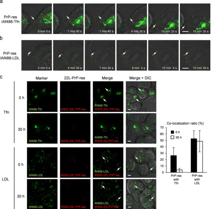
Time-lapse imaging showed that within 2 h after the initiation of incubation with PrP-res, a particle of Af555-22-L-PrP-res on the cell surface was internalized and merged with Af488-Tfn signals (Fig. 3a, between 1 min 40 s and 4 min 30 s) during transport to a peri-nuclear region, and thereafter, Af555-PrP-res and Af488-Tfn moved together back to a peripheral region of the cell (e.g., 10 min 20 s). We also observed internalization of an Af555-22-L-PrP-res particle from the cell surface that subsequently merged with an Af488-LDL-positive vesicle (Fig. 3b, between 7 min 50 s and 9 min 5 s), and thereafter, Af555-PrP-res and Af488-LDL moved together (e.g., 10 min 5 s).
To clarify the trafficking pathway of the inoculated PrP-res, cells incubated with Af555-22-L-PrP-res for 6 h were subsequently cultured for up to 30 h. Six hours after the incubation with PrP- res, some of the Af555-22-L-PrP-res signals co-localized with the Af488-Tfn signal not at the center of the cluster of the Af488-Tfn signal, but at a region around the cluster (Fig. 3c, Tfn, 0 h). On the other hand, a large portion of the Af555-22-L-PrP-res appeared to co-localize with Af488-LDL throughout the cells (Fig. 3c, LDL, 0 h).
Quantitative analysis of the co-localization revealed that Af555- 22-L-PrP-res co-localized more with Af488-LDL (53%) than AF488- Tfn (26%). The co-localization of Af555-22-L-PrP-res with Af488- LDL was still obvious (49%) 30 h after the inoculation, whereas, the co-localization of Af555-22-L-PrP-res with Af488-Tfn had appar- ently decreased (4%) (Fig. 3c, LDL and Tfn, 30 h). These results suggested that internalized PrP-res entered both the endocytic- recycling pathway and the endo-lysosomal pathway immediately following internalization, but a large portion of the inoculated PrP- res was eventually directed to the endo-lysosomal pathway.
The kinetics of inoculated PrP-res metabolism and generation of PrPSc
To determine the fate of the inoculated PrP-res and detectde novogeneration of PrPSc, the PrP-res level in cells inoculated with Af488-22-L-PrP-res was analyzed with immunoblotting. The level of inoculated Af488-22-L-PrP-res, which was detected with an anti-Alexa Fluor 488 antibody, was drastically decreased within 24 h post inoculation (hpi) (Fig. 4a). In contrast, the total PrP-res level, which was detected with an anti-PrP antibody reactive to both the inoculated PrP-res and newly generated PrPSc, was unchanged at 24 hpi but increased thereafter (Fig. 4a). The decrease in the inoculated PrP-res was also consistent with the decrease in the PrP-res signal observed by live-cell imaging of Af555-22-L-PrP-res over the same time period (Fig. 4b). The increase of the mono-glycosylated PrP-res on immunoblots of total PrP-res at 24 hpi suggested that thede novogeneration of PrPSctakes place within 24 hpi (Fig. 4a, arrowhead). Therefore, we analyzed the inoculated PrP-res and newly generated PrPScsimul- taneously in individual cells by IFA. We used Af555-22-L-PrP-res in combination with mAb 132-mediated specific detection of PrPScto distinguishde novoPrPSc, which could be detected only with mAb 132 (Fig. 4c, arrows), from exogenous PrP-res, which was labeled with both Alexa Fluor 555 and mAb 132 (Fig. 4c, arrowheads).
Signals from newly generated PrPScbecame detectable at 24 hpi, especially at a peri-nuclear region of the cells (arrows), and the
number of newly generated PrPSc granules and theirfluorescent intensities increased thereafter. In contrast to the increase of newly generated PrPSc, the inoculated PrP-res decreased with time (Fig. 4c, arrowheads).
Intracellular localization of inoculated PrP-res and newly generated PrPSc
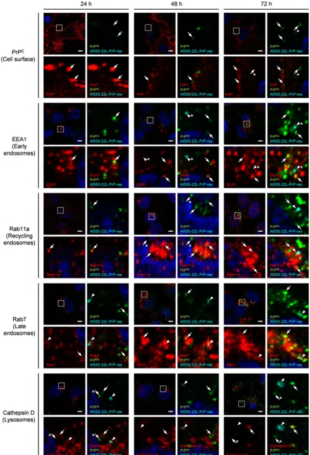
To identify the site of de novo generation of PrPSc, we next analyzed the intracellular localization of inoculated Af555-22-L- PrP-res and newly generated PrPSc with markers for endocytic compartments. At 24 hpi, somefluorescent signals from the newly generated PrPSc co-localized well with EEA1 (early endosomes)
and Rab7 (late endosomes), but were poorly co-localized with Rab11a (recycling endosomes) or cathepsin D (lysosomes) (Fig. 5).
Furthermore, faint signals from newly generated PrPSc could be detected at the cell surface at 24 hpi. The intensities of these PrPSc signals increased with time after inoculation (Fig. 5, from 24 to 72 hpi). In order to clarify the kinetics of distribution of the inoculated PrP-res and newly generated PrPSc, we quantitatively analyzed co-localization ratios of the inoculated PrP-res and newly generated PrPScco-localized with markers (Fig. 6). Consistent with the data from the live-cell imaging (Fig. 3), a large proportion of the inoculated PrP-res localized to late endosomes (40%) while a smaller portion was detected at the cell surface (22%) at 0 hpi.
The exogenous PrP-res at the cell surface almost disappeared Fig. 2.Intracellular localization offluorescent-dye-labeled, purified-PrP-res. N2a-3 cells grown on chambered coverglass were transfected with expression plasmids of EGFP- tagged wild-type of Rab4a, 5a, 7, 9, 11a or 22a. Medium (200μl) containing 10 ng of Af555-22-L-PrP-res was added to cells 72 h after transfection. Time series of images were acquired any of 15 min-duration (images were acquired every 5 s within 2 h after the initiation of incubation with Af555-22-L-PrP-res. Each image in a row shows a merged image of Af555-22-L-PrP-res (red) and EGFP-Rab GTPase (green) with differential interference contrast (DIC) at the indicated time point. Arrows indicate examples of Af555- 22-L-PrP-res that became co-localized with EGFP-Rab GTPases during the period of observation. Scale bar: 5μm.
during the subsequent 48 h and the remaining PrP-res localized to late endosomes (from 16% to 14%) and lysosomes (from 11% to 9%).
In contrast, at 24 hpi newly generated PrPSclocalized mainly at the cell surface (27%), in early endosomes (15%), and late endosomes (30%), but a minor portion localized to lysosomes (3%). Interest- ingly, the proportion of newly generated PrPSc localized to early endosomes or recycling endosomes increased (from 15% to 30%
and from 5% to 33%, respectively); in contrast, the amount of PrPSc at the cell surface decreased from 27% to 3% with time after inoculation. These data suggested that even though newly generated PrPSc was detected in late endosomes and lysosomes at 24 hpi, newly generated PrPSc remarkably appeared in the intracellular organelles on the endocytic-recycling pathway thereafter.
Fig. 3.Co-localization of Af555-22-L-PrP-res with Tfn or LDL. Time-lapse imaging of inoculated PrP-res and Af488-Tfn (a) or Af488-LDL (b). N2a-3 cells grown on chambered coverglass were incubated with Af555-22-L-PrP-res (red) and 10μg/ml of Af488-Tfn (green) as a marker of the endocytic-recycling pathway (a) or 4μg/ml of Af488-LDL (green) as a marker of the endo-lysosomal pathway (b). Time-lapse images were acquired any of 15 min-duration (images were acquired every 5 s) within 2 h after the initiation of incubation with PrP-res. Each image within a row shows a merged image of Af555-22-L-PrP-res (red) and Af488-Tfn or Af488-LDL (green) with the DIC at the indicated time point. Arrows indicate examples of Af555-22-L-PrP-res that became co-localized with Af488-Tfn (a) or Af488-LDL (b) in the period of observation. (c) Co- localization of Af555-22-L-PrP-res with Af488-Tfn or Af488-LDL. N2a-3 cells were incubated with Af555-22-L-PrP-res and Af488-Tfn or Af488-LDL for 6 h at 371C. After the removal of excess Af555-22-L-PrP-res, the cells were subjected to live-cell imaging (0 h). To monitor co-localization of PrP-res one day after inoculation, N2a-3 cells were incubated with Af555-22-L-PrP-res for 6 h. After the removal of excess Af555-22-L-PrP-res, the cells were further incubated for 24 h at 371C. The cells were then incubated for an additional 6 h in the presence of Af488-Tfn or Af488-LDL before imaging (30 h). The right-most column shows the merged images of Af488-Tfn (green) or Af488-LDL (green), Af555-22-L-PrP-res (red), and DIC. Arrows indicate examples of Af555-22-L-PrP-res co-localized with Af488-Tfn or Af488-LDL. Scale bar: 5μm. The graph on the right shows ratios of Af555-22-L-PrP-res signals co-localized with Af488-Tfn or Af488-LDL signals relative to sum of the Af555-22-L-PrP-res signals (methods for the calculation of the co-localization ratio are described in the Supplementary information). Mean and SD of the values acquired from 15fields of view are depicted. Total numbers of foci and cells used for co-localization statistics from the 15 viewfields were listed inTable S4.
Influence of the impairment of intracellular transport on de novo generation of PrPSc
To determine which pathway of intracellular transport is involved in de novogeneration of PrPSc, we analyzed the levels of newly generated PrPSc in cells in which trafficking between the endocytic compartments was selectively impaired by the
overexpression of wild-type or dominant-negative mutants of Rab GTPase proteins (Fig. 7). The impairment of the endocytic- recycling pathway by overexpression of wild-type Rab22a or a dominant-negative mutant of Rab11a, which are known to affect the transport from early endosomes to recycling endosomes (Magadan et al., 2006) or the transport from recycling endosomes to plasma membrane (Ren et al., 1998), respectively, reduced the Fig. 4.Kinetics of inoculated PrP-res andde novogeneration of PrPSc. (a) The levels of inoculated PrP-res and total PrP-res. N2a-3 cells grown on 12-well plates were inoculated with Af488-22-L-PrP-res. After the inoculation, the cells were cultured for the indicated period and then subjected to immunoblotting. As a control for detection of PrP-res, mock-infected cells were prepared (M). PK-untreated samples equivalent to 100μg of total protein per lane were loaded to detect Af488-22-L-PrP-res with anti- Alexa Fluor 488 antibody (Alexa), while PK-digested samples equivalent to 100μg of total protein were also loaded to monitor total PrP-res, which contained inoculated PrP- res and newly generated PrP-res with anti-PrP mAb 31C6 (PrP-res).β-actin was used as an internal control. Bands corresponding to monomeric and dimeric PrPSc(indicated by the square bracket) were quantified. The arrowhead indicates the mono-glycosylated form of PrP-res. The graph on the right shows the result of a quantitative analysis.
Black bars indicate the inoculated PrP-res levels relative to that at 0 h and gray line indicates the total PrP-res levels relative to that at 0 h. Mean and SD of 3 independent experiments are shown. (b) Live-cell image of Af555-22-L-PrP-res. N2a-3 cells grown on chambered coverglass were inoculated with Af555-22-L-PrP-res and then cultured for the indicated time. Merged images containing Af555-22-L-PrP-res (red) and DIC are shown. (c) Discrimination of newly generated PrPScfrom inoculated PrP-res in a single cell. The top panel shows the merged images of signals of Af555-22-L-PrP-res (red), PrPSc(green) and DAPI-stained nuclei (blue). The bottom panel shows the corresponding high-magnification images of the boxed region. Arrowheads indicate examples of inoculated PrP-res that was detected via both Alexa Fluor 555 (red, directly coupled to purified 22-L-PrP-res) and mAb 132 (green, indirect immuno-staining with Alexa Fluor 488-conjugated secondary antibody). As a result, inoculated PrP-res can be detected as yellow. Arrows indicate examples of newly generated PrPScthat was detected only with mAb 132 (green). Scale bar: 5μm.
Fig. 5.Kinetics of the intracellular localization of inoculated PrP-res and newly generated PrPSc. N2a-3 cells inoculated with Af555-22-L-PrP-res were cultured for the indicated time and subjected to double-staining of PrPScwith mAb 132 and an organelle marker molecule as indicated. For staining of PrPCat cell surface, cells were incubated with B103 antiserum prior to thefixation for PrPSc-specific detection. Alexa Fluor 647-conjugated secondary antibody was used to stain the marker molecules. The cell nuclei were counterstained with DAPI. The upper left image in each panel consists of four images shows a lower magnification view of a merged image of organelle marker molecule (red), PrPSc(green), Af555-22-L-PrP-res (cyan), and nuclei (blue). The other three images are the corresponding high-magnification image of the boxed region for the merged image of organelle marker and nuclei (bottom left), for the merged image of PrPSc, Af555-22-L-PrP-res and nuclei (upper right), and for the merged images of the organelle marker, PrPSc, Af555-22-L-PrP-res and nuclei (bottom right). Arrowheads indicate representative co-localization of Af555-22-L-PrP-res with organelle marker molecules, while arrows indicate
Sc
the control (Fig. 7). Additionally, the impairment of the endo- lysosomal pathway by the overexpression of a dominant-negative mutant of Rab7, which is known to affect the transport from early endosomes to late endosomes and/or lysosomes (Bucci et al., 2000; Feng et al., 1995), also reduced the amount of de novo generated PrPScat 48 hpi by 60% of the control (Fig. 7). A wild-type Rab9 is reported to be involved in the transport from late endosomes to TGN (Riederer et al., 1994). The overexpression of wild-type Rab9 reduced the amount ofde novogenerated PrPScby 71% of the control; however, it was not clear which intracellular transport pathway was actually influenced (Fig. 7). The level of endogenous PrPCin cells and cell-surface expression of PrPCwere not altered by overexpression of wild-type Rab9, wild-type Rab22a, a dominant-negative Rab7 mutant, or a dominant- negative Rab11a (Supplementary Fig. 1). Taken together, these results indicated that the inhibition ofde novogeneration of PrPSc was not caused by a change in the expression of PrPC. These data suggested that intracellular transport along the endocytic- recycling pathway as well as the endo-lysosomal pathway is involved in thede novo generation of PrPScafter the inoculation of PrP-res.
Discussion
The lack of a method that can distinguish newly generated PrPScfrom endogenous PrPCand from inoculum-derived PrPScwas one of the obstacles to the investigation of the cellular events that mediate thede novogeneration of PrPScin the early stage of prion infection. Therefore, the analysis of intracellular dynamics of PrPSc just after inoculation of prions has been limited to inoculum- derived PrPSc as descried previously (Jen et al., 2010; Magalhaes et al., 2005). Here, we solved this problem with a combination of the fluorescent-dye-labeled purified PrP-res and PrPSc-specific staining with mAb 132. This technique allowed us to distinguish
Fig. 7.Effect of overexpression of Rab GTPases on thede novogeneration of PrPSc. N2a-3 cells grown in a 24-well plate were inoculated with unlabeled 22-L-PrP-res and were incubated for 6 h. After washing the cells, an expression plasmid encoding an EGFP-tagged wild-type (Wt) Rab GTPase, Rab4a, Rab5a, Rab7, Rab9, Rab11a or Rab22a, or an EGFP-tagged dominant-negative mutant (DN) of a Rab GTPases, Rab4aS22N (Roberts et al., 2001), Rab5aS34N (Stenmark et al., 1994), Rab7T22N (Feng et al., 1995), Rab9S21N (Riederer et al., 1994), Rab11aS25N (Ren et al., 1998) or Rab22aS19N (Weigert et al., 2004), was introduced. The expression vector pEGFP-C1 (Clontech) was used as a control. At 48 h after transfection, the cells were processed for dot-blotting to monitor PrP-res. A representative image of a dot-blot is shown on the top, and the graph below shows the levels of PrP-res relative to that of control plasmid-transfected cells (EGFP). Mean and SD of 4 independent experiments are depicted. Asterisks indicate a significant decrease compared to the control (Student'st-test, po0.05).
Fig. 6.Co-localization statistics. Co-localization analysis of the images shown in Fig. 5was carried out as described in the Supplementary materials. (a) Ratio of the Af555-22-L-PrP-res signals co-localized with each marker to the sum of the Af555- 22-L-PrP-res signals. (b) Ratio of the newly generated PrPScsignals co-localized with each marker to the sum of newly generated PrPScsignals. Mean and SD of the value acquired in 5fields of view are shown. Total numbers of foci and cells used for co-localization statistics from the 5 viewfields were listed inTable S5.
newly generated PrPSc from the inoculated PrP-res within individual cells.
Considering that the inoculated PrP-res was mainly detected in late endosomes and lysosomes (Figs. 5 and 6) and that its levels decreased from 24 to 72 h after the inoculation (Fig. 4), the PrP-res directed to the endo-lysosomal pathway appeared to be degraded in late endosomes and/or lysosomes. Previous studies showed that inoculated PrP-res is transported to late endosomes/lysosomes after being taken up by SN56 cells or primary sensory neurons (Jen et al., 2010; Magalhaes et al., 2005). Purified PrP-res of the Obihiro strain, which cannot establish a persistent infection in N2a-3 cells, was also transported to late endosomes and lysosomes; the intracellular distribution of Alexa Fluor-labeled Obihiro strain- derived PrP-res was indistinguishable from that of the 22-L strain-derived PrP-res and the inoculated PrP-res of Obihiro strain was degraded similarly to 22-L strain-derived PrP-res (data not shown). Amyloidfibrils of the A
β
1-42 peptide were also reported to be transported via the endo-lysosomal pathway in SN56 cells (Magalhaes et al., 2005). Taken together, the trafficking of PrP-res via the endo-lysosomal pathway appears to be a general pathway for degradation of exogenously introduced macromolecules, rather than a pathway specific to PrPSc and the propagation of prions (Saftig and Klumperman, 2009).Recently, Goold et al. reported that the plasma membrane is one of the sites for conversion based on the results from experi- ments using PK-1 cells that express PrPCtagged with Myc-epitope at the C-terminus (Goold et al., 2011). In this report, PrPSc was primarily generated at the plasma membrane within 2 min after prion challenge. Contrary to this report, we found no evidence that de novogeneration of PrPSc primarily occurred at plasma mem- brane (Figs. 4–6). This discrepancy may be due to multiple factors, one of which includes clonal differences between these cell lines.
For example, the former study showed that newly generated PrPSc attained steady levels by 2 h after inoculation of prions and approximately 20% of PK-1 cells in the cultures became PrPSc- positive. However, in the N2a-3 cells used in our study, the levels of newly generated PrPScin the cells increased at least up to 72 hpi (Fig. 4), and most of the cells in the culture eventually became PrPSc-positive (data not shown). In addition, Goold et al. reported that PrPScwas detected at the cell membrane of prion-infected PK- 1 cells; however, PrPSc was only weakly detected at the plasma membrane of N2a-3 cells persistently infected with prions, even when formic acid pre-treatment that was used for PrPSc-specific detection by Goold et al. was employed (data not shown). Finally, the differences between the two studies might be accounted for the different inocula (purified PrP-res vs. brain homogenate) used to initiate infection.
Most PrPCis known to cycle between a peri-nuclear region of the cell and the plasma membrane via the endocytic-recycling pathway after being trafficked to the cell surface, but some portion of PrPCis also delivered to late endosomes and lysosomes (Morris et al., 2006; Peters et al., 2003; Shyng et al., 1993). Considering that the newly generated PrPScappeared in late endosomes, but was rarely observed in lysosomes at 24 hpi, at which time a large portion of the inoculated PrP-res was localized in late endosomes (Figs. 5 and 6), the initial conversion of PrPCto PrPScmay occur in late endosomes, at least when purified PrP-res is used as an inoculum. This idea is consistent with thefinding that overexpres- sion of the dominant-negative mutant of Rab7, which inhibits transport from early endosomes to late endosomes and/or lyso- somes (Bucci et al., 2000; Feng et al., 1995), partly inhibited the generation of PrPScafter the inoculation of PrP-res (reduced to 60%
of the control, Fig. 7). This finding raises the possibility that although most of the inoculated PrP-res was transported to and degraded in late endosomes/lysosomes, smaller PrP-res oligomers might be generated in late endosomes during the degradation
process. Such smaller PrP-res oligomers may initiate PrPCconver- sion because smaller oligomers have greater seeding activity for the conversion of PrPCand higher infectivity than larger PrP-res aggregates (Silveira et al., 2005).
Earlier studies suggested that in cells persistently infected with prions, PrPScis formed either on the plasma membrane or during endocytic trafficking (Borchelt et al., 1992; Caughey and Raymond, 1991; Taraboulos et al., 1992). In later years, Béranger et al. and Marijanovic et al. reported that overexpression of dominant- negative mutants of Rab4a or Rab11a, which are known to impair the transport from early endosomes to the plasma membrane (Roberts et al., 2001) or from recycling endosomes to the plasma membrane (Ren et al., 1998), respectively, raised the PrPSclevel (Beranger et al., 2002; Marijanovic et al., 2009). On the other hand, overexpression of wild-type Rab22a, which inhibits the transport from early endosomes to recycling endosomes (Magadan et al., 2006), reduced the PrPSclevel in cells persistently infected with prions (Marijanovic et al., 2009). Based on thesefindings, one of the sites for PrPSc formation in cells persistently infected with prions is thought to be in the transport pathway from early endosomes to recycling endosomes. Also in the early stage of prion infection, we confirmed the inhibition ofde novogeneration of PrPScby overexpression of a wild-type Rab22a (reduced to 69%
of the control,Fig. 7), consistent with the results of Marijanovic et al. in persistently prion-infected cells (Marijanovic et al., 2009).
However, unlike the findings in cells persistently infected with prions, the generation of PrPScafter the inoculation of PrP-res was partly inhibited by overexpression of a dominant-negative Rab11a mutant (reduced to 62% of the control,Fig. 7). These observations suggested that the initiation of PrPScgeneration shortly after PrP- res inoculation required the recycling pathway between recycling endosomes and the plasma membrane. Further studies will be required to explain the apparent inconsistency between these results. However, the newly generated PrPSc appeared at the plasma membrane and in early endosomes where the inoculated PrP-res was rarely detected at 24 hpi (Figs. 5 and 6), suggesting the involvement of the recycling pathway; therefore, either the exogenous PrP-res degraded to an undetectable level or PrPSc newly generated in late endosomes was recycled back to the plasma membrane and acted as a“seed”for conversion. Moreover, the marked increase in the newly generated PrPSc at early and recycling endosomes during the following 48 h (Figs. 5 and 6) suggested that efficient generation of PrPScoccurred once the PrPSc was transferred to the endocytic-recycling pathway.
The results in this study suggest that the transfer of the inoculated PrP-res and/or newly generated PrPScfrom the endo- lysosomal pathway to the endocytic-recycling pathway is impor- tant for efficient PrPSc formation after prion inoculation. One possible route of such transfer is the trafficking from the late endosomes to the plasma membrane through the TGN, a route by which cation-independent mannose-6-phosphate receptor (CIMPR) is transported (Maxfield and McGraw, 2004). CIMPR delivers hydrolase precursors from the Golgi apparatus to the endosomes and releases hydrolases into compartments in the process of late endosome formation; CIMPR is then recycled back from late endosomes to the TGN by retrograde transport. CIMPR in the TGN is also delivered to the plasma membrane (Ghosh et al., 2003). The overexpression of Rab9, which is involved in this retrograde transport of CIMPR (Riederer et al., 1994), was reported to decrease the levels of PrPScin cells persistently infected with prions (Gilch et al., 2009) as well as inhibit the generation of PrPSc after prion inoculation (this study), suggesting that the trafficking from late endosomes to the TGN may be involved in the genera- tion of PrPSc.
Direct transport from late endosomes to the plasma membrane might be an alternative route. This atypical transport was reported
ibility complex (MHC) and CD1 family molecules in antigen- presenting cells (Gelin et al., 2009; Neefjes et al., 2011). Further- more, MHC class II molecules are also known to be transported in the process of exosomes release (Berger and Roche, 2009; Von Bartheld and Altick, 2011). Considering the fact that PrPCis present on the membranes of both multivesicular bodies and intraluminal vesicles and that PrPScas well as PrPCare released from cells with exosomes (Fevrier et al., 2004), the inoculated PrP-res and/or newly generated PrPSc in late endosomes may be recycled back to the plasma membrane through multivesicular bodies via a pathway similar to the MHC class II molecules. This idea is consistent with thefinding thatde novogeneration of PrPScwas inhibited by the overexpression of the dominant-negative Rab11a mutant, which also inhibits the release of exosomes (Savina et al., 2002). The PrPSc recycled to the plasma membrane via these mechanisms may, in turn, contribute to de novo generation of PrPScin the compartments on the endocytic-recycling pathway.
The intracellular dynamics of PrPScin CNS neurons in the early stages after prion infection is largely unknown. It was reported that PrPSccould be detected in endosomal and lysosomal fractions prior to the detection of PrPScin the plasma membrane fraction after intracerebral inoculation of prions (Dearmond and Bajsarowicz, 2010), and that protease-sensitive PrPSc could be detected in early and recycling endosomes in neurons in the hippocampus during the preclinical stage of infected mice (Godsave et al., 2008). These facts suggest that there are simila- rities in prion propagation in neuroblastoma cells and neurons in CNS.
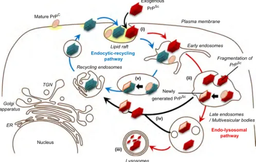
In this study, we showed the intracellular dynamics of inocu- lated PrP-res in prion-susceptible neuroblastoma cells. Our data suggest that transfer of inoculated PrP-res from the endo- lysosomal pathway to the endocytic-recycling pathway is involved in the initiation of efficientde novogeneration of PrPScin the early stage of infection (Fig. 8). However, further analyses are required for understanding the mechanisms of prion propagation in neu- rons in CNS. Experiments are underway to clarify the intracellular
staining with mAb 132.
Materials and methods
Antibodies, expression plasmids, and reagents
Anti-PrP mouse mAbs 31C6 and 132 and a rabbit polyclonal antibody B103 were used (Horiuchi et al., 1995; Kim et al., 2004).
The other commercially available primary and secondary antibo- dies that were used for immunoblotting and IFA are listed inTable S1. Alexa Fluor 488- and 555-conjugated Tfn and Alexa Fluor 488- conjugated LDL (Life Technologies) were used as markers for the endocytic pathway. Expression plasmids encoding EGFP-tagged Rab GTPases were prepared as described in Supplemental materi- als according to methods described elsewhere (Table S2 and S3) (Fukuda, 2003).
Cell culture
N2a-3 cells, a subclone of the mouse neuroblastoma cell line Neuro2a, were cultured as described previously (Uryu et al., 2007).
Purification of PrP-res
PrP-res was prepared from detergent-resistant membranes as described previously (Baron et al., 2011) with slight modifications (Supplementary materials). The purification procedure included a PK treatment, so from this point forward we use the term “PrP- res”to indicate the purified, PK-treated PrPSc.
Fluorescent-dye-labeling of PrP-res
PrP-res in PBS (10
μ
g in 50μ
l) containing 0.5% Zwittergent 3-14 was sonicated with a Cross Ultrasonic Protein Auto Activating Instrument, ELSTEIN NP070-GOT (Nepa Gene), by 4 cycles ofExogenous PrPSc Mature PrPC
Plasma membrane
Early endosomes
Fragmentation of PrPSc
Late endosomes / Multivesicular bodies
Endo-lysosomal pathway Golgi
ER
Nucleus
Lysosomes apparatus
Recycling endosomes
TGN
Newly generated PrPSc Lipid raft
Endocytic-recycling pathway
(i)
(ii) (v)
(iv)
(iii)
Fig. 8.Summary of intracellular dynamics of inoculated PrPScandde novogeneration of PrPScin early stage of prion infection. Exogenously introduced PrPSc(red parallelograms) is internalized into a cell (i). Although inoculated PrPScis transported throughout endocytic compartment immediately after internalization, most of the inoculated PrPSceventually directed to endo-lysosomal pathway (indicated as red arrows) and is delivered to lysosomes and degraded (iii). Conversion of PrPC(pink ellipse) to PrPSc(pink parallelogram) may be initiated by fragmented inoculated PrPSc(a smaller PrPScoligomer) that is generated during the transport on the endo-lysosomal pathway (ii). A smaller PrPScoligomer generated by fragmentation of inoculated PrPScand/or newly generated PrPScis transferred from the endo-lysosomal pathway to the endocytic-recycling pathway (iv). Once PrPScthat is capable of inducing conversion (blue parallelograms) is transferred to endocytic-recycling pathway (indicated as blue arrows) initiates efficient PrPScformation that leads to the establishment of prion infection. The sites where efficient conversion occurs are thought to be early endosomes and recycling endosomes (v).
15 min-sonication followed by 5 min-incubation at 41C prior to fluorescent-dye labeling. The PrP-res was then mixed with 200
μ
gof Alexa Fluor 488 succinimidyl ester or Alexa Fluor 555 succini- midyl ester (Life Technologies) dissolved in 5
μ
l of anhydrous dimethyl sulfoxide (Sigma). Fluorescent-dye labeling was per- formed under 16 cycles of 15 min-sonication followed by 30 min-incubation at 41C. To quench the excess reactive dye, 3 ml of 50 mM glycine in PBS was mixed with the PrP-res and the sample was centrifuged at 45,000 rpm for 30 min at 41C in a S80AT3 rotor (Hitachi). The pellet was washed twice with 50 mM glycine in PBS and each wash was followed by centrifugation; the final pellet was resuspended in 100μ
l of PBS with sonication.The fluorescent-dye-labeled PrP-res or unlabeled PrP-res was subjected to SDS-PAGE followed by fluorescent imaging with Typhoon FLA 9000 (GE Healthcare) or by silver-staining with 2D-SILVER STAIN-II (Cosmo bio CO.), respectively.
Inoculation of PrP-res
Purified PrP-res orfluorescent-dye-labeled PrP-res was diluted with Opti-MEM (Life technologies), sonicated with an ELSTEIN NP070-GOT sonicator for 5 min, and centrifuged at 10,000gfor 10 min to remove large PrP-res aggregates. The supernatant was diluted with Opti-MEM at 10 ng PrP-res/200
μ
l or 20 ng PrP-res/250
μ
l or 35 ng PrP-res/500μ
l for use on 8-well Lab-Tek II chambered coverglass (Thermo Scientific) or 24- or 12-well plates, respectively. The culture medium for the N2a-3 cells grown on a chambered coverglass or in the multi-well plates was replaced with Opti-MEM containing PrP-res, and the cells were then incubated for 2 or 6 h at 371C. After incubation, the cells were washed three times with pre-warmed PBS and then cultured in Opti-MEM containing 10% fetal bovine serum (FBS), 1% non- essential amino acids (NEAA; Gibco), and 1X penicillin- streptomycin solution (100 U/ml–100μ
g/ml, PS; Gibco).Immunofluorescence assay (IFA)
IFA, including PrPSc-specific staining, was carried out as described previously (Yamasaki et al., 2012) with some modifica- tions. Cells were grown on an 8-well Lab-Tek II chambered cover- glass and all the staining procedures were carried out without the removal of the media chamber. For double staining of cell surface PrPCand PrPSc, living cells were incubated with B103 (5
μ
g/ml)in Opti-MEM for 15 min at 371C. The cells were then immediately fixed with pre-warmed 4% paraformaldehyde with 4% sucrose in PBS for 10 min and blocked with 5% FBS in PBS. Cells were then incubated with secondary antibody, and the cells werefixed again with 4% paraformaldehyde in PBS for 10 min and were subjected to the PrPSc-specific staining. To counterstain cell nuclei, cells were incubated with 5
μ
g/ml of 4', 6-diamidino-2-phenylindole, dilac- tate (DAPI; Invitrogen) in PBS at room temperature (rt) for 30 min.Finally, the media chamber was filled with PBS and confocal fluorescent images were acquired with a 63objective lens on a Zeiss LSM700 inverted microscope and the ZEN 2009 software.
Z-series of the images were taken at every 0.8
μ
m steps from the top to bottom of the cells in the area.Transfection
N2a-3 cells seeded onto 8-well chambered coverglass or 24- well plates at 1:5 ratio were cultured in Dulbecco's modified Eagle's medium (DMEM; ICN Biomedicals) for 2 days prior to the inoculation of PrP-res and transfection. The cells were transfected with 2.8% Lipofectamine 2000 (Invitrogen) and 2
μ
g/ml expression plasmid in 250μ
l (8-well chamber) or 500μ
l (24-well plate) of Opti-MEM that contained 8% FBS, 0.8% NEAA, and 0.8X PS. Aftertransfection (24 h), the medium was replaced with fresh Opti- MEM containing 10% FBS, 1% NEAA and 1X PS and cultured until used for immunoblots or IFA.
Immunoblotting and dot-blotting
Immunoblotting and dot-blotting were performed to monitor PrP and other molecules as described elsewhere (Nakamitsu et al., 2010; Uryu et al., 2007). To monitor PrP-res via immunoblotting, the protein concentration of cell lysate was adjusted to 1 mg/ml and the samples were then treated with 1
μ
g/ml of PK for 20 min at 371C. Meanwhile, to monitor Alexa Fluor 488-labeled PrP-res, PK treatment was omitted. The cell lysates were incubated with 50μ
g/ml of DNase I (Roche) for 15 min at rt. Proteins were concentrated by incubating the samples with 0.3% phosphotungs- tic acid for 20 min at rt; this incubation was followed by centri- fugation at 20,500gfor 20 min at 41C.To monitor PrP-res via dot-blotting, cell lysate equivalent to 40
μ
g of total protein per well was transferred onto a polyvinyli- dene difluoride (PVDF) membrane by a dot-blotter (Bio-Rad). The PVDF membrane was treated with 20μ
g/ml of PK for 1 h at 371C and then incubated with 1 mM Pefabloc SC for 15 min at 41C. The membrane was treated with 50μ
g/ml of DNase I for 15 min at rt and then incubated in 3 M GdnSCN for 30 min at rt. Samples on each membrane were then subjected to immuno-detection with mAb 31C6 and HRP-conjugated secondary antibody. ECL Western Blotting Detection Reagents (GE Healthcare) and a LAS-3000 chemiluminescence image analyzer (Fuji Film) were used to visualize the immunoreactive proteins.Acknowledgments
T.Y. is supported by a Grant-in-Aid for JSPS Fellows (No.
224181). This work was supported by a Grant-in-Aid for Science Research (A) (Grant no. 23248050), a Grant from the Global COE Program (F-001) and the Program for Leading Graduate Schools (F01), and the Japan Initiative for Global Research Network on Infectious Diseases (J-GRID), from the Ministry of Education, Culture, Sports, Science, and Technology, Japan. This work was also supported by grants for TSE research (H23-Shokuhin-Ippan- 005) and Research on Measures for Intractable Diseases from the Ministry of Health, Labour and Welfare of Japan and in part by the Intramural Research Program, National Institute of Allergy and Infectious Diseases, National Institutes of Health. We also thank Zensho Co. Ltd. for BSL3 facility.
Appendix A. Supporting information
Supplementary data associated with this article can be found in the online version athttp://dx.doi.org/10.1016/j.virol.2013.11.007.
References
Baron, G.S., Hughson, A.G., Raymond, G.J., Offerdahl, D.K., Barton, K.A., Raymond, L.D., Dorward, D.W., Caughey, B., 2011. Effect of glycans and the glycophosphatidylinositol anchor on strain dependent conformations of scrapie prion protein: improved purifications and infrared spectra. Biochemistry 50, 4479–4490.
Beranger, F., Mange, A., Goud, B., Lehmann, S., 2002. Stimulation of PrP (C) retrograde transport toward the endoplasmic reticulum increases accumu- lation of PrP(Sc) in prion-infected cells. J. Biol. Chem. 277, 38972–38977.
Berger, A.C., Roche, P.A., 2009. MHC class II transport at a glance. J. Cell Sci. 122, 1–4.
Borchelt, D.R., Taraboulos, A., Prusiner, S.B., 1992. Evidence for synthesis of scrapie prion proteins in the endocytic pathway. J. Biol. Chem. 267, 16188–16199.
Bucci, C., Thomsen, P., Nicoziani, P., McCarthy, J., van Deurs, B., 2000. Rab7: a key to lysosome biogenesis. Mol. Biol. Cell 11, 467–480.
cell surface precursor that is both protease- and phospholipase-sensitive. J. Biol.
Chem. 266, 18217–18223.
Dearmond, S.J., Bajsarowicz, K., 2010. PrPSc accumulation in neuronal plasma membranes links Notch-1 activation to dendritic degeneration in prion diseases. Mol. Neurodegener 5, 6.
Feng, Y., Press, B., Wandinger-Ness, A., 1995. Rab 7: an important regulator of late endocytic membrane traffic. J. Cell Biol. 131, 1435–1452.
Fevrier, B., Vilette, D., Archer, F., Loew, D., Faigle, W., Vidal, M., Laude, H., Raposo, G., 2004. Cells release prions in association with exosomes. Proc. Natl. Acad. Sci.
U.S.A 101, 9683–9688.
Fukuda, M., 2003. Distinct Rab binding specificity of Rim1, Rim2, rabphilin, and Noc2. Identification of a critical determinant of Rab3A/Rab27A recognition by Rim2. J. Biol. Chem. 278, 15373–15380.
Gauczynski, S., Nikles, D., El-Gogo, S., Papy-Garcia, D., Rey, C., Alban, S., Barritault, D., Lasmezas, C.I., Weiss, S., 2006. The 37-kDa/67-kDa laminin receptor acts as a receptor for infectious prions and is inhibited by polysulfated glycanes. J. Infect.
Dis. 194, 702–709.
Gelin, C., Sloma, I., Charron, D., Mooney, N., 2009. Regulation of MHC II and CD1 antigen presentation: from ubiquity to security. J. Leukoc. Biol. 85, 215–224.
Ghosh, P., Dahms, N.M., Kornfeld, S., 2003. Mannose 6-phosphate receptors: new twists in the tale. Nat. Rev. Mol. Cell Biol. 4, 202–212.
Gilch, S., Bach, C., Lutzny, G., Vorberg, I., Schatzl, H.M., 2009. Inhibition of cholesterol recycling impairs cellular PrP(Sc) propagation. Cell. Mol. Life Sci.
66, 3979–3991.
Godsave, S.F., Wille, H., Kujala, P., Latawiec, D., DeArmond, S.J., Serban, A., Prusiner, S.B., Peters, P.J., 2008. Cryo-immunogold electron microscopy for prions:
toward identification of a conversion site. J. Neurosci. 28, 12489–12499.
Goold, R., Rabbanian, S., Sutton, L., Andre, R., Arora, P., Moonga, J., Clarke, A.R., Schiavo, G., Jat, P., Collinge, J., Tabrizi, S.J., 2011. Rapid cell-surface prion protein conversion revealed using a novel cell system. Nat. Commun. 2, 281.
Greil, C.S., Vorberg, I.M., Ward, A.E., Meade-White, K.D., Harris, D.A., Priola, S.A., 2008. Acute cellular uptake of abnormal prion protein is cell type and scrapie- strain independent. Virology 379, 284–293.
Hijazi, N., Kariv-Inbal, Z., Gasset, M., Gabizon, R., 2005. PrPSc incorporation to cells requires endogenous glycosaminoglycan expression. J. Biol. Chem. 280, 17057–17061.
Horiuchi, M., Yamazaki, N., Ikeda, T., Ishiguro, N., Shinagawa, M., 1995. A cellular form of prion protein (PrPC) exists in many non-neuronal tissues of sheep.
J. Gen. Virol. 76 (Pt 10), 2583–2587.
Horonchik, L., Tzaban, S., Ben-Zaken, O., Yedidia, Y., Rouvinski, A., Papy-Garcia, D., Barritault, D., Vlodavsky, I., Taraboulos, A., 2005. Heparan sulfate is a cellular receptor for purified infectious prions. J. Biol. Chem. 280, 17062–17067.
Ikonen, E., 2008. Cellular cholesterol trafficking and compartmentalization. Nat.
Rev. Mol. Cell Biol. 9, 125–138.
Jen, A., Parkyn, C.J., Mootoosamy, R.C., Ford, M.J., Warley, A., Liu, Q., Bu, G., Baskakov, I.V., Moestrup, S., McGuinness, L., Emptage, N., Morris, R.J., 2010. Neuronal low- density lipoprotein receptor-related protein 1 binds and endocytoses prion fibrils via receptor cluster 4. J. Cell Sci. 123, 246–255.
Kim, C.L., Umetani, A., Matsui, T., Ishiguro, N., Shinagawa, M., Horiuchi, M., 2004.
Antigenic characterization of an abnormal isoform of prion protein using a new diverse panel of monoclonal antibodies. Virology 320, 40–51.
Magadan, J.G., Barbieri, M.A., Mesa, R., Stahl, P.D., Mayorga, L.S., 2006. Rab22a regulates the sorting of transferrin to recycling endosomes. Mol. Cell. Biol. 26, 2595–2614.
Magalhaes, A.C., Baron, G.S., Lee, K.S., Steele-Mortimer, O., Dorward, D., Prado, M.A., Caughey, B., 2005. Uptake and neuritic transport of scrapie prion protein coincident with infection of neuronal cells. J. Neurosci. 25, 5207–5216.
Marijanovic, Z., Caputo, A., Campana, V., Zurzolo, C., 2009. Identification of an intracellular site of prion conversion. PLoS Pathog. 5, e1000426.
Maxfield, F.R., McGraw, T.E., 2004. Endocytic recycling. Nat. Rev. Mol. Cell Biol. 5, 121–132.
McKinley, M.P., Taraboulos, A., Kenaga, L., Serban, D., Stieber, A., DeArmond, S.J., Prusiner, S.B., Gonatas, N., 1991. Ultrastructural localization of scrapie prion proteins in cytoplasmic vesicles of infected cultured cells. Lab. Invest. 65, 622–630.
Morris, R.J., Parkyn, C.J., Jen, A., 2006. Traffic of prion protein between different compartments on the neuronal surface, and the propagation of prion disease.
FEBS Lett. 580, 5565–5571.
Cell density-dependent increase in the level of protease-resistant prion protein in prion-infected Neuro2a mouse neuroblastoma cells. J. Gen. Virol. 91, 563–569.
Neefjes, J., Jongsma, M.L., Paul, P., Bakke, O., 2011. Towards a systems understanding of MHC class I and MHC class II antigen presentation. Nat. Rev. Immunol. 11, 823–836.
Paquet, S., Daude, N., Courageot, M.P., Chapuis, J., Laude, H., Vilette, D., 2007. PrPc does not mediate internalization of PrPSc but is required at an early stage for de novo prion infection of Rov cells. J. Virol. 81, 10786–10791.
Peters, P.J., Mironov Jr., A., Peretz, D., van Donselaar, E., Leclerc, E., Erpel, S., DeArmond, S.J., Burton, D.R., Williamson, R.A., Vey, M., Prusiner, S.B., 2003.
Trafficking of prion proteins through a caveolae-mediated endosomal pathway.
J. Cell Biol. 162, 703–717.
Pimpinelli, F., Lehmann, S., Maridonneau-Parini, I., 2005. The scrapie prion protein is present inflotillin-1-positive vesicles in central- but not peripheral-derived neuronal cell lines 21, 2063–2072Eur. J. Neurosci. 21, 2063–2072.
Prusiner, S.B., 1998. Prions. Proc. Natl. Acad. Sci. U.S.A 95, 13363–13383.
Ren, M., Xu, G., Zeng, J., De Lemos-Chiarandini, C., Adesnik, M., Sabatini, D.D., 1998.
Hydrolysis of GTP on rab11 is required for the direct delivery of transferrin from the pericentriolar recycling compartment to the cell surface but not from sorting endosomes. Proc. Natl. Acad. Sci. U.S.A 95, 6187–6192.
Riederer, M.A., Soldati, T., Shapiro, A.D., Lin, J., Pfeffer, S.R., 1994. Lysosome biogenesis requires Rab9 function and receptor recycling from endosomes to the trans-Golgi network. J. Cell Biol. 125, 573–582.
Roberts, M., Barry, S., Woods, A., van der Sluijs, P., Norman, J., 2001. PDGF-regulated rab4-dependent recycling of alphavbeta3 integrin from early endosomes is necessary for cell adhesion and spreading. Curr. Biol. 11, 1392–1402.
Saftig, P., Klumperman, J., 2009. Lysosome biogenesis and lysosomal membrane proteins: trafficking meets function. Nat. Rev. Mol. Cell Biol. 10, 623–635.
Savina, A., Vidal, M., Colombo, M.I., 2002. The exosome pathway in K562 cells is regulated by Rab11. J. Cell Sci. 115, 2505–2515.
Shyng, S.L., Huber, M.T., Harris, D.A., 1993. A prion protein cycles between the cell surface and an endocytic compartment in cultured neuroblastoma cells. J. Biol.
Chem. 268, 15922–15928.
Silveira, J.R., Raymond, G.J., Hughson, A.G., Race, R.E., Sim, V.L., Hayes, S.F., Caughey, B., 2005. The most infectious prion protein particles. Nature 437, 257–261.
Stenmark, H., 2009. Rab GTPases as coordinators of vesicle traffic. Nat. Rev. Mol. Cell Biol. 10, 513–525.
Stenmark, H., Parton, R.G., Steele-Mortimer, O., Lutcke, A., Gruenberg, J., Zerial, M., 1994. Inhibition of rab5 GTPase activity stimulates membrane fusion in endocytosis. EMBO J. 13, 1287–1296.
Taraboulos, A., Raeber, A.J., Borchelt, D.R., Serban, D., Prusiner, S.B., 1992. Synthesis and trafficking of prion proteins in cultured cells. Mol. Biol. Cell 3, 851–863.
Taraboulos, A., Serban, D., Prusiner, S.B., 1990. Scrapie prion proteins accumulate in the cytoplasm of persistently infected cultured cells. J. Cell Biol. 110, 2117–2132.
Uryu, M., Karino, A., Kamihara, Y., Horiuchi, M., 2007. Characterization of prion susceptibility in Neuro2a mouse neuroblastoma cell subclones. Microbiol.
Immunol. 51, 661–669.
Veith, N.M., Plattner, H., Stuermer, C.A., Schulz-Schaeffer, W.J., Burkle, A., 2009.
Immunolocalisation of PrPSc in scrapie-infected N2a mouse neuroblastoma cells by light and electron microscopy. Eur. J. Cell Biol. 88, 45–63.
Vey, M., Pilkuhn, S., Wille, H., Nixon, R., DeArmond, S.J., Smart, E.J., Anderson, R.G., Taraboulos, A., Prusiner, S.B., 1996. Subcellular colocalization of the cellular and scrapie prion proteins in caveolae-like membranous domains. Proc. Natl. Acad.
Sci. U.S.A 93, 14945–14949.
Von Bartheld, C.S., Altick, A.L., 2011. Multivesicular bodies in neurons: distribution, protein content, and trafficking functions. Prog. Neurobiol. 93, 313–340.
Vorberg, I., Raines, A., Priola, S.A., 2004. Acute formation of protease-resistant prion protein does not always lead to persistent scrapie infection in vitro. J. Biol.
Chem. 279, 29218–29225.
Wang, F., Wang, X., Yuan, C.G., Ma, J., 2010. Generating a prion with bacterially expressed recombinant prion protein. Science 327, 1132–1135.
Weigert, R., Yeung, A.C., Li, J., Donaldson, J.G., 2004. Rab22a regulates the recycling of membrane proteins internalized independently of clathrin. Mol. Biol. Cell 15, 3758–3770.
Yamasaki, T., Suzuki, A., Shimizu, T., Watarai, M., Hasebe, R., Horiuchi, M., 2012.
Characterization of intracellular localization of PrP(Sc) in prion-infected cells using a mAb that recognizes the region consisting of aa 119–127 of mouse PrP.
J. Gen. Virol. 93, 668–680.
Published Ahead of Print 30 July 2014.
2014, 88(20):11791. DOI: 10.1128/JVI.00585-14.
J. Virol.
Takehiro Nakagaki, Katsuya Satoh and Noriyuki Nishida Kazunori Sano, Ryuichiro Atarashi, Daisuke Ishibashi,
Quaking-Induced Conversion Protein Fibrils in Real-Time
Can Be Transmitted to Recombinant Prion
http://jvi.asm.org/content/88/20/11791
Updated information and services can be found at:
These include:
REFERENCES
http://jvi.asm.org/content/88/20/11791#ref-list-1 at:
This article cites 53 articles, 26 of which can be accessed free
CONTENT ALERTS
more»
articles cite this article),
Receive: RSS Feeds, eTOCs, free email alerts (when new
http://journals.asm.org/site/misc/reprints.xhtml Information about commercial reprint orders:
http://journals.asm.org/site/subscriptions/
To subscribe to to another ASM Journal go to: