緒 言
小児の硬膜動静脈瘻は稀な疾患で1-6,8-10),dural sinus malformation(DSM),infantile dural arteriovenous shunt(infantile dural AV shunt),adult type of dural
AV shuntの3つに分類される8).DSMはAV shuntを伴
う巨大なvenous pouchが特徴で,新生児期に心不全で発 症することが多い1-10).治療は呼吸・循環管理とともに 血管内治療が行われるが1,2,4-6,8),予後不良のことが多 い4,8,10).経動脈的塞栓術と経静脈的塞栓術を組み合わせ て段階的に行い,良好な結果を得た新生児のDSMの1 例を報告する.
症例呈示
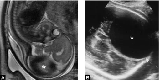
症例:日齢0日,男児. 主訴:心不全. 家族歴:特記事項なし. 現病歴:母親は28歳の経産婦で,患児は第2子である. 妊娠25週3日の超音波検査で胎児後頭部に31×19mm大 の囊胞性病変を認めた.妊娠28週6日の胎児MR検査で 病変はgalenic cisternではなく表在性の硬膜部に認めて おり,この時期に診断される疾患としてDSMが最も可 能性が高いと考えた.妊娠35週3日には後頭部のvenous pouchは61×88mm大に拡大していた(Fig. 1A, B).妊娠1) Department of Neurosurgery, Osaka City General Hospital 2) Department of Pediatric Neurosurgery, Osaka City General Hospital 3) Department of Pediatric Cardiology, Osaka City General Hospital 4) Department of Neonatology, Osaka City General Hospital 5) Department of Neurosurgery, Fukui Prefectural Hospital
●Abstract● Objective: A case of dural sinus malformation (DSM) in a neonate is reported.
Case presentation: A boy with a prenatal diagnosis of DSM was born by cesarean section at a gestational period of 36 weeks. Birth weight was 2741g and head circumference was 38cm. Chest x-ray showed cardiomegaly and cardiac ultrasound examination demonstrated congestive heart failure. To improve his symptoms, endovascular treatment was performed on Day 0. Angiograms showed a huge venous pouch in the left parieto-occipital region, fed by numerous meningeal arteries. Transarterial embolization using N-butylcyanoacrylate and detachable platinum coils was performed via several meningeal feeders. This resulted in marked improvement of his heart failure. As there was Kasabach-Merritt phenomenon caused by the progression of thrombosis in the venous pouch, repeated blood transfusions were required. Subsequently, transarterial glue embolization and transvenous embolization were performed at the age of 5 and 8 months, respectively. Finally, the venous pouch was occluded with coils. These procedures resulted in a marked reduction of the arteriovenous shunts and shrinkage of the venous pouch. At the age of 21 months, the patient presented with mild developmental delay, but without any neurological deficits.
Conclusion: Staged endovascular treatment by transarterial and transvenous embolization was effective for this case of neonatal DSM. ●Key Words●
dural sinus malformation, embolization, neonate
(Received October 8, 2008:Accepted October 29, 2008) 1)大阪市立総合医療センター 脳神経外科
2)同 小児脳神経外科 3)同 小児循環器内科 4)同 新生児科 5)福井県立病院 脳神経外科
36週1日に全身麻酔下での予定帝王切開で出生した.出 生時体重2741g,Apgar scoreは5/7点で,血圧45/36mmHg, 脈拍170回/分であった.頭囲は38cmで大泉門は緊満して いたが,明らかな神経脱落症状はなかった.また外表上, 異常所見は認めなかった.直ちに気管内挿管を行い,人 工呼吸管理を開始し,呼吸窮迫症候群に対して肺サーフ ァクタントの気管内投与を行った.胸部単純写で心胸郭 比は66%と心拡大を認めていた.心臓超音波検査では心 駆出率は70%と低下はなかったが,2.6mmの下大静脈径 に比し上大静脈は9.3mmと著明に拡張しており,高度の 右心負荷が認められた.また心拡張期に大動脈から頸動 脈 へ の 逆 流 現 象 も 認 め ら れ た.Neonatal evaluation
score7)は13点であったが,venous pouchによる脳圧排が
強いことや右心負荷が今後さらに増大することが予想さ れたため,出生4時間後に全身麻酔下で血管内治療を施 行した.麻酔導入時よりカテコラミンの持続投与を開始 した.出生時に臍帯動静脈の確保を試みたが,確保でき なかった. 第1回血管内治療:4Fr シースを大腿動脈に留置した. 血管撮影では拡張した中硬膜動脈や後頭動脈などの多数 の髄膜動脈がvenous pouchの壁にAV shuntを形成してお り,特に左後頭動脈からは大きなdirect shuntが認めら れた.AV shuntの血流は多いが,venous pouch内の血流 は非常に遅く,内部で渦を巻いている状態であった.左 内頸動脈は相対的に狭小化していた.またvenous pouch と正常脳静脈灌流との関係は判別できなかった(Fig. 2A, B). ま ず 左 後 頭 動 脈 に Rapidtransit(Cordis Neurovascular Inc., Miami Lakes, FL, USA)を誘導し,
A B
Fig. 1 Antenatal T2-weighted MR image (A) at a gestational period of 28 weeks and ultra- sonogram of the head (B) at 35 weeks, indicating a huge venous pouch (asterisk) in the left posterior portion of the fetal head.
Fig. 2 Left common carotid angiograms (CAGs) (lateral views in early (A) and late phases
(B)) before the first intervention showing the huge venous pouch (asterisks) fed by numerous meningeal arteries. The left internal carotid artery (arrows) is small.
4日に抜管し,日齢5日にカテコラミンを中止した.ま た上大静脈の径も徐々に縮小し,日齢6日には2.7mmと なった.しかしvenous pouchの血栓化により日齢1日に 血小板91000/mm3,PT-INR 1.61,APTT 155.7秒,フィ ブリノーゲン 44mg/dl,FDP 48.8μg/mlと凝固異常を 認めたため新鮮凍結血漿を輸血した.さらに日齢3日に はHb 8.4g/dlと貧血も進行し,赤血球濃厚液も輸血した. 日齢9日のMR検査ではvenous pouch内の大部分に血栓 化を認めていた(Fig. 3).輸血は日齢12日まで繰り返 し必要とし,その後は貧血,凝固異常とも徐々に改善し ていった.最終的には生後3ヵ月時に頭囲45cmと拡大 を認めるものの,明らかな神経脱落症状がない状態で自 きた. 第3回血管内治療:生後8ヵ月時に経静脈的塞栓術を行 った.Venous pouchは主に左後頭部に存在し,一部正中 近くにも認めていたが,静脈洞交会にAV shuntはなか った.左S状静脈洞は描出されず,AV shuntからの血流 は静脈洞交会を介して右横-S状静脈洞へ流出していた (Fig. 5A).正常脳静脈は上矢状洞から静脈洞交会を介 して右横-S状静脈洞,上矢状洞から頭皮静脈,海綿静 脈洞から下錐体静脈洞などの流出路により頭蓋外へ灌流 していた.右内頸静脈は頸部で低形成であったので(Fig. 5B),4Fr Heartcath(Terumo Corporation, Tokyo, Japan) を 大 腿 静 脈 経 由 に 右 外 頸 静 脈 か らmastoid
Fig. 3 T1-weighted MR image 9 days after the first intervention demonstrating the grossly thrombosed venous pouch.
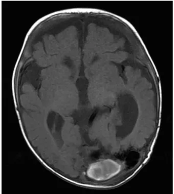
Fig. 4 T1-weighted MR image at the age of 5 months demonstra-ting marked shrinkage of the venous pouch. Mild cerebral atrophy and ventricular dilatation are also observed.
emissary vein を 介 し て 右 横 静 脈 洞 に 留 置 し た. Rapidtransit を venous pouch 内 に 誘 導 し GDC 18と MicruSphere 18,HeliPaq 18(Micrus Endovascular Corporation, San Jose, CA, USA)を計19本560cm使用 して,左側から正中部分のvenous pouchを塞栓した.髄 膜動脈からのAV shuntだけでなくpial AV shuntも著明 に減少した(Fig. 5C, D).術後は問題なく経過した. 1歳9ヵ月時点で頭囲は55cmと拡大しており軽度の発 達遅延を認めているが,神経脱落症状なく成長している. 脳室拡大が脳委縮によるものだけか水頭症が合併してい るかの判断が困難であるが,頭囲拡大傾向はなく,また 発達遅延の進行もないため残存するAV shuntとともに 経過観察している.
考 察
DSMは小児の硬膜動静脈瘻の1つで1-6,8-10),頭蓋内動 静 脈 奇 形 の1.9%と 稀 な 疾 患 で あ る1).DSMは さ ら に DSM with giant pouchとDSM of the jugular bulbとに分 けられる1,8).DSMの大部分はDSM with giant pouchで, 本症例のように横静脈洞,S状静脈洞や静脈洞交会とい ったposterior sinusが巨大なpouch状に拡張し,その壁 に slow flow の AV shunt が 形 成 さ れ て い る.VenousFig. 5 Left CAGs (anteroposterior (AP) view in early (A) and late (B) phases) at the age of 8 months, before the third intervention, showing marked shrinkage of the venous pouch and pial arteriovenous (AV) shunts of the middle cerebral artery, as well as dural AV shunts. Hypoplasia of the right internal jugular vein at the cervical portion (single arrow) and enlargement of the right mastoid emissary vein (double arrows) are also observed.
C:After the third intervention, left CAG (AP view) showing a marked reduction of the AV shunts.
D:Skull x-ray (AP view) demonstrating the deposited coils in the venous pouch as well as glue cast in the meningeal feeders.
D C
B A
頭静脈洞,辺縁静脈洞が代わりに発達しており,正常脳 静脈灌流の流出路として機能している11).DSMは横静 脈洞のballooningが何らかの原因で病的に進行・残存し, 出生後も認めているものと考えられている1,2,8).また venous pouchの内皮は未発達で,さらに血流が遅いこと もあり,venous pouch内は血栓化を起こしやすい2,3,8). DSMは新生児期から幼児期に認められるが1,8),その 症状は他の小児動静脈シャント疾患と同様に,発症時の 年齢に応じて心不全,巨頭症,水頭症,痙攣,発達遅延 などを呈する1-6,8,10).またvenous pouch内に血栓化が進 行することで,血小板減少や凝固異常,溶血性貧血とい っ たKasabach-Merritt現 象 を 来 す こ と も あ る1,2,6,8). Venous pouch内の血栓化などにより正常脳静脈灌流が著 しく障害されると,静脈鬱血から脳内出血や静脈性梗塞, さらには白質を中心とした急速な脳実質の破壊(melting brain syndrome)へと進行する1,6-8).Lasjauniasらは症 例に応じてヘパリンを持続投与し,venous pouch内の血 栓化の進行予防を行っている1,8).本症例では初回塞栓 術後にKasabach-Merritt現象を呈したが,輸血による対 症療法を約2週間行い,重篤な脳静脈灌流障害を来すこ となく改善した.抗凝固療法の開始時期やその期間に関 して一定の見解はなく,今後の課題である. DSMなどの新生児脳血管奇形の治療適応はLasjaunias らが提唱するneonatal evaluation scoreを指標とするこ とが多い.これは脳機能,心機能,肝機能(凝固系を含 む),呼吸機能,腎機能を点数化したもので,21点中13 点以上を経過観察,8から12点を緊急の血管内治療,7 点以下を治療適応なしとしている7).本症例では出生時 は13点で,Lasjauniasらの方針では内科的治療で経過観 察となる.しかしvenous pouchによる脳圧排が強いこと や別症例の経験から右心負荷がさらに増大し心不全が増 悪することが予想されたため,出生当日に血管内治療を 場合には,venous pouch内にコイルを少なめに留置して 血流をある程度減らすことが有効なこともある5,6). DSMの予後は悪いことが多く4,8,10),Barbosaら1)は29 例のDSMに対して内科的治療または血管内治療を行い, 17例(58.6%)で神経症状なし,1例(3.5%)で重度の 神経症状あり,11例(37.9%)で死亡であった.特に venous pouchが正常脳静脈灌流に関与している場合や正 常脳静脈灌流が障害された場合の予後は不良であった. 本症例はvenous pouchが正常脳静脈灌流に関与している か当初は判別困難であった.2回の経動脈的塞栓術で AV shuntが減少し,それに関与していないことが明ら か に な っ た. そ の 後 に 経 静 脈 的 塞 栓 術 を 行 い,AV shuntをさらに減らすことができたことで良好な結果が 得られた. DSMの治療目標はAV shuntを減少させ,患児を正常 な発達に導くことである1,2,8).そのためには経動脈的塞 栓術と経静脈的塞栓術を組み合わせて段階的に治療にあ たる必要があると考えられた. 文 献
1)Barbosa M, Mahadevan J, Weon YC, et al: Dural sinus malformations (DSM) with giant lakes, in neonates and infants. Review of 30 consecutive cases. Intervent Neuroradiol 9:407-424, 2003.
2)de Haan TR, Padberg RD, Hagebeuk EE, et al: A case of neonatal dural sinus malformation: clinical symptoms, imaging and neuropathological investigations. Eur J Paediatr Neurol 12:41-45, 2008.
3)Goddard AJ, Phatouros CC, Khangure MS: Spontaneous regression of an infantile dural sinus fistula with pial recruitment. AJNR 23:1482-1484, 2002.
4)Kincaid PK, Duckwiler GR, Gobin YP, et al: Dural arteriovenous fistula in children: endovascular treatment and outcomes in seven cases. AJNR 22:1217-1225, 2001.
5 ) K o m i y a m a M , N i s h i k a w a M , K i t a n o S , e t a l : Transumbilical embolization of a congenital dural arteriovenous fistula at the torcular herophili in a neonate. Case report. J Neurosurg 90:964-969, 1999. 6)Komiyama M, Matsusaka Y, Ishiguro T, et al:
Endovascular treatment of dural sinus malformation with arteriovenous shunt in a low birth weight neonate. case report. Neurol Med Chir (Tokyo) 44:655-659, 2004. 7)Lasjaunias P, Ter Brugge K, Berenstein A: Introduction
and general comments regarding pediatric arteriovenous shunts: Surgical Neuroangiography, vol.3 Clinical and interventional aspects in children, Berlin, Springer-Verlag, 2006, 27-104.
8)Lasjaunias P, Ter Brugge K, Berenstein A: Dural arteriovenous shunts: Surgical Neuroangiography, vol.3
Clinical and interventional aspects in children, Berlin, Springer-Verlag, 2006, 389-453.
9)Monges JA, Galarza M, Sosa FP, et al: Direct surgical approach of a congenital dural arteriovenous fistula at the torcular herophili in a neonate. Case illustration. J Neurosurg 102 (4 suppl):440, 2005.
10)Morita A, Meyer FB, Nichols DA, et al: Childhood dural arteriovenous fistulae of the posterior dural sinuses: three case reports and literature review. Neurosurgery 37:1193-1199, 1995.
11)Okudera T, Huang YP, Ohta T, et al: Development of posterior fossa dural sinuses, emissary veins, and jugular bulb: Morphological and radiologic study. AJNR 15:1871-1883, 1994.
JNET 2:222-227, 2008
要 旨
【目的】新生児dural sinus malformation(DSM)の1例を報告する.【症例】男児.妊娠28週にDSMと診断され,妊娠36週 に帝王切開で出生した.出生時から心不全を認めたため,出生当日に血管内治療を行った.多数の髄膜動脈が後頭部の巨 大なvenous pouchに動静脈シャントを形成しており,複数の流入血管を介して経動脈的塞栓術を行った.術後,心不全は改 善した.その後は5ヵ月と8ヵ月時に経動脈的および経静脈的塞栓術をそれぞれ行い,動静脈シャントは著明に減少し, venous pouchも縮小した.1歳9ヵ月時点で軽度の発達遅延を認める以外に神経脱落症状なく成長している.【結語】DSM の新生児例に対し経動脈的塞栓術と経静脈的塞栓術を段階的に行い良好な結果が得られた.