Recent Development in Antineoplastic
Biopolymers
from Marine Organisms1
Yoshiaki Kamano and
•
Ilui pi ng
4pp r
Zhang' 2
Kanagawa University
Department of Biological Science, Faculty of (iliratsuka,.Kanagawa, 259-12, JAPAN)
Science
*1 This article was presented by one
"Researt Institute for Information University " November 15, 1991.
*2 Leaving from "Shan Dong Institute
(YK) of author at the forum of and knowledge, Iliratsuka, Kanagawa
of Marne Materia Medica" in China.
- 49 -
Intrudoction
Marine organisms comprise over a half millon species and inhabit very unusual circumstances as compared with terrestrial organisms. Some orga- nisms, such as marine algae, marine sponges, tunicates, nudibranchs, and bryozoans, lack any physical defence means in thier structures to avoid the menace of predators and the infection of pathogenic microorganisms.
In order to keep alive and adapt in these severe environment, marine orga- nisms seem to develop suitable bio-defence systems with characteristic me- tabolisms which are significantly different from those known in terrestri- al organisms.
In this relation, the chemical metabolites of marine organisms have been paid much attention in recent years and an increasing number of marine na-
tural products have been discovered. Those substances are not only diverse in their chemical structures, but also shown to exhibit varieties of bio- logial activities, e.g. growth inhibitions against pathogenic microorga- nisms, physiological functions (pheromone, allelochemic and synomene), pharmacological actions, cytotoxicities and anti-cancer activities, and toxici ties.
In this mean, the effort to discover new anti-cancer substances from ma- rine organisms has been also undertaken. The resulting several interest
biopolymers, for example the antineoplastic glycoproteins such as sphyrna- stains from the hummerhead shark (Sphyrna lemini), stronglostains from the sea urchin (Stronglocentrolus drobachiensis), aplysianins from the sea hare (Aplysia kurodai) and dolabellanins from another sea hare (Dolabella auricularia) have been isolated recently. On the other hand, many novel.
antineoplastic compounds having usual molecular weight (e.g. macrolides, peptides,alkaloids, terpenoids and others) have been isolated from many
marine organisms.
In this article, the important chemically clarified biopolymers, having strongly cytostatic and potent antineoplastic activities, from marine or- ganisms were reviewed.
— 50 —
1. Example of The Biopolymer Screening
Recently, Sasaki and Endo" have discovered the important antineoplastic polymer fractions from the water extracts of 25 marine organisms, in which the popular marine animals as Japanese foods, e.g. Awabi (Haliotis discus hannai), Sazae (Butillus cornutus), Hotategai (Aequipecten irradians),
etc.are including.
The molecular weights and antineoplastic (in vivo) activities against sarcoma-180 of fractionated polymers were summarized in Table 1. Each po- lymer fraction was yielded by usual fractionation with Sephadex seies.
The in vivo activity against Sarcoma-180 (mice) for polymer fractions was obtained by the systematic imjection (i.p. 400mg/Kg/dose) to ICR mice
(female). From the data in Table 1, it was found that many marine organis- ms produced the orginal antineoplastic polymers. In them, the polymers
from Haliotis diversicolor aquatilis (Tokobushi), Butillus cornutus (Sazae ), Placopecten magellanicus (Hotategai), Mercenaria mercenaria (Honbinosu- gai), Crassostrea virginica (Americagaki) and Ecteinascidia turbinata ( Guntaiboya) showed the remarkale antineoplastic activities over 80% effect , respectively.
However, unfortunately, the exteuded purication and identification of these biopolymer fractions are not completed at this time.
Table 1. An ti tumor the Water
Activity of Extracts of
the Polymers isolated from Marine Organisms 1)
Japanese Name
Scientific Name
Molecular Weight of Polymers
Antitumor Activity
(%) a)
Rate of Perfect Recovery Ezoawabi
Tokobushi Sazae Hotategai
Hotategai
1laliotis discus hannai
II. diversicolor aquatilis Butillus coruulus Placopeclen magellanicus Aequipecten irradians
MW>10 MW<10 MW>50 50>MW>10 300>MW>100 300>MW>100
MW >300 300> MW > 100
MW >300 300>MW>100
61.6 7.5 87.6 57.3 83.5 96.7 47.6 81.6 56.3 76.7
0/6 0/6 0/6 0/6 3/6 1/6 1/6 1/6 2/6 3/6
— 51 —
Table 1 ( continued )
Japanose Name
Scientific Name
Molecular Weight of Polymers
Antitumor Activity
(%) o..
Rate of Perfect Recovery
Honbinosugai (Quahog) Honbinosugai (Little necked clam) Honbinosuagi (Cherry stone clam)
Ohonogai Igai
Aneri cagaki Begaika Ezobafununi Kitamura
sakiuni Literhoya
Shiroboya Yurelboya Guntalboya
Ak.afu ji - tsubo Erako
kani Madaragani
Mercenaria inercenaria M. mercenaria M. mercenaria Mya arena ria Mytilus edulis Crassost rea virginica Loligo paelici Strongylocentrotus
inlennedius S. nudus
Halocynthia hilgendorfi f. ritteri
Styela plicata Ciona inteslinalis
Ecteinascidia turbinata
Megabalanus rosa
Pseudopotarnilla accelata
Geryon quinquedens Canceraeta
irroratus
MW>300 300>MW>100 MW >300 300>MW>100
MW>300 300>MW>100
MW>300 300>MW>100
MW>300 300>MW>100
MW>300 300>MW>100
MW>300 300 > M W > 300
MW>10 MW<10 MW>10 MW>300 300>MW>50
50>MW>10 MW>10 10>MW>0.5
50 % EtOH extract MW>10 10>MW>0.5
50% EtOH extract MW>10
body fluid MW>50 50>MW>10 MW>10
MW>300 300>MW>100
MW >300 300>MW>100
61.7 20.4 86.0 27.1 50.5 75.7 23.4 77.6 12.1 4.7 29.0 82.2 35.5 50.5 43.8 34.9 74.0 56.3 76.7 20.4 74.8 56.3 54.4
61.2 17.5 45.6
92.2 41.1 63.0 65.1 28.1 47.7 47.7 56.1 52.3
0/6
1/6 0/6 0/6 0/6 0/6 0/6 0/6 0/6 0/6 0/6 0/6 0/6 0/6 0/6 0/6 0/6 0/6 0/6
0/6 0/6
0/6 0/6 0/6 3/6 0/6 0/6 0/6 0/6 0/6 0/6 0/6 0/6
a) Antitumor in vivo Activity against Sarcoma-180 (abdominal dropsy cell) was estimated by the followed formula.Activity (%)=(A-B)/Ax100
A: Avarage of tumor weight on control B: Avarage of tumor weihgt on treatment
In the experiment, ICR mice (female, ca. 23g weight) was used . - 52 -
2 . Sphyrnastatins and Strongylostatins
In 1977, Pettit and Ode reported that two antineoplastic glycoproteins, designated sphyrnastatin 1 and sphyrnastatin 2, were isolated from the water extract of blood and body fluids of the hurnmerhead shark Sphyrna lemini by chromatography on macroreticular resin followed by gel permeation chromatography.
Sphyrnastatins 1 and 2 were glycoproteins of 22% and 26% carbohydrate, respectively, with apparent molecular weights of at least 40x106 Amino acid analyses indicated a minimum protein segment of 274 amino acid units for sphyrnastatin 1 and 380 amino acid units for sphyrnastatin 2. Upon biological evaluation, both compounds
produced a 30-40%(at 11-13 mg/kg dose) life extension in mice inoculated with P-388 murine lymphocytic leukemia. 2
In 1979, Pettit and co-workers reported that a new glycoprotein antineoplastic agent designated strongylostatin 1 was isolated from the water extract of the well-known green
sea urchin Stronglocentrollcs drobaclziensis (Muller) of the Strongylocentrotideae family.
A series of gel permeation chromatography was used for separation. The apparent
molecular weight of this compound appeared to be over 40x 106. The results of amino
acid analyses suggested a minimum protein segment of 546 amino acid units.
Strongylostatin 1 (approximately 1.6% yield based on the aqueous extract) was found to
produce 35-53% life extension in the P388 murine lymphocytic leukemia system. 3'
In 1981, Pettit and co-workers described that the same green sea urchin was found to
contain a second protein anticancer constitutent designated strongylostatin 2. It was
found to be a glycoprotein with a unit/subunit molecular weight of at least 65,000. An
amino acid analysis showed Gly to be most prominent, and the carbohydrate portion was
estimated to represent 22% of the glycoprotein. Treatment of the murine P388 lyrnphocytic leukemia with strongylostatin 2 resulted in 39-42% life extension at a dose
of 4.5mg/kg. "
3 , Aplysianins
Recently, Kamiya and co-workers reported that three antitumor factors, aplysianins A, E and P, inducing tumor lysis were isolated from the sea hare Aplysia kurodai.
Aplysianin E was purified to apparent homogeneity from the supernatant of a homogenate of eggs of Aplysia kurodai. Purified aplysianin E was a 250kDa glycoprotein containing
— 53 —
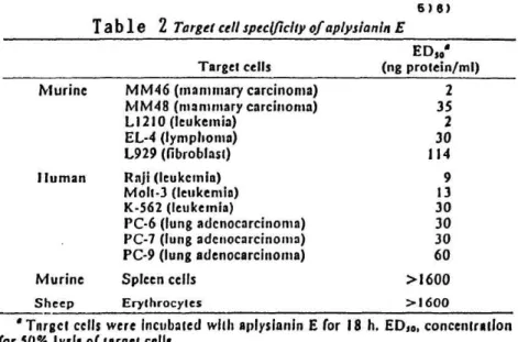
three different subunits. This factor was half-maximally active at 2-114 ng protein/ml and lysed all the tumor cells tested but did not lyse normal white or red blood cells.
Aplysianin E was labile on treatments with heat, low pH, urea, guanidine, sodium lauryl sulfate, and periodatc, but not with proteases or organic solvents. Aplysianin E completely inhibited the syntheses of DNA, RNA, and protein by tumor cells within 2 h and caused their complete cytolysis within 15 h. Tumor lysis by aplysianin E was inhibited by N-acetylneuraminic acid, suggesting that recognition of the sugar moiety is a key step in cytolysis induced by aplysianin E. Aplysianin E also prolonged the survival of mice bearing syngeneic MM46 ascites or solid tumors. 5) 6 )
The cytolytic activity against various target cells was examined as shown in Table 2. All the murine and human tumor cells tested were lysed
by purified aplysianin E in the concentration range of 2-114 ng protein/ml . In contrast, normal spleen cells and erythrocytes were resistant to this cytolytic factor. These results indicate that tumor cells are relatively susceptible to aplysianin E.
6)8)
Table 2 Target cell specificity of aplysianin E
Target cells
EDs•
(ng protein/ml) Murinc
Iluman
Murine Sheep
MM46 (mammary carcinoma) MM48 (mammary carcinoma) L1210 (leukemia)
EL-4 (lymphoma) L929 (fibroblast) Raji (leukemia) Molt-3 (leukemia) K-562 (leukemia)
PC-6 (lung adcnocarcinoma) PC-7 (lung adenocarcinoma) PC-9 (lung adenocarcinoma) Spleen cells
Erythrocytes
2 35 2 30 114
9 13 30 30 30 60
>1600
>1600
• Target cells were Incubated with aplysianin E for 18 h. EDso, concentration for 50% lysis of target cells.
Table 3 shows effect against challenge with
that aplysianin the MM 46 tumor.
a lethal dose fo
E as well as mitomycin Some mice that survived
fresh tumor cells.
C had
were
a protective resistant to
— 54 —
T a bl e 3 Neutralizing effects of aplysianin E on AMA(46 tumor cells 5)6)
Neutralization' Challenge'
Treatment
Dose/ Survival Survival Survival Survival
mouse ratio days ratio days
None Mitornycin C Aplysianin E Aplysianin E Aplysianin E
10 pg 0.1 unit'
I unit 10 units
0/5 5/5 1/5 4/5 4/5
16.2 ± 0.8`
>45 17.0 ± 0.0
23 25
0/5 0/5 1/l 2/4 3/4
18.8 ± 1.6 15.2 ± 2.5
•• 55 18.0 ± 1.4
19
Murine MM46 tumor cells were pretreated with mitomycin C or aplysianin E in vitro at 37.0 for 60 min. Then the cells (5 x 10' cells/mouse) were inoculated i.p. into C311/11e mice. Data are representative of two similar experiments.
6 Fresh MM46 tumor cells (5 x 10' cells/mouse) were inoculated i.p. into surviving mice 50 days after neutralization.
Mean ± SD.
g One unit equals 8.2 pg of protein.
Aplysianin A was isolated from the albumen gland of Aplysia kurodai.. The aplysianin A obtained showed similar physiological activity to that of aplysianin E, an active factor in eggs. It showed a 50% inhibition of 13. subtilis growth at a concentration of 411g protein/ml. The concentration required for 50% lysis of murinc MM46 tumor cells was as low as 14ng protein/ml. Physicochemical properties were, on the other hand, quite different from those of aplysianin E. Aplysianin A, which has a molecular weight of approximately 320,000 and contains four Mr 85,000 subunits, is larger than aplysianin E. However, these two factors have a common antigen and a similar amino acid
composition, suggesting that aplysianin A is a precursor of aplysianin E. 7 )
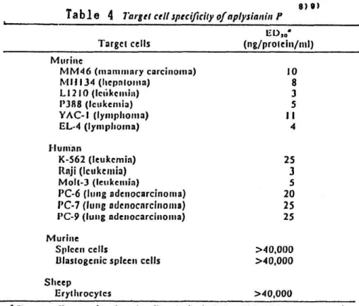
Aplysianin P was purified from the purple fluid of Aplysia kurodai. purified aplysianin P was a single polypeptide of m.w. 60,000. This factor was half-maximally active at 3-25 ng protein/ml and lysed all the tumor cells tested but did not lyse normal WBC or RBC. Aplysianin P was labile on treatments with heat, low pH, urea, and periodate, but not with Pronase. The factor completely inhibited the syntheses of DNA, RNA, and protein by tumor cells within 2 h and caused their complete cytolysis within 18 h. Tumor lysis by aplysianin P was also inhibited by N-acetylneuraminic acid, suggesting that recognition of the sugar moiety is a key step in the cytolysis induced by aplysianin P. The factor also prolonged the survival of mice bearing syngeneic MM46 ascites. It did not resemble previously isolated antineoplastic glycoproteins from the eggs(aplysianin E) or albumen gland (aplysianin A) of A. kurodai in terms of molecular size, antigenicity, or amino acid composition. 8' 9 )
— 55 —
The cytolytic activity againgst various target cells was examined, as shown in Table 4 all the murine and human tumor cells tested were lysed
purified aplysianin P in the concentration range of 3-25 ng protein/ml.
contrast, normal spleen cells and erythrocytes were resistant to this cy tolytic factor. These results indicate that tumor cells are relatively
susceptible to aplysianin P.
by In
Table 4 Target cell specificity of aplysianin f
8) 9)
Target cells
ED„' (ng/prolcin/m1) Murinc
MM46 (mammary carcinoma) M11134 (hepatuina)
L1210 (Icukcmia)
P388 (leukemia) YAC-I (lymphoma)
EL-4 (lymphoma) 1-Iunian
K-562 (leukemia) Raji (leukemia)
Molt-3 (leukemia)
PC-6 (lung adenocarcinoma) PC-7 (lung adenocarcinoma)
PC-9 (lung adenocarcinoma) Murine
Spleen cells
Ulastogenic spleen cells Sheep
Erythrocytes
10 8 3 5 11 4
25 3 5 20 25 25
>40,000
>40,000
>40,000
Target cells were Incubated wtih aplysianin P for 18 Ii. ED,e, concentration for 50% lysis of target cells.
4. Dolabellanins
The Indian and the Pacific Ocean sea hare, Dolabella auricularia, is a large shell-less opisthobranch mollusk which is found intertidally feeding on brown algae. Dolabella auricularia contain cytotoxic and/or antineo- plastic subustances such as dolastatins, as shown in the section of pep-
tides. These substances are low molecular weight compounds. Recently, Kam iya and co-workers found high moleular weight compounds with antineoplas- tic and antibacterial activities in the body fluid, albumen gland and purple fluid of Dolabella auricularia. These active substances of Dolabel- la auricularia were also isolated as glycoproteins, which differed from aplysianins in Aplysia kurodai.le)-13)
An antineoplastic factor, dolabelanin C, including tumor lysis activitiy was purified to apparent homogeneity from the body fluid of the sea hare Dolabella auricularia. Purified dolabellanin C is a glycoprotein of 215
— 56 —
kDa cotainig 3 subunits of 70 kDa. The amino acid sequence of the amino terminal region was also determined. This factor was active even at 0.38ng protein/ml, but did not lyse normal white or red blood cells.1)
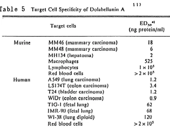
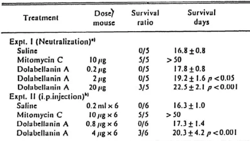
Dolabellanin A was purified to apparent homogeneity from the albumen gland of the sea hare Dolabella auricularia. Dolabellanin A was a glyco- protein of 250 kDa cotaining 4 subunits. The amino acid composition and the N-terminal amino acid sequence of the factor were determined. By dola- bellanin A, all the murine and human tumor cells tested were lysed at the concentration range of 1-18 ng protein/ml (Table 5). The in vivo antitumor activity of dolabellanin A was tested by incubating mixtures of tumor cells and dolabellanin A in vitro and then injecting them intraperitoneal- ly into mice. (Table 6). Mitomycin C was used as a positive control. Dola-
bellanin A had protective effect against the MM46 tumor (P>0.001) and also some animals survived after injection of 4 ng/mouse daily for 6 days
(see Table 6). 11) 12)
11)
Table 5 Target Cc II Specificity of D olahcllunin A
Target cells EDs„'I
(ng protcin/ml) Murine
Human
MM46 (mammary carcinoma) MM48 (mammary carcinoma) M11134 (hepatoma)
Macrophages Lymphocytes Rcd blood cells A549 (lung carcinoma) LS 174T (colon carcinoma) T24 (bladder carcinoma) WiDr (colon carcinomn) TIG-I (fetal lung) I M R-90 (fetal lung) W1-38 (lung diploid) Red blood cells
18 6
2 525 I x 10'
>2x 10' 1.2
3.4 1.2 0.9 62 68 120
>2x 10' a) Target cells were incubated with dolabcllunin A for I R h.
for 5O' lysis of target cells.
ED«. concentration
— 57 —
12)
Tabl e 6 Effect of Dolabellanin A on Survival of Mice Bearing Ascitic MM46 Tumor Cells
Treatment
Dose)
mouse
Survival ratio
Survival days Expt. I (Neutralization)"
Saline
Mitomycin C 10 pg Dolabellanin A 0.2 pg Dolabellanin A 2 pg Dolabellanin A 20 pg Expt. II (i.p.injection)")
Saline0.2 ml x 6
Mitomycin C 10 pg x 6 Dolabcllanin A 0.8 pg x 6 Dolabellanin A 4 pg x 6
0/5 5/5 0/5 0/5 3/5 0/6 5/5 0/6 3/6
16.8 ± 0.8
> 50 17.8 +0.8
19.2±1.6p<0.05 22.5±2.1 p<0.001 16.3± 1.0
> 50 17.3 ± 1.4
20.3±4.2 p <0.001 a) /Amine MM46 tumor cells were treated with mitomycin C or dulabellarrin A in vitro. at 37"C For 31) min. Thcn the cclls (5 x 1U1 cclls/mouse) were injected i.p. into ('311/l le mice. h) C311/Ile mice received ;iscitic MM46 cclls i.p. on day 0 and were treated i.p. with saline. nsitontycin C or dolabellanin A daily for 6d from day 0.
Third cytolytic factor, dolabellanin P, was purified to apparent homogeneity from the purple fluid of Dolabella auricularia. Purified dolabellanin P is a single polypeptide of 60 kDa. Examination of the amino acid composition of dolabellanin P revealed that it contained a relatively large amount of .Asx and Gix. The sequence of the amino terminal region of dolabellatnin P was shown:
Val-( )-Arg-Glu-Asp-Gln-Lys-Trp-Lys-Leu-Ser-Phe-Asp-Lys-Glu---
This factor nonspecifically lysed all the cell tested at 50-200ng protein/ml.
Dolabellanin P caused completed cytolysis within 2h. l 3) , as shown in Table 7.
13)
Table 7 Target Cell Specificity of Dolabcllanin P
Target cells EDso') (ug protein/Inl)
Murine
.I luman
Murine
MM46 (mammary adenocarcinoma) MM48 (mammary adcnocarcinoma) MI1134 (lung adcnocarcinoma) A 549 (lung adenocarcinoma) 1' 24 (urinary bladder carcinoma) SK LL'U-I (lung adcnocarcinoma) Spleen cells
Thymus cclls
100 80 60 100 50 200 67 150 a) Target cells were incubated with dolabellanin P for 18 h. EDso:
giving 50% lysis of target cclls.
concentration
— 58 —
5 , Halitoxin
Baslow et al. reported that the aqueous extracts of the sponge Haliclona viridis was
14)
toxic for mice. Ten years later (1978), from several species of the same genus Haliclona, a cytotoxic metabolite called halitoxin (1) was isolated by Schmitz and co- workers. The structure proposed for halitoxin was a polymer consisting of various combination of different pyridinium units of two general structural types, as shown in Chart.12) Halitoxin was hemolytic and toxic to fish and mice. The cytotoxity of fractionated halitoxin R (m.w. 500-1000) was reported as ED50 2.8 µg/m1 in KB cell culture. 1 5 )
Me
6. Polypeptides
(CH2)n
((CH2)3 -- (CH2)m
(CH2)2
`^ (CH
2)n n=6,7,8,9
Chart 1
N ~
(CH2)2 (CH2)m
(CH2)3 N
=3,5
Me
Mucus secreted from the integument of the marine heteronemertine Cerebratulus lacteus (Leidy) contains two major types of polypeptide neurotoxin. The A toxins (11,000 daltons) which are lethaI to a variety of animal species including mammals. The B toxins (6,000 daltons) which appear to be selectively toxic for crustaceans. BOth types of toxin were readily obtained from live worms by stimulation and collection of mucus with 1%
acetic acid followed by batch adsorption of the basic polypeptide fraction on CM- cellulose. 1 6 )
A toxins were homogeneous by gel electrophoreses and amino acid composition analyses, and by NH2 -terminal and COOH-terminal partial sequence analyses.
— 59 —
As shown in Chart 2, each of three A toxins(2) consists of a single basic polypeptide chain of 93 to 99 residues, cross-linked by three or four disulfide bonds, lacking reducing sugar and cyteinyl residues. The three A toxins rapidly lysed human red cells and Ehrlich ascites tumor cells at 1 to 10 µg/m1 concentration.' 7'
A-1:
A-2:
A-3:
la- Ala Ala
POOH -COON .COON Tyr-Pro-Gly
Tyr-Pro-GI) Tyr-Pro-GI}
-Ser-Gln- '-Ser-Glu -Asp-AI
aly~
Gly^
GIN
Ile---Ala Ile...Val -Thr---Let H2N-Ile
H2N-II H2N-Vat
Ser-Trp-Pro- Ser-Trp-Pro Ser-Trp-Pro
Ala- -Ser -Ala
Chart 2
? . Antineoplastic Polymers from Marine Bacteria
An active polysaccharide, named marinactan was obtained by Umezawa and co- workers by a screening of antitumor activity against sarcoma 180 solid tumor in mice from the marine bacterium Flavobacterium uliginosum. Marinactan is a novel heteroglycan consisting of glucose, mannose and fructose in a ratio of approximately 7:2:1. Marinactan, 10-50mg/kg per day for 10 days i.p., produced 70-90% inhibition of the growth of solid sarcoma 180. Complete regression of the tumor was observed in some treated mice. Its administrations before and after tumor transplantation showed almost the same inhibitory effect. Marinactan prolonged markedly the survival period of mice bearing ascites sarcoma 180.18'
Antirumor activity of the marine bacterium, Vibrio anguillarum P-B-1 against Ehrlich carcinoma cells in ddY mice, was investigated by Shimizu and co-workers. The aqueous layer obtained by the hot phenol-water procedure exhibited more antitumor activity than did the middle layer or the phenol layer. This finding indicated that lipopolysaccharide (LPS) derived from V. anguillarum exhibited significant antitumor activity. In fact, mice injected with LPS obtained by ultracentrifugation and treatment with RNase had a longer mean survival period than the control mice. V. anguillarum LPS also inhibited the growth of syngeneicfibrosarcoma induced by 3-methylcholanthrene in C57BL/6 mice.9'
By Creech and Breuninger, the antitumor properties of polysaccharide fractions, obtained by high speed centrifugation of aqueous extracts of the cells of Serratia
- 60 -
marcescens followed by phenol extraction and trypsin digestion
, were investigated. The polysaccharide fractions were highly effective in causing complete regression of well established solid sarcoma 37 in Swiss mice . The products derived primarily from the cell wall were more potent than the polysaccharides of extracellula
r and cytoplasnic origins.2e'
8. Mieellaneous
Boiled Scallop Extract.2 ' ) Sasaki and co-workers reported that a fraction from boiled scallop extract modified by a sonication procedure exhibited marked antitumor activity when it was injected intradermally into either ICR or C3I-I/I-Ie mice that had been given SC implants of sarcoma 180 and Ehrlich carcinoma. However, this fraction was almost ineffective against L1210 cells. In a smaller dose (100 mg/kg) in in vivo sarcoma 180 test, this fraction was still effective. It had an inhibition ratio of 74.2% and caused complete tumor regression in 2 of 5 mice. The inhibition ratio observed with Ehrlich carcinoma was 73.5%. From the result of a further fractionation and purification, this fraction was found to contain two active principles -one was probably a heat-stable protein (mol. wt., —21,000 daltons) and the other, a heat-unstable peptidoglycan (mol.
wt., — 100,000 daltons).
Mercenaria extract. 2 2' In 1964, Schmeer reported that a polymeric fraction, which was obtained from the water extract of Mercenaria mercenaria by partial purification with sephadex G-25, inhibited both sarcoma 180 and Krebs 2 carcinoma in Swiss albino mice. Unfortunately, further investigation has not been done.
Marine Animal Lecti,i? 3' By Kamiya and co-workers, several lectins from marine animals, such as Balanus roseus hemagglutinin, B. balanoides hemagglutinin, Tetraclita squamosa japonica hemagglutinin and Aplysia kurodai agglutinin, were tested
for induction of tumor lysis mediated by macrophages. Among them, B. roseus and B.
balanoides lectins agglutinated several murineitumor cells and induced binding of macrophages to tumor cells. Binding of these cells was inhibited by galacturonic acid, suggesting that carbohydrate moieties on the cell membrane of the two types of cells are recognized by these lectins. B.roseus lectin was also effective in vivo.
- 61 -
References
1) T. Sasaki and Y.Endo, "Bioactive Marine Natural Products" (Nippon
Suisan Gakkai Series 65), Koseishakoseikaku, 1987, p.21.
2) G.R. Pettit and R.N. Ode; J.Pharm. Sci., 66, 757 (1977)
3) G.R. Pettit, C.L.Herald, and L.D.Vanell; J. Nat. Prod., 42, 407 (1979) 4) G.R. Pettit, J. A. Hasler, K.D.Paull, and C.L.Herald; J. Nat. Prod., 44
, 701 (1981)
5) J. Kisugi, H. Kamiya, and M. Yamazaki; Cancer Research, 47, 5649 (1987) 6) J. Kisugi, H. Ohye, H. Kamiya, and M. Yamazaki; Chem. Pharm. Bull., 37, 3050 (1989)
7) H. Kamiya, K.Muramoto, and M. Yamazaki; Experirntia, 42 1065 (1986) 8) M. Yamazaki, K.Kimura, J. Kisugi, and H. Kamiya; FEBS Letters, 198, 25
(1986)
9) M. Yamazaki, K.Kimura, J. Kisugi, K.Muramoto, and H. Kamiya; Cancer Research, 49 3834 (1989)
10) J. Kisugi, H. Kamiya and M. Yamazaki; Develop. and Comparat Immunology , 13, 3 (1989)
11) J. Kisugi, M. Yamazaki, Y. Ishii, S. Tansho, K. Muramoto, and H. Kamiya; Chem. Pharm. Bull., 37, 2773 (1989)
12) M. Yamazaki, J. Kisugi, and H. Kamiya; Chem. Pharm. Bull., 37, 3343 (1989)
13) M. Yamazaki, S. Tansho, J. Kisugi, K. Muramoto, and H. Kamiya; Chem.
Pharm. Bull., 37, 2179 (1989)
14) M. H. Baslow, "Marine Pharmacology", Williams & Wikins, Baltimore (1969)
15) F. J. Schmitz, K. H. Hollenbeak and D. C. Campbell; J. Org. Chem., 43, 3916 (1978)
16) W. R. Kem; J. Biol. Chem., 251, 4184 (1976)
17) W. R. Kem and K. M. Blumenthal; J. Biol. Chem.,' 253, 5752 (1978)
18) H. Umezawa, Y. Okami, S. Kurasawa, T. Ohnuki, M. Ishizika, T. Takeuchi , T. Shiio and Y. Ygari; J. Antibiot., 36, 471 (1983)
19) T. Shimizu, Y. Itoh, I. Mifuchi, T. Iguchi, S. Kondo, and K. Hisatsune
; Gann, 74, 279 (1983)
20) H. J. Creech, and E. R. Breuniger; Can. J. Biochem, 42, 593 (1964) 21) T. Sasaki, N. Takasuka, and N. Abiko; J. Natl. Cancer Inst., 60 1409 (1978)
22) H. R. Schmeer; Science, 144, 413 (1964)
23) M. Yamazaki, M. Esumi-Kurisu, D. Mizuno, K. Ogata, and H. Kamiya; Gann , 74, 405 (1083)
— 62 —