Expression of MMP-2, MMP-9 and TIMP-1 in the Wall of Abdominal Aortic
Aneurysms
Kengo Nishimura, Shigetsugu Ohgi and Eiji Nanba*
Second Department of Surgery, Faculty of Medicine, Tottori University and *Gene Research Center, Tottori University, Yonago 683-0826 Japan
An impaired mechanism of regulatory feedback by matrix metalloproteinases (MMPs) and tissue inhibitors of metalloproteinases (TIMPs) has been implicated in the develop-ment of abdominal aortic aneurysms (AAAs). This study examined the pathogenesis of AAAs with respect to pathological characteristics and expressions of MMP-2, MMP-9 and TIMP-1. Their expressions were evaluated by immunohistochemistry, competitive polymerase chain reaction (PCR) and Western blotting in a total of 23 consecutive AAAs. The AAA specimens were obtained by surgery, while control specimens were ob-tained at autopsy. Specimens consisted of 6 patients with small-diameter AAAs (30–45 mm), 17 with medium-large-diameter AAAs (> 45 mm) and 11 controls (17–25 mm). Immunohistochemistry showed MMP-2- and TIMP-1-positive cells mainly in the intima, and MMP-9-positive cells in the intima and adventitia. Competitive PCR showed a significantly higher expression of MMP-2 messenger RNA (mRNA) in the small-diameter AAAs, and higher expressions of MMP-9 mRNA in the small-diameter and medium-large-diameter AAAs than in the controls. The mRNA levels significantly correlated between TIMP-1 and MMP-9, and between MMP-2 and MMP-9 in the AAAs, especially in the medium-large-diameter AAAs. Western blotting revealed the expression of MMPs and TIMP-1 variably in all the specimens examined. These results indicated that MMP-2 and MMP-9 might act cooperatively and play a crucial role in the development of AAAs, and that TIMP-1 inhibits MMP-9 in the AAAs, especially in those medium-large-diameter AAAs.
Key words: abdominal aortic aneurysm; competitive polymerase chain reaction; immuno-histochemistry; matrix metalloproteinase; tissue inhibitor of metalloproteinase
Abbreviations: AAA, abdominal aortic aneurysm; cDNA, complementary DNA ESDNA, external standard DNA; GAPDH, glyceraldehyde-3-phosphate dehydrogenase; MMP, matrix metalloproteinase;mRNA, messenger RNA; PBS, phosphate-buffered saline; PCR, polymerase chain reaction; SAB, streptavidin-biotin-peroxidase; SCDNA, specimen cDNA;TIMP, tissue inhibitor of metalloproteinase
The pathologic hallmark of aortic aneurysm is considered to be the remodeling of the aortic wall, involving fragmentation and decrease of elastic fibers in the tunica media (Baxter et al., 1992; Gandhi et al., 1994). True aneurysms formed in Marfan’s syndrome (Chikumi et al., 2000) were found to be due to a mutation in the fibrillin gene (Dietz et al., 1991; Tsipouras et al., 1992), indicating that aneurysms may be caused by genetic disorders affecting 2 or more components of the aortic wall. Recent hypo-theses have indicated that matrix
metallo-proteinases (MMPs) involved in the metabo-lism of elastin in the wall of aortic aneurysms may be one of the etiologic factors in the de-velopment of abdominal aortic aneurysm (AAA) (Campa et al., 1987; Tilson et al., 1988; Baxter et al., 1992; Freestone et al., 1995; Newman et al., 1994; Thompson et al., 1995; McMillan et al., 1995, 1995, 1997; Tamarina et al., 1997; Davis et al., 1998; Elmore et al., 1998). However, the precise roles of monocytes, macro-phages and vascular smooth muscle cells which are considered to be involved in the early changes
of vascular remodeling, have not been ade-quately elucidated. It is important to examine the entire wall as a functioning organ using molecular-biological techniques, to compare the pathological changes of the individual layers including the intima, tunica media and adventitia. AAAs might result from impair-ment of the mechanism of regulatory feedback by MMPs and tissue inhibitors of metallo-proteinases (TIMPs). A comparative analysis of anatomic changes and tissue metabolism in AAAs small in diameter without obvious struc-tural destruction may provide a clue to the patho-genesis.
With this background, the present study examined the metabolism of elastin in the hu-man aortic wall during the process of expansion of aortic diameter with respect to the expression of MMP-2, MMP-9 and TIMP-1.
Materials and Methods Specimens
The subjects in this study consisted of 23 con-secutive patients with AAA who had undergone an elective excision with graft replacement be-tween November 1998 and December 1999. Eleven specimens obtained at autopsy perform-ed between February 1999 and March 2000 served as controls. Table 1 lists clinicopatho-logical profiles of the AAA patients and the autopsy cases.
Measurement of aortic diameter and stag-ing of AAA
During surgery, the maximal diameter of the aorta distal to the renal artery was measured di-rectly using the divider. The aortic diameter in the control group was measured at autopsy. In this study, an aorta with a diameter more than 30 mm was defined as an AAA. An aorta with a diameter between 30 mm and 45 mm was de-fined as a small-diameter AAA, and that with a diameter over 45 mm as a medium-large-diameter AAA.
Method for collection of aortic wall
After approval by the Ethics Committee of Tottori University Faculty of Medicine, surgi-cal specimens of the aortic wall were collected from the patients after obtaining informed con-sent. During surgery, a rectangular strip of the anterior wall was resected from the site of max-imal diameter to the aneurysmal-normal junc-tion throughout the wall. At autopsy, a secjunc-tion was obtained from the abdominal aorta with a grossly atheroma-free intima throughout the wall. Immediately after resection, part of each specimen was fixed in 10% neutral buffered formalin for 48 h, and the rest was frozen in liquid nitrogen and stored at –80˚C.
Histopathologic analysis
Three-micron-thick sections were cut from paraffin-embedded human AAA walls and ab-dominal aortas from autopsy materials. The sections were stained with hematoxylin-eosin staining and elastica van Gieson. For the local-ization of MMP-2, MMP-9 and TIMP-1, the streptavidin-biotin-peroxidase complex (SAB) method was used for immunohistochemical staining. As the first antibody, purified mouse monoclonal anti-human MMP-2 antibody (diluted 1:200), purified mouse monoclonal anti-human MMP-9 antibody (diluted 1:400), purified mouse monoclonal anti-human TIMP-1 antibody (diluted TIMP-1:200) (Fuji Chemical Inc., Takaoka, Japan), and purified mouse mono-clonal anti-human HAM56 antibody (DAKO, Carpenteria, CA) were used. As the 2nd anti-body and enzyme reagents, a Histofine SAB-PO kit (Nichirei, Tokyo, Japan) was used. En-dogenous peroxidase activity was blocked with methanol containing 0.3% hydrogen peroxide at room temperature for 30 min. For the retriev-al of the antigens, retriev-all slides were placed in 10 mmol/L citrate buffer (pH 6.0) and heated at 94˚C for 15 min in a microwave oven. Non-specific staining was prevented by treatment with phosphate-buffered saline (PBS) contain-ing 2% fetal calf serum. The sections were treated with diaminobezidine for color
develop-ment, and counterstained with 3% methyl green. The degree of tissue staining was judged under a light microscope at 4 × 3.3 magnifica-tion for elastica van Gieson staining and at 20 × 5 magnification for immunohistochemical staining, and then the expression was analyzed.
Competitive polymerase chain reaction (PCR)
The frozen tissue samples were homogenized in guanidium isothiocyanate using a Polytron homogenizer (Kinematica, Luzern, Switzerland). After homogenization, total RNA was isolated by CsCl in ultracentrifugation at 100,000 g for 20 h. Total RNA was further treated with gribo-nuclease-free deoxyribonuclease I (Nippon gene, Tokyo) and extracted with phenol/chloro-form/isoamylalcohol (50:49:1) and precipitated with ethanol. Reverse transcription of 1.5-µg total RNA was performed to synthesize com-plementary DNA (cDNA) using 2 µL of
500-µg/mL Random Hexamer (Promega, Madison,
WI) and 2 µL of 200-units/µL Moloney murine leukemia virus reverse transcriptase (Gibco BRL Products, Life Technologies, Rockville, MD). Control reactions omitting the reverse transcriptase (reverse transcriptase negative) were set up for each RNA sample.
The primers of human glyceraldehyde-3-phosphate dehydrogenase (GAPDH), MMP-2, MMP-9 and TIMP-1 were designed based on Tamarina’s report (Tamarina et al., 1997) by using the Genetyx-Mac Macintosh software (Software Development Co., Ltd., Tokyo) (Table 2). We designed the external standard DNA (ESDNA) for each studied gene that lacked from 10 to 20 base pairs (bp) so that the ESDNA differed in size from the specimen cDNA (SCDNA) obtained by the previous method (Ho et al., 1989). The identity of the SCDNA and ESDNA was verified by DNA se-quence analysis with ABI310 (Applied Bio-systems, Tokyo).
The PCR reaction mixture (12 µL final vol-ume) contained 0.64 µL of each cDNA, 0.5
Table 1. Patient Characteristics
Variable AAA Small Medium-large Control P value
AAA† AAA‡ Number of patients Total 23 6 17 11 Male 22 5 17 7 Female 1 1 0 4 Age (year) Mean ± SD 72.4 ± 6.8 70.3 ± 3.7 73.1 ± 7.8 66.1 ± 14.1 NS Range 62–85 66–77 62–85 47–92 Diameter (mm) Mean ± SD 53.3 ± 10.8 40.8 ± 5.6 57.5 ± 8.6 20.4 ± 2.4 < 0.01 Range 30–80 30–45 48–80 17–25 History of smoking 16 (69.6) 5 (83.3) 11 (64.7) 7 (63.6) NS Chronic obstructive pulmonary disease 8 (34.8) 1 (16.7) 7 (41.1) 1 ( 9.1) NS Hypertension 14 (60.9) 4 (66.7) 10 (58.8) 2 (18.2) NS Diabetes mellitus 5 (21.7) 2 (33.3) 4 (23.5) 2 (18.2) NS Hyperlipidemia 6 (26.1) 2 (33.3) 4 (23.5) 2 (18.2) NS Ischemic heart disease 10 (43.5) 1 (16.7) 9 (52.9) 0 ( 0 ) < 0.01 Arteriosclerosis obliterans 7 (30.4) 2 (22.2) 5 (29.4) 1 ( 9.1) NS Cerebrovbascular disease 3 (13.0) 1 (16.7) 2 (11.8) 3 (27.3) NS Malignant tumor 5 (21.7) 3 (50.0) 2 (11.8) 8 (72.7) < 0.05 ( ), %.
AAA, abdominal aorataic aneurysm; NS, not significant. † Aneurysmal diameter, 30–45 mm.
units Taq polymerase (Gene Amp Taq Gold, Perkin Elmer, Branchburg, NJ), 12 pmol of for-ward primer, 6 pmol of reverse primer conju-gated with Cy5 (Amersham Pharmacia Biotech, Buckinghamshire, England) and 6 pmol of re-verse primer. For each sample, 4 or 5 equal aliquots were prepared with a dilution series of the ESDNA, and the PCR mixture was spiked into these aliquots. Amplification was perform-ed in the thermal cycler (Labosystems Japan Co., Tokyo) for 35 cycles: denaturation at 95˚C for 1 min; annealing for 1 min at 55˚C for GAPDH, MMP-9 and TIMP-1 and at 60˚C for MMP-2; and extension at 72˚C for 1 min.
As the reverse primer was fluorescently labeled with Cy5, PCR products were detected with an ALF red DNA Sequencer (Amersham Pharmacia Biotech). After competitive PCR was completed, the products corresponding to the SCDNA and the ESDNA were electro-phoresed on a 6% polyacrylamide and 6 mol/L urea gel on the Sequencer. The peaks were detected and analyzed with the Allele Links software (Amersham Pharmacia Biotech). The specific SCDNA template in each starting cDNA sample was equimolar to ESDNA when that ESDNA area/SCDNA area was equal to 1 (Fig. 1). The RNA amount is presented as a
percentage to glyceraldehy3-phosphate de-hydrogenase (GAPDH).
Western blotting
Frozen specimens of human AAA walls or autopsied abdominal aortic walls were homo-genized in a homogenizer, and the homogenate was dissolved in lysis buffer containing 0.1% nonylphenoxy polyethoxy ethanol (NP-40). The solution was centrifuged in an ultracentri-fuge at 15,000 revolutions per minute for 10 min, and the supernatant was used as a test sample. The concentration of protein was measured with a Protein Assay Kit (Bio-Rad, Hercules, CA). Test samples of the same protein content (375 µg) were electrophoresed on a 5% sodium dodecyl-sulfate-polyacrylamide gel at a con-stant voltage of 150 V, and the proteins were transferred onto a nitrocellulose membrane at 80 V for 150 min. Markers (Bio-Rad Labs., Richmond, CA) were dissolved in a Coomassie brilliant blue solution and used for electro-phoresis. After blocking nonspecific reaction with 10% skim milk in PBS, the membrane was reacted at 4˚C overnight with one of the follow-ing first antibodies: purified mouse monoclonal anti-human MMP-2 antibody (diluted 1:50),
Table 2. Primers used for competitive PCR
Gene PCR Length (bp) Primer
MMP-2 SP 398 5’=CAGGCTCTTCTCCTTTCACAAC 3’=AAGCCACGGCTTGGTTTTCCTC ESP 379 5’=ACTCCAGACCCCTGGCTTTT 3’=CCAGGGGTCTGGAGTTGTCCCACTGCCCTGTGCCA MMP-9 SP 172 5’=TGGGCTACGTGACCTATGACAT 3’=GCCCAGCCCACCTCCACTCCTC ESP 162 5’=AAGGAGCCAGTTTGCCGGAT 3’=GCAAACTGGCTCCTTGGTCCCAGTGGGGATTTACA TIMP-1 SP 279 5’=GGGGCTTCACCAAGACCTACAC 3’=AAGAAAGATGGGAGTGGGAACA ESP 267 5’=TCTGAAAAGGGCTTCCAGTC 3’=GAAGCCCTTTTCAGACTGGTCCGTCCACAAGCAAT GAPDH SP 216 5’=TGCCTCCTGCACCACCAACTGC 3’=ATGACCTTGCCCACAGCCTT ESP 205 5’=TGGAAGGACTCATGACCACA 3’=TCATGAGTCCTTCCATTGTCATGGATGACCTTGGC bp, base pair; ESP, external standard primer; GAPDH, glyceraldehyde-3-phosphate dehydrogenase; MMP, matrix metalloproteinase; PCR, polymerase chain reaction; SP, specimen primer; TIMP, tissue inhibitor of metalloproteinase.
purified mouse monoclonal anti-human MMP-9 (diluted 1:50) and purified mouse monoclonal anti-human TIMP-1 (diluted 1:50) (Fuji Chemi-cal Inc., Takaoka). Subsequently, the mem-brane was reacted with purified goat polyclonal peroxidase-conjugated anti-mouse immuno-globulin G antibody (diluted 1:1,000) as the 2nd antibody at room temperature for 1 h. The membrane was washed with PBS, and treated with enhanced chemiluminescence Western blotting solution. The enhanced chemilumines-cence of protein bands was detected by expos-ing X-ray film to the membrane, and the expres-sion of MMP-2, MMP-9 and TIMP-1 proteins was evaluated.
Statistical analysis
Using the StatView (Abacus Concepts, Inc., Berkeley, CA) software, the difference between 2 groups was tested by the Mann-Whitney U test. Differences among multiple groups were tested by the Kruskal-Wallis test, and the groups with significant differences were tested by the Tukey test (nonparametric test). The correlation between 2 groups was tested with Spearman’s correlation coefficient by rank. A value of P < 0.05 was considered significant.
Results Staging of AAA
The 23 patients with AAA (≥ 30 mm) consisted of 22 men and 1 woman. The mean age (± SD) was 72.4 ± 6.8 years, and the mean diameter (± SD) of aneurysms was 53.3 ± 10.8 mm. Of them, 6 patients (5 males and 1 female) had small AAAs (30–45 mm): their mean age was 70.3 ± 3.7 years and the mean diameter of aneu-rysms, 40.8 ± 5.6 mm (small-diameter AAA group). The remaining 17 patients (all male) had medium or large AAAs (> 45 mm): their mean age was 73.1 ± 7.8 years and the mean diameter of aneurysms, 57.5 ± 8.6 mm (medium-large-diameter AAA group).
The control group of 11 autopsy cases con-sisted of 7 males and 4 females aged 66.1 ± 14.1 years: the mean aortic diameter was 20.4 ± 2.4 mm. There were no significant differences among the 3 groups in clinicopathological pro-files, except for aortic diameter, history of is-chemic heart disease and malignant disease (Table 1). 2.5 2.0 1.5 1.0 0.5 0.0 –0.5 –1.0 –1.5
SCDNA (a)
ESDNA (b)
–12 –11 –10 –9 –8 –7 –6 –5 –4 –3A
B
log(b/a) log(5Eχ) Y = 3.376 + 0.409X r 2= 0.997Fig. 1. A: Polymerase chain reaction (PCR) products were detected with an ALF red DNA Sequencer (Amersham Pharmacia Biotech), as the reverse primer was fluorescently labeled with Cy5. The Peaks were detected and analyzed with the Allele Links software (Amersham Pharmacia Biotech). B: The specific specimen cDNA (SCDNA) template in each starting cDNA sample was equimolar to the external standard DNA (ESDNA) when that ESDNA area/SCDNA area was equal to 1.
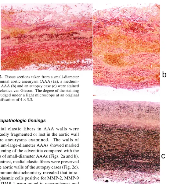
Fig. 2. Tissue sections taken from a small-diameter abdominal aortic aneurysm (AAA) (a), a medium-large AAA (b) and an autopsy case (c) were stained with elastica van Gieson. The degree of the staining was judged under a light microscope at an original magnification of 4 × 3.3.
Histopathologic findings
Medial elastic fibers in AAA walls were markedly fragmented or lost in the aortic wall of the aneurysms examined. The walls of medium-large-diameter AAAs showed marked thickening of the adventitia compared with the walls of small-diameter AAAs (Figs. 2a and b). In contrast, medial elastic fibers were preserved in the aortic walls of the autopsy cases (Fig. 2c). Immunohistochemistry revealed that intra-cytoplasmic cells positive for MMP-2, MMP-9 and TIMP-1 were noted in macrophages and lymphocytes as well as vascular smooth muscle cells. MMP-2-positive cells were present in the intimal plaques and thickened intimas in the AAA walls (Fig. 3a). MMP-9-positive cells were present in the outer layer of the tunica media, adventitia and intimal plaques in the AAA walls (Fig. 3b). TIMP-1-positive cells were noted in the intimal plaques (Fig. 3c). HAM56-positive cells were seen in similar lo-cations to MMP-9-positive cells in the AAA walls (Fig. 3d). A few cells positive for MMP-2, MMP-9 and TIMP-1 were also demonstrated in the control aortic walls.
Competitive PCR
Of the 34 specimens, messenger RNA (mRNA) could be obtained from 10 controls, 6 small AAAs and 14 medium-large AAAs. Figure 4 shows the expression ratios of MMP-2, MMP-9 and TIMP-1 to the internal control GAPDH. Table 3 summarizes the mean expression ratios of MMP-2, MMP-9 and TIMP-1 in the 3 groups. The MMP-2 expression ratio was the highest in the small AAAs, the value being significantly higher than that of the 10 controls (P < 0.05). The MMP-9 expression ratio was higher in the small AAAs and the medium-large
a
b
Fig. 3. AAA tissue shows a positive immunoreaction for matrix metalloproteinase (MMP)-2 (a), MMP-9 (b), tissue inhibitor of metalloproteinase (TIMP)-1 (c) and HAM56 (d). The degree of immunohistochemical staining was judged under a light microscope at an original magnification of 20 × 5.
AAAs than in the control group, P values being less than 0.01 and 0.05, respectively. On the other hand, there was no significant difference among the 3 groups on the TIMP-1 expression ratio, although the expression ratio tended to be higher in the small AAAs. Overall, the highest expression was noted in MMP-2, followed by TIMP-1 and MMP-9, in the order given.
The medium-large AAAs were divided into 2 subgroups by diameter; medium type < 65 mm and large type ≥ 65 mm. The mean expres-sion ratio of MMP-9 was 0.026 ± 0.051 in the medium-type AAA [n = 11] and 0.006 ± 0.009 in the large-type AAA [n = 3], the value being higher in the former than in the latter without significant differences. The mean expression ratios of MMP-2 and TIMP-1 were 0.045 ± 0.073 and 0.334 ± 0.521 in the medium type and 0.281 ± 0.419 and 0.128 ± 0.190 in the large type, respectively. There was no statistical difference.
Table 4 shows the results of Spearman’s coefficient by rank to examine the relationship of the expressions among MMP-2, MMP-9 and TIMP-1. The expression levels of MMP-9 and TIMP-1 signficantly correlated in all the AAAs and the medium-large AAAs (P < 0.01, Fig. 5), but not in the small AAAs and controls. Moreover, the expression levels of MMP-2 and MMP-9 also significantly correlated in a similar fashion (P < 0.05, Table 4). No significant correlation of expression was noted between MMP-2 and TIMP-1.
Western blotting
Protein could be obtained from a total of 9 specimens, 3 each from the 3 groups. MMP-2, MMP-9 and TIMP-1 were variably expressed as shown in Fig. 6. Of these, an obviously higher expression of MMP-2 and TIMP-1 was
a
c
d
b
15 25 35 45 55 65 75 85 (mm) 16 14 12 10 8 6 4 2 0 Diameter MMP-2 MMP-2/GAPDH 2.9 2.4 1.9 1.4 0.9 0.4 0 TIMP-1/GAPDH 0.16 0.14 0.12 0.10 0.08 0.06 0.04 0.02 0 MMP-9/GAPDH MMP-9 TIMP-1 15 25 35 45 55 65 75 85 (mm) Diameter 15 25 35 45 55 65 75 85 (mm) Diameter
noted in 2 of the small AAAs. A higher expres-sion of MMP-2 was also noted in 1 of the 3 aneurysms of the control group. On the other hand, MMP-9 showed lower or almost no ex-pressions in all the specimens examined.
Discussion
This study analyzed the relationship between the development of AAAs and the expression of MMP-2, MMP-9 and TIMP-1 in the aortic walls. The AAAs were subclassified into 2 categories due to the diameter of AAAs; small type being 45 mm or less, and medium-large type being greater than 45 mm. This classifica-tion is based on a previous observaclassifica-tion, which confirmed that AAAs with diameters greater than 45 mm showed a higher frequency of aneu-rysmal rupture (Kanaoka et al., 1999). Maeda et al. (1996) described 3 stages of AAA pro-gression; development, growth and rupture. They corrected the diameter of AAAs followed by body height as a congenital factor and by age as an acquired factor, and found that small and medium-large AAAs corresponded well to the stages of growth and rupture, respectively.
The present study confirmed the highest expression of MMP-2 mRNA as well as protein levels among the 3 molecules. The expression was significantly higher in the small-diameter AAAs than in the controls, suggesting the in-volvement of MMP-2 in the growth of AAAs. This is partially consistent with the report by Freestone et al. (1995), who found that the production of MMP-2 protein was higher in the AAAs of 40 to 55 mm in diameter. Moreover, it is well known that MMP-2 is expressed only in small amounts in obstructive arteriosclerotic lesions (Freestone et al., 1995; Davis et al., 1998). Thus, it is conceivable that MMP-2 might play a crucial role in the development and growth, i.e., an early stage of AAAs.
Fig. 4. Scatter diagrams show the expressions of matrix metalloproteinase (MMP)-2, MMP-9 and tissue inhibitor of metalloproteinase (TIMP)-1 mRNA in small abdominal aortic aneurysms (AAAs) [ n = 6] and medium-large AAAs [n = 14] and in control specimens [n = 10]. The RNA amount is presented as a percentage to glyceraldehyde-3-phosphate dehydrogenase (GAPDH).
Table 3. mRNA expressions of MMP-2, MMP-9 and TIMP-1 in the small and medium-large AAAs and controls
MMP-2 MMP-9 TIMP-1 Controls [10] 0.02 ± 0.03 * 0.001 ± 0.002 * ** 0.01 ± 0.03 Small AAAs [ 6] 3.17 ± 6.04 0.03 ± 0.03 0.58 ± 0.98 Medium-large AAAs [14] 0.10 ± 0.20 0.02 ± 0.05 0.29 ± 0.47 [ ], number of specimens
AAA, abnormal arotic aneurysm; MMP, matrix metalloproteinase; mRNA, messenger RNA; TIMP, tissue inhibitor of metalloproteinase.
*P < 0.05; **P < 0.01.
Both small and medium-large AAAs show-ed higher expressions of MMP-9 than controls. When the medium-large AAAs were divided into 2 subgroups by size, the mean expression ratio of MMP-9 mRNA was higher in the
medium-type AAAs than in the large-type AAAs, although the difference was not sig-nificant. Similar results were reported by McMillan et al. (1997), who divided AAAs into 3 groups; small (less than 49 mm), medium
Table 4. Correlation of MMP-2, MMP-9 and TIMP in the small, medium-large and total AAAs and controls
Small AAA Medium-large AAA AAA Controls
MMP-2 and MMP-9 –0.20 [6] 0.62 [15]* 0.46 [21]* 0.06 [10]
MMP-2 and TIMP-1 0.26 [6] 0.42 [14] 0.30 [20] 0.39 [10]
MMP-9 and TIMP-1 0.77 [6] 0.75 [15]** 0.70 [21]** 0.52 [11]
[ ], number of specimens
AAA, abnormal arotic aneurysm; MMP, matrix metalloproteinase; TIMP, tissue inhibitor of metallo-proteinase.
Spearman’s correlation coefficient by rank (*P < 0.05, **P < 0.01).
MMP-9/GAPDH 0.15 0.13 0.11 0.09 0.07 0.05 0.03 0.01 0 0 0.45 0.95 1.45 1.95 2.45
AAA
TIMP-1/GAPDH 0.15 0.13 0.11 0.09 0.07 0.05 0.03 0.01 0 MMP-9/GAPDHMedium-large AAA
MMP-9 and TIMP-1
0 0.4 0.8 1.2 TIMP-1/GAPDHFig. 5. Correlations between matrix metalloproteinase (MMP)-9 and tissue inhibitor of metalloproteinase (TIMP)-1. There is a significant correlation between TIMP-1 and MMP-9 in AAAs [n = 21, r = 0.70, P < 0.01] (left), especially in medium-large AAAs [n = 15, r = 0.75, P < 0.01] (right). The RNA amount is presented as a percentage to glyceraldehyde-3-phosphate dehydrogenase (GAPDH).
(50–69 mm) and large (more than 70 mm). They found a significantly higher expression in the medium type than in the small or large type. In other words, the MMP-9 expression obvious-ly decreased in the large AAAs. Moreover, im-munohistochemistry confirmed a diffuse dis-tribution of MMP-9-positive cells in the entire aneurysmal wall, in contrast to mainly intimal localization of MMP-2- and TIMP-1-positive cells. In spite of the expression ratio of MMP-9 being lower than that of MMP-2, the results indicated that MMP-9 might influence the dis-order of elastin metabolism and participate in the growth stage of AAAs, as suggested by McMillan et al. (1997).
It is of interest that the MMP-2 and MMP-9 expressions well correlated in the medium-large AAAs, but not in the small AAAs. The simultaneous lower expressions of MMP-2 and MMP-9 could imply that the gene expression might be regulated by the aneurysmal stroma or the cell-stromal interaction in the remodeled aneurysmal wall.
TIMP-1 has been shown to inhibit the activ-ity of MMPs and to prevent the degeneration of elastic fibers, as well as development and rup-ture of AAAs (Birkedal-Hansen et al., 1993; Allaire et al., 1998). In the present study, the TIMP-1 expression significantly correlated with MMP-9 in the medium-large AAAs, sug-gesting that TIMP-1 might inhibit MMP-9 in a reactive or protective manner. In fact, there was no significant correlation between the ex-pressions of TIMP-1 and MMP-9 in the small AAAs and control specimens. Although there
was no statistical correlation between the ex-pressions of TIMP-1 and MMP-2, there was a tendency for the TIMP-1 expression to increase along with the MMP-2 expression.
In conclusion, this study demonstrated that MMP-2 and MMP-9 play crucial roles in the development or growth of AAAs, and that TIMP-1 inhibits their proteinase activity. The precise roles of other MMP and TIMP family molecules, such as TIMP-2, await further clarif-ication.
Acknowledgments: We are grateful to Professor
Hisao Ito, First Department of Pathology and Profes-sor Tadashi Terada, Second Department of Pathol-ogy, Faculty of Medicine, Tottori University, for their helpful advice and suggestions. We are also grateful to Associate Professor Hitoshi Ohshiro, De-partment of Public Health, Faculty of Medicine, Tottori University, for his helpful statistical advice. Finally, we would like to thank Ms. Etsuko Ueta, Mr. Toshiyuki Yamamoto and Ms. Ami Inata, Ms. Kaori Adachi and Ms. Makiko Tamura, Gene Research Center, Tottori University, for their technical help.
References
1 Allaire E, Forough R, Clowes M, Starcher B, Clowes AW. Local overexpression of TIMP-1 prevents aortic aneurysm degeneration and rup-ture in a rat model. J Clin Invest 1998;102:1413– 1420.
2 Baxter BT, McGee GS, Shively VP, Drummond IA, Dixit SN, Yamauchi M, Pearce WH. Elastin content, cross-links, and mRNA in normal and aneurysmal human aorta. J Vasc Surg 1992;16: Fig. 6. Western blot analysis for the expression of matrix metalloproteinase (MMP)-2, MMP-9 and tissue inhibitor of metalloproteinase (TIMP)-1 in amall-diameter abdominal aortic aneurysms (AAAs) (lanes 1–3), medium-large-diameter AAAs (lanes 4–6) and in control specimens (lanes 7–9): n = 3 per group.
1 2 3 4 5 6 7 8 9 MMP-2 MMP-9 TIMP-1 68 kDa 97 kDa 29 kDa
14 McMillan WD, Patterson BK, Keen RR, Shively VP, Cipollone M, Pearce WH. In situ local-ization and questification of mRNA for 92-kD type IV collagenase and its inhibitor in aneurys-mal, occlusive, and normal aorta. Arterioscler Thromb Vasc Biol 1995;15:1139–1144. 15 McMillan WD, Patterson BK, Keen RR, Pearce
WH. In situ localization and quantification of seventy-two kilodalton type IV collagenase in aneurysmal, occulusive and normal aorta. J Vasc Surg 1995;22:295–305.
16 McMillan WD, Tamarina NA, Cipollone M, Johnson DA, Parker MA, Pearce WH. Size mat-ters the relationship between MMP-9 expression and aortic diameter. Circulation 1997;96:2228– 2232.
17 Newman KM, Jean-Claude J, Li H, Scholes JV, Ogata Y, Nagase H, Tilson MD. Cellular local-ization of metalloproteinases in the abdominal aortic aneurysm wall. J Vasc Surg 1994;20:814– 820.
18 Tamarina NA, McMillan WD, Shively VP, Pearce WH. Expression of metalloproteinases and their inhibitors in aneurysms and normal aorta. Surgery 1997;122:264–272.
19 Thompson RW, Holmes DR, Mertens RA, Liao S, Botney MD, Mecham RP, et al. Production and localization of 92-kilodalton gelatinase in ab-dominal aortic aneurysms: an elastolytic metal-loproteinase expressed by aneurysm-infiltrating macrophages. J Clin Invest 1995;96:318–326. 20 Tilson ND. Histochemistry of aortic elastin in
patient with non-specific aortic aneurysmal disease. Arch Surg 1988;123:503–505.
21 Tsipouras P, Del Mastro R, Sarfarazi M, Lee B, Vitale E, Child AH, et al. Genetic linkage of the Marfan syndrome, ectopia lentis, and congenital contractural arachnodactyly to the fibrillin genes on chromosomes 15 and 5. N Engl J Med 1992; 326:905–909.
Received December 18, 2000; accepted December 22, 2000
Corresponding author: Dr. Kengo Nishimura
192–200.
3 Birkedal-Hansen H, Moore WGI, Bodden MK, et al. Matrix metalloproteinases: a review. Crit Rev Oral Biol Med 1993;4:197–250.
4 Campa JS, Greenhalgh RM, Powell JT. Elastin degeneration in abdominal aortic aneurysms. Atherosclerosis 1987;65:13–21.
5 Chikumi H, Yamamoto T, Ohta Y, Nanba E, Nagata K, Ninomiya H, et al. Fibrillin gena (FBN1) mutations in Japanese patients with Marfan syndrome. J Hum Genet 2000;45:115– 118.
6 Davis V, Persidskaia R, Baca-Regen L, Itoh Y, Nagase H, Persidsky Y, et al. Matrix metallo-proteinase-2 production and its binding to the matrix are increased in abdominal aortic aneu-rysms. Arterioscler Thromb Vasc Biol 1998;18: 1625–1633.
7 Dietz HC, Cutting GR, Pyeritz RE, Maslen CL, Sakai LY, Corson GM, et al. Marfan syndrome caused by a recurrent de-novo missense mutation in the fibrillin gene. Nature 1991;352:337–339. 8 Elmore JR, Keister BF, Franklin DP, Youkey JR,
Carey DJ. Expression of metalloproteinases and TIMP in human abdominal aortic aneurysms. Ann Vasc Surg 1998;12:221–228.
9 Freestone T, Turner RJ, Coady A, Higman DJ, Greenhalgh RM, Powell JT. Inflammation and matrix metalloproteinases in the enlarging ab-dominal aortic aneurysm. Arterioscler Thromb Vasc Biol 1995;15:1145–1151.
10 Gandhi RH, Irizarry E, Cantor JO, Keller S, Nackman GB, Halpern VJ, Newman KM, Tilson. Analysis of elastin cross-linking and the connec-tive tissue matrix of abdominal aortic aneurysms. Surgery 1994;115:617–620.
11 Ho SN, Hunt HD, Horton RM. Site-directed mutagenesis by overlap extension using the poly-merase chain reaction. Gene 1989;77:51–59. 12 Kanaoka Y, Ohgi S and Mori T. Quantitative
evaluation of abdominal aortic aneurysm. Vasc Surg 1999;33:59–66.
13 Maeda T. Clinical study on the growing process of abdominal aortic aneurysms. Yonago Igaku Zasshi 1996;47:150–159 (in Japanese with Eng-lish abstract).


![Fig. 4. Scatter diagrams show the expressions of matrix metalloproteinase (MMP)- (MMP)-2, MMP-9 and tissue inhibitor of metalloproteinase (TIMP)-1 mRNA in small abdominal aortic aneurysms (AAAs) [ n = 6] and medium-large AAAs [n = 14] and in control speci](https://thumb-ap.123doks.com/thumbv2/123deta/5787011.1028408/8.892.145.440.190.954/scatter-diagrams-expressions-metalloproteinase-inhibitor-metalloproteinase-abdominal-aneurysms.webp)


