419
Performance Status 低下の経過中に発病したと考えられる
肺結核 3 例の臨床的検討
高原 誠
緒 言 当院は神経内科が中心で,神経筋疾患および脳血管障 害を有する患者の受診が多い。患者の多くが高齢で,病 状悪化により Performance Status(PS)1)が低下し,誤嚥 性肺炎を合併しやすい。 平成 17 年,18 年脳梗塞疑いで当院に入院した患者が活 動性肺結核と判明した2)ため,以後入院時肺結核を除外 できない症例は個室隔離とし,痰抗酸菌培養検査,イン ターフェロンγ遊離試験(Interferon-gamma release assay : IGRA)3)を行う方針とした。ところが初診時胸部 CT 上石 灰化や線維化という治癒病変主体の場合,IGRA 陽性で も陳旧性肺結核=結核治癒病変と考えやすい。その患者 が気道感染症状を繰り返す場合,誤嚥性肺炎と診断し, 肺結核の発見が遅れる事例が多い4)。この度われわれも 同様な入院患者を経験したので,院内感染対策上の注意 喚起のため報告する。 症 例 平成 22 年 1 月∼25 年 6 月の期間,当院入院中の患者 3 例が PS 低下の経過中に肺結核を発病,専門病院へ転院し た。男性 2 例,女性 1 例,年齢 60 歳以上でうち 2 例は 80 歳以上だった。IGRA は平成 22 年までは QuantiFERON® TB-2G(QFT-2G),23年以降はQuantiFERON® TB-3G(QFT-3G)を用いて測定した。 症例 1 は 93 歳女性。27 歳時結核性胸膜炎の既往があ る。89 歳で脳梗塞に罹患し 92 歳で再発,寝たきりで経 鼻胃管が必要な PS4 になった。平成 21 年 5 月右下葉の 肺炎に罹患し,胸部 X 線,CT で右側の石灰化を伴う胸膜 肥厚,右下葉の線維化病変が見られ,陳旧性肺結核と診 断された(Fig. 1 A, B)。以後も気道感染を繰り返した が,平成 22 年 1 月 38℃台の発熱が出現,一般抗生剤が 効かず血痰も伴った。胸部画像上右上葉に空洞を伴う浸 潤影,左上葉に tree-in-bud appearance5)が出現,左胸水が 増加したため,結核性肺炎,結核性胸膜炎と診断された (Fig. 1 C, D)。痰抗酸菌検査で塗抹陽性,Polymerase Chain Reaction(PCR)では結核菌群陽性,IGRA は QFT-2G の うち ESAT-6 が 5.25 IU/mL と陽性だった。ただし,Fig. 1 に示していないが,平成 21 年 5 月時点で CT 上左胸水が 存在し,結核性胸膜炎の反対側再発の可能性は,完全に は除外できない(痰抗酸菌培養検査もこのとき未施行)。 症例 2 は 62 歳男性。32 歳時若年性パーキンソン病を 発病,診断を受けた施設から陳旧性肺結核の指摘も受け ていた。平成 24 年 11 月,左下葉の誤嚥性肺炎に罹患し た(Fig. 2 A, B, C)。以後経口摂取不能となり,介助を要 する車椅子での移動,ポータブルトイレでの排泄,とKekkaku Vol. 92, No. 4 : 419_422, 2017
国立病院機構新潟病院内科 連絡先 : 高原 誠,国立病院機構新潟病院内科,〒 945 _ 8585
新潟県柏崎市赤坂町 3 _ 52 (E-mail : takahara@niigata-nh.go.jp)
(Received 10 Aug. 2016 / Accepted 3 Feb. 2017)
要旨:平成 22 年 1 月∼25 年 6 月の期間,当院入院中の 3 例が PS 低下に伴い肺結核を発病した。男性 2 例,女性 1 例で 60 歳以上だった。全例が結核発病数カ月前から誤嚥性肺炎を繰り返し,胸部 CT で 線維化病変,胸膜肥厚,胸膜や肺実質の石灰化が存在した。1 例は結核性胸膜炎の既往があり,他の 2 例は発病前 IGRA を検査し,1 例が判定保留,1 例が陽性だった。発病後は胸部 CT 上浸潤影,tree-in-bud appearance,小葉中心性結節影,粟粒結節,空洞病変等が出現した。高齢 PS 低下患者が誤嚥性 肺炎を繰り返す場合,結核発病を鑑別するため,胸部 CT 検査,複数回の痰抗酸菌検査が必要である。 キーワーズ:Performance status,胸部 CT,インターフェロンγ遊離試験,誤嚥性肺炎
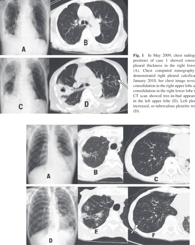
Fig. 1 In May 2009, chest radiograph (supine position) of case 1 showed consolidation and pleural thickness in the right lower lung field (A). Chest computed tomography (CT) scan demonstrated right pleural calcification (B). In January 2010, her chest image revealed cavitary consolidation in the right upper lobe and improved consolidation in the right lower lobe (C, D). Chest CT scan showed tree-in-bud appearance (arrow) in the left upper lobe (D). Left pleural effusion increased, so tuberculous pleuritis were suspected (D).
Fig. 2 In November 2012, chest radiograph and chest computed tomography (CT) scan of case 2 showed consolidation in the left lower lobe and fibrous tubercle in the right upper lobe which was unconnective to pneumonia (consolidation) (A, B, C). In March 2013, his chest radiograph revealed multiple nodular shadows in the whole lung field and improved consolidation in the left lower lobe (D). Chest CT scan demonstrated bronchogenic spread of centrilobular nodules and tree-in-bud appearance (arrow) (E, F). Small cavities in the right upper lobe were also present (E).
420 結核 第 92 巻 第 4 号 2017 年 4 月 PS3 になった。胸部 CT で右上葉に肺炎と無関係な線維 化結節があり(Fig. 2 B),胃瘻手術に際し肺結核を検索 したが,痰抗酸菌培養陰性 × 3 ,IGRA は QFT-3G が 0.15 と判定保留だった。胃瘻造設後も肺炎を繰り返し,4 カ 月後の胸部 CT 再検査では両側肺にびまん性に小葉中心 性 結 節 影 が 出 現(Fig. 2 D, E, F),右 下 葉 に tree-in-bud appearance5)(Fig. 2 F),右上葉に空洞病変が出現した(Fig.
2 E)。痰抗酸菌検査で塗抹陽性,PCRは結核菌群陽性で, 専門病院に転院した。QFT-3G の再検査結果は,0.42 と 陽性だった。
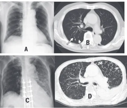
Fig. 3 In July 2009, chest radiograph of case 3 showed several small nodules in the right middle and lower lung field (A). Chest computed tomog-raphy (CT) scan demonstrated micronodules (arrows) of 2 _ 3mm in size, some of which were calcified (B). In June 2013, his chest radiograph (supine position) revealed bilateral multiple mi-cronodular shadows and bolts of metal for tho-racic and lumbar vertebral fracture (C). Chest CT scan revealed micronodules of 2 _ 4 mm in size randomly distributed in the both lungs, so he was diagnosed with miliary tuberculosis (D). However, small cavities were seen in the left upper lobe (D). Namely, bronchogenic lesions were also present. Left pleural effusion was supposed to be caused by congestive heart failure (C, D).
Getting TB during Decreased PS / M. Takahara 421
状構造と tree-in-bud appearance5)と 5 ∼ 8 mm の結節病変 の 3 つの経気道性散布病変が有意に多く存在した。非活 動性結核患者では線維化,気道血管系の構造変化,気腫 性変化と陳旧性病変が有意に多かった。 小葉中心性結節影は tree-in-bud appearance5)とほぼ同義 語で,Murata ら7)が提唱した。細気管支内腔,その周囲 または連続する肺胞領域での壊死性肉芽腫性炎症を示 す。Im ら5)によると,3 週間以内に診断された初期肺結 核患者 29 例中 28 例の画像で認められた。残り 1 例は, 28 歳女性の粟粒結核症例だった。 症例 1 ,2 では発病前に線維化病変が見られ,発病後 は経気道性散布に特徴的な早期病変が見られた。一方, 症例 3 では発病前石灰化病変が見られ,発病後粟粒結核 様の血行性散布のほか,経気道性散布病変も見られた。 最近の粟粒結核は非若年者の二次結核に伴うタイプが多 い8)。症例 3 のような 2 mm の肉芽腫では計算上内部に 10 個の肺胞を含み,細気管支と交通しやすく,経気道 性散布も起こりうる。藤田8)は上肺野で経気道性散布, 下肺野で血行性散布を示す画像を紹介している。 今回の 3 例は,胸部 CT 検査を追加したことで肺結核 発病後比較的早期に発見できたと思われたが,症例 2 に 関し,2 人の職員が潜在性結核感染症の治療を要した。 院内感染対策上,既往歴の有無にかかわらず,肺結核の 早期発見が重要である。 結 語 高齢 PS 低下患者が誤嚥性肺炎を繰り返す場合,結核 発病を鑑別するため,胸部 CT 検査,複数回の痰抗酸菌 検査が必要である。 症例 3 は 89 歳男性。85 歳時(平成 21 年),市の胸部検 診で異常を指摘され,胸部 CT 検査で一部石灰化を伴う 径 2 ∼ 3 mm の小結節が右中下肺野に数個存在した(Fig. 3 A, B)。痰は出ず抗酸菌培養検査未施行だが,QFT-2G のうち ESAT-6 が 0.42 IU/mL と陽性,陳旧性結核と診断 された。平成 22 年心不全に罹患,平成 24 年交通事故で 胸腰椎破裂骨折,他院で手術された。同年 12 月誤嚥性 肺炎で当院入院,寝たきりで経管栄養が必要な PS4 にな った。翌 25 年 4 月から発熱を繰り返し,6 月当院へ再入 院した。連日 38℃台で一般抗生剤が無効で,胸部 CT で 2 ∼ 4 mm 大の小結節がランダム分布し,粟粒結核と診 断した(Fig. 3 C, D)。但し,左上葉に空洞病変,右中葉に 小葉中心性結節影(Fig. 3 に非表示)と経気道性散布病 変も存在した。左胸水は心不全によるものだった。痰抗 酸菌培養検査で結核菌陽性と判明,専門病院へ転院した。 考 察
PS は Eastern Cooperative Oncology Group(ECOG)が提 唱した癌患者の管理指標で1),0 から 4 まで 5 段階ある。
今回報告の 3 例は 3 または 4 と重症だったため,PS が 低下した経過の中で内因性再燃した二次結核と考察され る4)。
Imら5)によると,tree-in-bud appearance は CT 上で 1 つの
茎から同じ口径の芽が多数生えたように見え,剖検例と の比較で呼吸細気管支や肺胞管の内腔壁にできた肉芽が 乾酪化したものである。それらが芽に該当し,茎は Miller の二次小葉に入る終末細気管支に該当する。 Hatipoğlu ら6)は活動性結核患者の高分解能 CT 所見を 非活動性結核患者と比較,小葉中心性結節または分枝線
結核 第 92 巻 第 4 号 2017 年 4 月 422 稿を終えるに当たり,胸部 CT の読影をされた新潟大 学医歯学総合病院放射線診断科の先生方,結核患者を受 け入れて下さった長岡赤十字病院結核病棟のスタッフの 方々に謝辞を述べたい。 本報告の内容は,第 54 回日本呼吸器学会学術講演会 (2014 年 5 月,大阪)で発表した。 著者の COI(Conflicts of interest)開示:本論文発表内 容に関して特に申告なし。 文 献
1 ) Sørensen JB, Klee M, Palshof T, et al.: Performance status assessment in cancer patients. An inter-observer variability study. Br J Cancer. 1993 ; 67 : 773 775. 2 ) 高原 誠, 三吉政道, 小澤哲夫, 他:一般病院入院後活 動性肺結核と判明した 2 症例の臨床的検討. 日胸. 2009 ; 68 : 234 244. 3 ) 日本結核病学会予防委員会:インターフェロンγ遊離 試験使用指針. 結核. 2014 ; 89 : 717 725. 4 ) 赤川志のぶ:高齢者の結核の現状と治療の実際. 日老 医誌. 2010 ; 47 : 165 173.
5 ) Im JG, Itoh H, Shim YS, et al.: Pulmonary Tuberculosis : CT findings̶Early Active Disease and Sequential Change with Antituberculous Therapy. Radiology. 1993 ; 186 : 653 660.
6 ) Hatipoğlu ON, Osma E, Manisali M, et al.: High resolution computed tomographic findings in pulmonary tuberculosis. Thorax. 1996 ; 51 : 397 402.
7 ) Murata K, Itoh H, Todo G, et al.: Centrilobular Lesions of the Lung: Demonstration by High-Resolution CT and Patho-logic Correlation. Radiology. 1986 ; 161 : 641 645. 8 ) 藤田次郎:粟粒結核. 呼吸. 2013 ; 32 : 1064 1071.
Abstract Between January 2010 and June 2013, three
hos-pitalized patients developed pulmonary tuberculosis during decreased performance status. They consisted of two men and one woman, and were over 60 years old. All of them developed repeated aspiration pneumonia frequently several months before the onset. Chest computed tomography (CT) scan demonstrated fibrotic changes, pleural thickness, and calci-fication of lung parenchyma or pleura before the onset. One case had previous history of tuberculous pleuritis. Two cases without previous history of tuberculosis underwent interferon-gamma release assay (IGRA) before the onset. One case was indeterminant and one case was positive. After they developed tuberculosis, consolidation, tree-in-bud appearance, centri-lobular nodoles, miliary nodules, and cavitary lesions were shown by chest CT scan. If elder patients with decreased performance status repeat aspiration pneumonia, chest
com-puted tomography and multiple sputum mycobacterium culture are necessary for excluding pulmonary tuberculosis diagnosis.
Key words : Performance status (PS), Chest computed
to-mography (CT), Interferon-gamma release assay (IGRA), Aspiration pneumonia
Department of Internal Medicine, National Hospital Orga-nization Niigata National Hospital
Correspondence to: Makoto Takahara, Department of Internal Medicine, National Hospital Organization Niigata National Hospital, 3_52, Akasaka-cho, Kashiwazaki-shi, Niigata 945_ 8585 Japan. (E-mail: takahara@niigata-nh.go.jp)
−−−−−−−−Case Report−−−−−−−−