Title A neurophysiological evidence of capsaicin-sensitive nervecomponents innervating interscapular brown adipose tissue( 本文 (Fulltext) )
Author(s) SHINOZAKI, Keiichi; SHIMIZU, Yasutake; SHIINA, Takahiko;NISHIJIMA, Kazutoshi; ATOJI, Yasuro; NIKAMI, Hideki; NIIJIMA, Akira; TAKEWAKI, Tadashi
Citation [Autonomic Neuroscience-Basic & Clinical] vol.[119] no.[1]p.[16]-[24]
Issue Date 2005-04-29
Rights Elsevier Science Bv
Version 著者最終稿 (author final version) postprint
URL http://hdl.handle.net/20.500.12099/31886
The neurophysiological evidence of heterogeneity of nerve components
innervating interscapular brown adipose tissue
Keiichi Shinozaki1 Yasutake Shimizu1 Takahiko Shiina1 Kazutoshi Nishijima2 Yasuro Atoji2
Akira Niijima3
Tadashi Takewaki1
1Department of Physiology and Pathogenetic Veterinary Sciences, The United Graduate School, Gifu University, 1-1 Yanagido, 501-1193 Gifu, Japan 2Department of Anatomy,
The United Graduate School, Gifu University, 501-1193, Gifu, Japan 3Department of Physiology, Niigata University, Niigata, Japan
Key Words: Brown adipose tissue; Thermogenesis; Sympathetic nervous system; Sympathetic efferent nerve activity
Abstract
Neurophysiological basis for the heterogeneity of the nerve components in the brown adipose tissue (BAT) was examined in this experiment. Efferent nerve signals were recorded from the central cut end of the small nerve filament dissected from the sympathetic nerve innervating the interscapular BAT (IBAT). By focusing on qualitative aspects of observed compound action potentials (spikes) we observed two distinctive types of spikes exhibited by the intercostal nerves innervating IBAT. The spikes mainly appeared upon sympathetic stimulations (cold-stimulation and glucose-administration) were characterized by short amplitudes with relatively narrow duration and their sensitivity to the ganglion blocker, hexamethonium (C6). On the other hand, the spikes seen throughout the experiments were characterized by tall amplitude with wide duration and their insensitivity to C6. Since BAT is activated by cold and feeding via sympathetic nervous system, the short spikes seem to be exhibited by sympathetic fibers. Even though the functional link between IBAT and C6 insensitive fibers remain unanswered, our result suggests IBAT is under control of various nervous system and substances in addition to the control of sympathetic nervous system and NE.
Introduction
Brown adipose tissue (BAT) plays a major role in nonshivering heat production and contributes to regulating body temperature (Smith and Horwitz 1969) as well as energy balance (Rothwell and Stock 1979; Rothwell et al. 1982). BAT is richly innervated by sympathetic nerves and the thermogenic response of BAT has been shown to be under control of the sympathetic nervous system (Seydoux and Girardier 1977). BAT thermogenesis is activated mainly in two completely different physiological conditions: 1) during cold exposure to maintain normal body temperature (cold-induced thermogenesis) (Smith 1969) and 2) during a meal to dissipate part of excess energy intake (diet-induced thermogenesis) (Rothwell and Stock 1979) through the same sympathetic efferent pathways.
Activation of BAT thermogenesis is physiologically mediated via the action of norepinephrine (NE) being released from sympathetic nerve endings (Horwitz et al. 1969; Horwitz et al. 1972). Of the cellular events associated with this activation, the interaction of NE with adrenoceptor β3 subtype is mainly attributed (Arch et al. 1984; Emorine et al. 1989). It is clear that NE released from the sympathetic nerve endings, through the activation of β-receptors of BAT, stimulates a cascade of reactions leading to thermogenesis and cell proliferation. At the same time, however, several lines of evidences have suggested that neural factor(s) other than NE is also involved in controlling the function of BAT. In particular, peptidergic innervations have been morphologically identified along with noradrenergic fibers within BAT (Lever et al.
1988; Norman et al. 1988), and they are thought to be involved in a trophic response as well as cell proliferation of BAT (Cui et al. 1990). In addition, Minokoshi et al have shown that synthesis of fatty acids in BAT is promoted by an electrical stimulation of the sympathetic nerves entering the tissue, but the effect of nerve stimulation is not altered by combined α- and β-adrenoceptor blockers (Minokoshi et al. 1990). These observations clearly indicate the presence of non-adrenergic components in the nerves innervating BAT.
Nevertheless of these facts, neurophysiological basis for the heterogeneity of the nerve components in BAT is not so far understood. In this study, we utilized computer software to focus on more qualitative aspects of observed action potentials in interscapular BAT (IBAT) rather than quantitative information. By using electrophysiological method, we tried to identify the electrical nerve activity expressed by various types of nerve fibers innervating IBAT.
Materials and Methods
Subjects. Male Wister rats, 8-10 weeks in age, were obtained from Nihon SLC,
Shizuoka, Japan. All rats were housed in plastic cages at 24 1 ℃ with a 12:12-h light-dark cycle (light on 0700-1900 h). They were given free access to laboratory chow (LABO MR Stock, Nihon-Nosan, Kanagawa, Japan) and water.
animals. Body temperature of one group was maintained at 37 ℃ throughout the experiment. A second group was exposed to cold and body temperature was lowered to 33 ℃ . For a third group, glucose solution (150 mg/kg) was administered intravenously. Each experimental subject was anesthetized by an intraperitoneal administration of α -chloralose-urethan solution (50 mg/kg and 500 mg/kg, respectively). Prior to electrical nerve recording, an intravenous cannula was placed to the right jugular vein of the subjects for intravenous administration of experimental solutions. The animal was placed on the DC heating pad (Homeothermic Blanket System, Harvard Apparatus, Massachusetts, USA) and colonic temperature was maintained at 37 0.2 ℃ throughout the experiment, except during cold stimulation.
Electrical Nerve Recording. Spontaneous efferent discharge of the nerves was measured by multi-fiber recordings (Niijima et al. 1984). The rat was placed in the prone position. A small incision was made above the scapula and interscapular BAT (IBAT) was partially separated from the muscle below. Five intercostal nerves, which contain sympathetic nerves entering IBAT, were identified and one of the five nerve branches was dissected as distally as possible. When isolating the nerve, care was taken to make sure the nerve was not associated with blood vessels or passing through BAT to the skin. The isolated nerve was placed on a pair of silver-silver chloride wire electrodes while the nerve branch was kept in mineral oil to prevent dehydration.
The original signal of efferent mass discharges was amplified and filtered (low cut at 150 Hz; high cut at 10k Hz) using a Bioelectric Amplifier MEG-1200 (Nihon Kohden,
Tokyo, Japan). The amplified signal was converted to digital signal by Power Lab (ADInstruments, Colorado, USA) and it was then recorded on a computer through the recording software (Chart V5.0.1, ADInstruments, Colorado, USA). The sampling rate was set at 20k Hz. Spikes above a threshold voltage level set just above background were counted by Spike Histogram (version 5, ADInstruments, Colorado, USA). Because signal-to-noise ratio depend on the number of nerve filaments and the condition of contact between nerve and electrodes, the background noise was corrected by measuring postmortem recordings. Throughout the recording, the quality of nerve recording was continuously monitored by an oscilloscope. The amplification, filtration, and sampling rate settings were held constant across all studies.
Experimental Design. We recorded and examined the effect of cold stimulation and intravenous administration of glucose solution on sympathetic nerve activity of IBAT. After prepared nerve samples were placed on the electrodes, animals were allowed to stabilize for 30 min. The baseline activity was recorded for 20 min prior to stimulus protocol. The cold stimulation was applied after 20 min of baseline recording and continued until body temperature reached 33 0.2 ℃. The nerve recording was continued for 30 min after body temperature reached 33 ℃. Likewise, glucose solution was administered intravenously after 20 min of baseline recording and the observation was carried out for another 30 min.
In order to examine if we could discriminate and classify the compound action potentials obtained from the interscapular nerve bundle, we administered the ganglion blocker, hexamethonium bromide (C6) intravenously. To determine the
effective concentration for blocking ganglionic transmission, we tested the effect of C6 on splenic nerve activity before actually testing it on intercostal nerve activity. For recording spontaneous efferent nerve activity of the spleen, the rat was placed on its back and the spleen was exposed. The terminal branch of coeliac sympathetic plexus, which run along with the terminal branch of splenic artery, was confirmed. The nerve was carefully detached from the artery and connective tissue and cut at the surface of the spleen. The isolated nerve was placed on a pair of silver-silver chloride electrodes as described. In order to prevent drying, the nerve was covered with glycerol-mineral oil mixture. The data recording method was the same as described in BAT nerve preparation.
Once the dosage of C6 (20 mg/kg dissolved in 0.9 % NaCl solution) was determined, its effect was examined on IBAT nerve activity in three different occasions. In one group, C6 was administered at basal condition. In other groups (hypothermic group and hyperglycemic group), C6 was administered after previously described protocols were carried out. The nerve recording was monitored and recorded for 30 min after C6 administration.
Data Analysis. Results were expressed as mean SE. Statistical significance was examined by an ANOVA, with post-hoc testing by means of Fischer’s PLSD. To further examine compound action potentials (spikes), we used spike discriminator (Spike Histogram, ADInstruments, Colorado, USA). This software enables us to visualize and quantify the spikes obtained from the experiments. After subtracting background noise, obtained spikes were plotted on the graph. The spikes
were plotted according to its duration of the peak at half its maximum amplitude (X-axis) and amplitude (Y-axis: see Fig. 1). According to the graph, suitable spikes were picked randomly and the shapes were compared.
Histology. Once the data was recorded, morphology of the nerves was examined. The nerve filaments were cut as distally as possible and kept in the prefix solution (2% formalin and 2% glutaraldehyde dissolved in 0.1 M phosphate buffer). Samples were sliced in 2-3μm by microtome and hematoxyline-eosine (HE) stained for microscopic examination.
Results
The compund action potentials (spikes) were recorded from the intercostal nerve
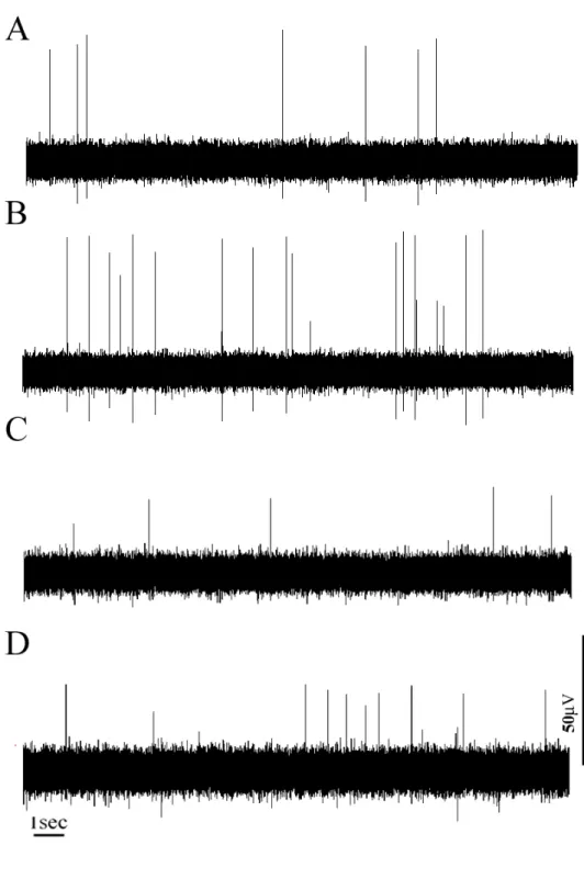
branches innervating IBAT. Fig. 1 represents 20-second excerpts of representative recordings of spontaneous efferent activity in IBAT. Both reduced body temperature and i.v. administration of glucose caused increased sympathetic nervous outflow to IBAT. At the basal condition, the mean rate of efferent mass discharge was 0.66 0.26 Hz in cold-stimulated group and 0.72 0.15 Hz in glucose-administered group . When cold stimulation was given and body temperature was lowered to 33 ℃, efferent nerve activity was significantly activated that spike frequency was raised to 1.74 0.83 Hz compared to the basal activity (P < 0.05, Fischer’s PLSD). Intravenous administration of glucose solution also activated efferent nervous activity significantly
to 1.19 0.26 Hz compared to the basal level (P < 0.05, Fischer’s PLSD).
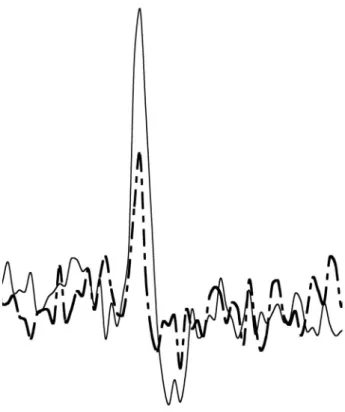
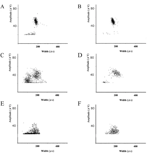
The shape of a typical biphasic spike is shown in Fig.2. Two criteria were measured for spike-to-spike comparison. One was amplitude of first peak and the other was duration of first peak at half its maximum amplitude (b). By plotting spike duration of first peak at half its maximum amplitude on X-axis and its spike amplitude on Y-axis, we examined spike distribution. Figure 3 represents distribution of observed spikes (in 20 minutes) above noise threshold in one subject from both cold-stimulated group (Fig. 3A-B) and glucose-administered group (Fig. 3C-D). Under the normothermic condition, spikes were mostly found in a cluster (duration: 200 μsec, amplitude 50 μV) in cold-stimulated group (Fig 3A). When cold stimulation was given, an additional cluster of spikes (duration: 100 μsec, amplitude 20 μV) appeared (Fig. 3B), being distinguishable from that of normothermic condition. Figure 3C to D represents the spike distribution of a glucose-administered subject and the figures show similar trend to the cold-stimulated example. In addition to the spikes characterized by high amplitudes (40 μV) with relatively long duration (200 μs), lower spikes (15-25 μV) with relatively narrow duration (120 μs) near noise threshold became more evident as stimulus was added (Fig. 3d). In both cases, at the basal state, majority of spikes were clustered above 30 μV while some lower spikes were grouped around noise threshold on the spike distribution graph (Fig. 3A and C). Typical examples of shapes of those obtained spikes are shown in Fig. 4. The spike characterized by high amplitude was seen regardless of the subjects’ state and its duration tended to be relatively wider than those obtained from the spikes with low
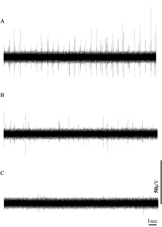
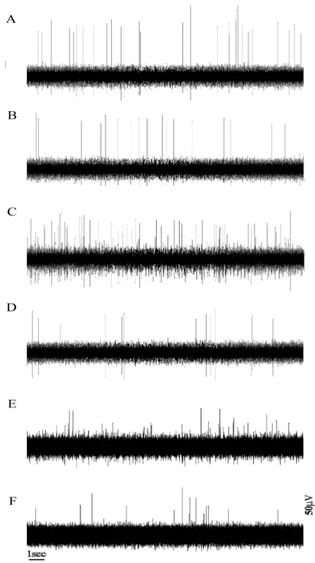
amplitude (Fig. 4 continuous line). On the other hand, the low spike (Fig. 4 dotted line) was mainly seen when the stimuli were applied to the subjects (Fig. 3B and D). Hexamethonium bromide (C6) solution was used for discriminating electrical nerve activity. To decide an appropriate concentration, we examined the effect of C6 on electrical nerve activity of the splenic nerve. The reason why we chose splenic nerve was that it is well recognized as sympathetic nerve component. Fig. 5 represents the efferent nerve activity of a splenic nerve. Upon C6 administration (20 mg / kg), both amplitude and frequency of the spikes were decreased greatly as time progress in splenic nerve and almost no spike was observed by 6 min (Fig. 5B and C). Then we tested C6 of equal concentration for IBAT nerve recordings (Fig. 6). Firstly, we administered C6 at basal condition and almost no inhibition was seen (Fig. 6b). The rate of firing at the basal condition was 0.89 0.3 Hz while ganglion blockade resulted spike frequency of 0.81 0.27 Hz, indicating no significant difference. On the other hand, when C6 was administered after cold stimulation, there was significant inhibition of electrical nerve activity (1.74 0.83 Hz versus 0.99 0.53, P < 0.05, Fischer’s PLSD: Fig. 6C and D). It should be noted that even though C6 administration caused significant inhibition of efferent nerve activity, it did not result in total inhibition of electrical nerve activity. For the glucose-administered group, there was significant difference in electrical nerve activity between before and after C6 administration. (1.19
0.26 Hz versus 0.54 0.24, P < 0.05, Fischer’s PLSD).
Fig. 7 represents more detailed information about obtained spikes. While it is hard to point out the effect of C6 at basal state (Fig. 7B), it is clear that the spikes appeared
upon cold-stimulation and glucose-administration were clearly erased by C6 administration (Fig. 7D and F). The plotted images after C6 administration looked very similar to the control state (data not shown).
Finally, the nerves used for the recordings were detached for morphological examination. The nerves were cross-sectioned in 2-3 μm and HE stained. As shown in Fig. 8A, nerve bundle innervating IBAT consisted of myelinated and unmyelinated fibers. The splenic nerve, on the other hand, was composed exclusively of unmyelinated fibers (Fig 8B).
Discussion
Electrophysiological as well as pharmacological properties of the nerve bundle connected to IBAT was examined in this experiment and we have confirmed that there are at least two different types of nerve fibers within a nerve bundle. Electrophysiologically, intercostal nerves innervating IBAT exhibit: 1) the spike characterized by high amplitude with relatively long duration (large spike) and 2) the spike characterized by low amplitude with relatively narrow duration (small spike). Pharmacologically, the nerves contain: 1) the fibers that are sensitive to the ganglion blocker, hexamethonium and 2) the fibers that are insensitive to it. Morphological examination also revealed two distinctive nerve types within a nerve bundle: 1) thick myelinated fibers and 2) fine unmyelinated fibers. These results demonstrate that BAT is under control of multiple nerve types.
To estimate cellular activity of BAT, electrophysiological method has been widely practiced by measuring efferent mass discharge rate of an intercostal nerve (Niijima et al. 1984; Sakaguchi and Yamazaki 1988; Kurosawa 1991; Kawate et al. 1994). However, qualitative information such as the shape and the duration of spikes has been ignored even though such experiments were carried out in whole-nerve method. Since the shape of spikes in the whole-nerve method is modulated by various aspects such as synchronicity and type of nerve fibers in action, we felt qualitative information of nerve recordings should also be considered. As shown in Fig. 4, magnification of spikes obtained from nerve recordings exhibited drastically different shapes and we tried to examine further detail of such electrophysiological characters of intercostal nerve activity and heterogeneity of neuronal regulation of IBAT.
The thermogenic response of BAT is triggered by the cold (Smith 1969) and feeding (Rothwell and Stock 1979). In deed, hypothermia and hyperglycemia caused facilitated nerve activity as shown in Fig. 1B and D. These results indicate that BAT was activated to maintain body temperature and to dissipate excess energy. Furthermore, in association with the alterations of physiological state, the electrophysiological character of the intercostal nerve innervating IBAT exhibited specific tendency. The number of the small spikes, not so evident at resting state, got drastically increased after stimuli were given while the number of tall spikes seemed not be affected by much (Fig. 3B and D). Considering BAT is richly innervated by sympathetic nerves (Seydoux and Girardier 1977) and both cold-induced and food-induced BAT thermogenesis are activated via sympathetic nervous system, these
short spikes seem to represent the activity of sympathetic nerve fibers. On the other hand, the neuronal origin of the large spikes remained to be unanswered. In order to investigate if it is possible to isolate these different spikes pharmacologically, we examined the effect of ganglionic blockade.
The concentration of hexamethonium (C6) to be administered was decided from the result we obtained from splenic nerve and we confirmed that 20 mg/kg would completely block the transmission at ganglions (Fig. 5). Despite the result we obtained from splenic nerve, administration of C6 did not cause significant inhibition of electrical nerve activity in normothermic group (Fig. 6B). Administration of C6 caused significant inhibition of electrical nerve activity in hypothermic group and hyperglycemic group, but unlike splenic nerve, C6 did not completely erase the activity (Fig. 6D). In Fig. 7, these results can be seen in more detail and it is apparent that the small spikes prevailed upon stimulations were clearly suppressed by C6 administration while the large spikes were not clearly affected by it. This observation not only indicates the nerve bundle innervating the IBAT is composed of C6 sensitive and insensitive fibers, but also represents the small spikes, presumably discharged by sympathetic fibers, are interrelated to these C6 sensitive fibers. Our result is further supported by the observations that C6 inhibits BAT thermogenesis induced by chemical stimulation (Amir 1990) and facilitated NE turnover in BAT induced by electrical stimulation (Saito et al. 1987). Therefore, these data together reflects the evidence that the spikes characterized by tall amplitude was exhibited by the nerve fibers that were not of sympathetic origin. The functional link between IBAT and C6 insensitive fibers,
however, remain unanswered. By focusing on specific characters exhibited by electrical nerve activity and the effect of a ganglionic blocking agent, we were able to separate two distinctive types of compound action potential pharmacologically.
Morphologically, it has been proven the nerve bundles entering IBAT consist at least two types of nerves. Immunohistochemical staining of the nerves innervating the IBAT revealed that along with an enzyme tyrosine hydroxylase (TH), the neuropeptides such as neuropeptide Y (NPY), substance P (SP), calcitonin gene-related peptide (CGRP) are widely distributed within IBAT (Norman et al. 1988). In addition, SP and CGRP are identified in thick myelinated fibers while NYP is co-stored with NE in thin unmyelinated fibers (Cannon et al. 1986; De Matteis et al. 1998). These neuropeptides in the peripheral nerves are characteristic of sensory fibers. In present study, we indeed found thick myelinated fibers and thin unmyelinated fibers in an IBAT nerve bundle (Fig. 8A). In comparison, the nerve bundle innervating spleen, whose activity was sensitive to C6, was only composed of thin unmyelinated fibers (Fig. 8B). These observations suggest C6 sensitive fibers found in IBAT nerve bundle were sympathetic nerve fibers and the thick myelinated fibers seen within IBAT nerve bundles in our study were more than likely the sensory fibers.
The major function of sensory fibers is to transmit information received by various sensory receptors located within peripheral tissues to central nervous system. However, sensory fibers are also involved in efferent functions in many tissues including BAT. In fact, administration of capsaicin, the pungent principle in hot peppers known for impairing the function of a subpopulation of sensory fibers (Holzer
1991) greatly alters cellular content of BAT (Cui et al. 1990). If the subject was treated with capsaicin, BAT becomes atrophied (Cui and Himms-Hagen 1992) and CGRP distribution gets scarce (Giordano et al. 1998). In addition, the content of the uncoupling protein, responsible for thermogenesis in BAT (Cui et al. 1990) is greatly reduced even though NE concentration is left unchanged (Himms-Hagen 1990). Since the unmyelinated efferent fibers of the sympathetic nervous system are not directly sensitive to capsaicin (Cervero and McRitchie 1982), it can be considered that even if noradrenergic sympathetic fibers are alive, impairment of efferent sensory nerves result in BAT to dysfunction. Taken together, we suspect a cluster of the spikes that were constitutively present regardless of C6 administration was produced by the efferent sensory nerve fibers.
Even though specific function of efferent sensory fibers innervating the BAT is not fully understood, they are probably involved in trophic response of BAT (Cui et al. 1990; Himms-Hagen 1990; Giordano et al. 1998). In addition, efferent sensory fibers in IBAT may take a role in protecting the tissue from overheating because direct application of CGRP on IBAT attenuate thermogenesis induced by NE (Osaka et al. 1998). If the efferent sensory fibers innervating IBAT are actually involved in thermoregulation, the functional link between IBAT and C6 insensitive fibers can be proposed that identified thick myelinated fibers presumably insensitive to C6 and containing CGRP and SP act as inhibitory fibers against stimulatory noradrenergic fibers (Cui et al. 1990; Osaka et al. 1998).
structure (Geloen et al. 1992). In contrast to a typical multilocular structure of brown adipocytes found in normal rats, the denervated rats’ BAT was largely composed of adipocytes filled with large lipid droplets, some of which appeared unilocular (Minokoshi et al. 1986). These histological changes were reflected in the gross appearance of the tissue: the denervated one was much paler than the control and looked like white adipose tissue (Minokoshi et al. 1986). This suggests that even when thermogenesis is not needed for survival, the intact nerves are needed for maintaining the characteristic feature of BAT. Hence the fact that the hexamethonium-insensitive nerves are constitutively active even when the thermogenesis in BAT is not activated explains hexamethonium-insensitive nerves may play a role in conserving the tissue as brown adipocytes.
In summery, by focusing on qualitative parameters represented by electrical nerve activity, we were able to see the difference that had not been understood by just looking at the change in mean discharge rate. Previously, purpose of measuring electrical nerve discharge was to evaluate either given stimulus facilitates or suppresses nervous activity. However, our experiment showed that examination of electrophysiological properties could serve as a bridge over phenomenology and morphology. In present experiment, we have confirmed that the nerve filament innervating IBAT is composed of at least two different types of fibers. Pharmacologically, one type of nerve fibers was sensitive to C6 while another type of fibers was insensitive to it. Physiologically, one type of nerve fibers create tall and wide spikes while the other create short and narrow spikes. And morphologically, the
nerve bundles innervate the BAT are composed of thick myelinated fibers and thin unmyelinated fibers. Our result suggests IBAT is under control of various nervous system and substances in addition to the control of sympathetic nervous system and NE.
Amir, S. 1990. Activation of brown adipose tissue thermogenesis by chemical stimulation of the posterior hypothalamus. Brain Res 5341-2, 303-8.
Arch, J. R., A. T. Ainsworth, M. A. Cawthorne, V. Piercy, M. V. Sennitt, V. E. Thody, C. Wilson and S. Wilson 1984. Atypical beta-adrenoceptor on brown adipocytes as target for anti-obesity drugs. Nature 3095964, 163-5.
Cannon, B., J. Nedergaard, J. M. Lundberg, T. Hokfelt, L. Terenius and M. Goldstein 1986. 'Neuropeptide tyrosine' (NPY) is co-stored with noradrenaline in vascular but not in parenchymal sympathetic nerves of brown adipose tissue. Exp Cell Res 1642, 546-50.
Cervero, F. and H. A. McRitchie 1982. Neonatal capsaicin does not affect unmyelinated efferent fibers of the autonomic nervous system: functional evidence. Brain Res 2391, 283-8.
Cui, J. and J. Himms-Hagen 1992. Rapid but transient atrophy of brown adipose tissue in capsaicin-desensitized rats. Am J Physiol 2624 Pt 2, R562-7.
Cui, J., G. Zaror-Behrens and J. Himms-Hagen 1990. Capsaicin desensitization induces atrophy of brown adipose tissue in rats. Am J Physiol 2592 Pt 2, R324-32. De Matteis, R., D. Ricquier and S. Cinti 1998. TH-, NPY-, SP-, and
CGRP-immunoreactive nerves in interscapular brown adipose tissue of adult rats acclimated at different temperatures: an immunohistochemical study. J
Neurocytol 2712, 877-86.
Emorine, L. J., S. Marullo, M. M. Briend-Sutren, G. Patey, K. Tate, C.
Delavier-Klutchko and A. D. Strosberg 1989. Molecular characterization of the human beta 3-adrenergic receptor. Science 2454922, 1118-21.
Geloen, A., A. J. Collet and L. J. Bukowiecki 1992. Role of sympathetic innervation in brown adipocyte proliferation. Am J Physiol 2636 Pt 2, R1176-81.
Giordano, A., M. Morroni, F. Carle, R. Gesuita, G. F. Marchesi and S. Cinti 1998. Sensory nerves affect the recruitment and differentiation of rat periovarian brown adipocytes during cold acclimation. J Cell Sci 111 ( Pt 17)2587-94. Himms-Hagen, J., J. Cui, and S. L. Sigurdson 1990. Sympathetic and sensory nerves in
control of growth of brown adipose tissue: effects of denervation and of capsaicin. Neurochem. Int. 17271-279.
Holzer, P. 1991. Capsaicin as a tool for studying sensory neuron functions. Adv Exp Med Biol 2983-16.
Horwitz, B. A., J. R. Detrick and R. E. Smith 1972. Norepinephrine-induced thermogenesis: effect of interscapular brown fat. Experientia 283, 284-6. Horwitz, B. A., J. M. Horowitz, Jr. and R. E. Smith 1969. Norepinephrine-induced
depolarization of brown fat cells. Proc Natl Acad Sci U S A 641, 113-20. Kawate, R., M. I. Talan and B. T. Engel 1994. Sympathetic nervous activity to brown
adipose tissue increases in cold-tolerant mice. Physiol Behav 555, 921-5. Kurosawa, M. 1991. Reflex changes in thermogenesis in the interscapular brown
adipose tissue in response to thermal stimulation of the skin via sympathetic efferent nerves in anesthetized rats. J Auton Nerv Syst 331, 15-23.
Lever, J. D., S. Mukherjee, D. Norman, D. Symons and R. T. Jung 1988. Neuropeptide and noradrenaline distributions in rat interscapular brown fat and in its intact and obstructed nerves of supply. J Auton Nerv Syst 251, 15-25.
Minokoshi, Y., M. Saito and T. Shimazu 1986. Metabolic and morphological alterations of brown adipose tissue after sympathetic denervation in rats. J Auton Nerv Syst 153, 197-204.
Minokoshi, Y., M. Saito and T. Shimazu 1990. Adrenergic blockade paradoxically increases lipogenic response of brown adipose tissue to sympathetic nerve stimulation. Neurosci Lett 1093, 341-6.
Niijima, A., F. Rohner-Jeanrenaud and B. Jeanrenaud 1984. Role of ventromedial hypothalamus on sympathetic efferents of brown adipose tissue. Am J Physiol 2474 Pt 2, R650-4.
Norman, D., S. Mukherjee, D. Symons, R. T. Jung and J. D. Lever 1988. Neuropeptides in interscapular and perirenal brown adipose tissue in the rat: a plurality of innervation. J Neurocytol 173, 305-11.
Temperature- and capsaicin-sensitive nerve fibers in brown adipose tissue attenuate thermogenesis in the rat. Pflugers Arch 4371, 36-42.
Rothwell, N. J. and M. J. Stock 1979. A role for brown adipose tissue in diet-induced thermogenesis. Nature 2815726, 31-5.
Rothwell, N. J., M. J. Stock and D. Stribling 1982. Diet-induced thermogenesis. Pharmacol Ther 172, 251-68.
Saito, M., Y. Minokoshi and T. Shimazu 1987. Ventromedial hypothalamic stimulation accelerates norepinephrine turnover in brown adipose tissue of rats. Life Sci 412, 193-7.
Sakaguchi, T. and M. Yamazaki 1988. Hepatic portal injection of glucose elevates efferent sympathetic discharges of interscapular brown adipose tissue. Exp Neurol 1013, 464-9.
Seydoux, J. and L. Girardier 1977. Control of brown fat thermogenesis by the sympathetic nervous system. Experientia 339, 1128-30.
Smith, R. E. and B. A. Horwitz 1969. Brown fat and thermogenesis. Physiol Rev 492, 330-425.
Smith, R. E. a. H., B.A. 1969. Brown fat and thermogenesis. Physiological Review 49330-425.
Fig. 1. Typical recordings of spontaneous efferent nerve activity of IBAT. (A) At
normothermic state (37℃). (B) At hypothermic state (33℃). (C) At normothermic state. (D) At hyperglycemic state. Both cold stimulation and glucose administration caused facilitated efferent nerve activity.
Fig. 2. Diagram showing the spike shape measurements taken in this study. (a),
Fig 3. Spike distribution of cold-induced subject (A-B) and glucose-induced subject
(C-D). The spikes are plotted according to its duration of the peak at half its maximum amplitude (X-axis) and amplitude (Y-axis). (A) At basal body temperature. Two groups of spikes can be seen. Majority of spikes are clustered around axis of 200 μs and 50 μV (large spikes) while small number of spikes are seen about 100 μs and 20 μV (small spikes. (B) At hypothermic state. More small spikes are evident compared to the basal state. (C) At basal state. (D) After glucose administration. More small spikes are evident near noise threshold and they started to synchronize their activity, resulting in elevated amplitude.
Fig. 4. Typical compound action potentials (spikes) recorded from the IBAT nerve.
Examples of two spikes recorded from the same nerve filament. Dashed line represents the small spike. It is characterized by short amplitude and narrow duration. It should be noted that depending on an individual, duration of the short spikes was greatly varied. Continuous line represents the large spike. It has much taller amplitude and its duration is relatively long.
Fig. 5. Spontaneous efferent nerve activity of splenic nerve at basal body temperature (37℃). (A) Before C6 administration. (B) 3 minutes after C6 administration. (C) 6 minutes after C6 administration. Note that by 6 minutes, nerve activity is completely diminished.
Fig. 6. Effects of ganglionic blockade on normothermic state (A-B), hypothermic
state (C-D), and hyperglycemic state (E-F). At normothermic state, hexamethonium (C6) did not drastically inhibit efferent nerve activity (B). At hypothermic state, C6 significantly inhibited efferent nerve activity. However, some spikes are still seen after 6 minutes (D). At hyperglycemic state, efferent nerve activity was somewhat inhibited (F).
Fig. 7. Changes induced by ganglion blockade on spike distribution. At basal state
(A-B), hypothermic state (C-D), and hyperglycemic state (E-F). Hexamethonium (C6) clearly inhibited the small spikes while large spikes were largely left unchanged.
Fig. 8. (a) HE-stained IBAT nerve filament (x 200). The nerve contains myelinated
fibers (represented by ▼) and clusters of unmyelinated fibers. (b) HE-stained splenic nerve. The nerve contains predominantly unmyelinated fibers.