INTRODUCTION
Subpubic cartilaginous cyst was first reported in 1996 by Algucial -Garcia and Littman (1). It is a rare tumor - like mass that contains degenerative cartilaginous tissues arising from the pubic symphy-sis (1). A few case reports have described the cyst to be stable in size (2) (3) or to reduce only slightly in size over time (4), with no reports of cases where the tumor regressed. This report presents a first case of subpubic cartilaginous cyst that showed complete spontaneous regression.
CASE REPORT
A 59 year old woman presented to her gynecologist with a 2 -year history of a vulvar mass with dull pain. Ultrasonography re-vealed a tumor in the pubic symphysis and she was subsequently referred to our department for further examination.
Physical examination revealed an immobile vulvar mass. Plain radiography showed slight sclerotic changes around the pubic symphysis. Magnetic resonance imaging (MRI) revealed a 1.5
!
1.0!
1.0 cm mass located anterior to the middle pubic symphysis (Figure 1). The lesion presented as a low intensity area on T1 -weighted images and as a homogenously high - intensity area on T2 - weighted images. The lesion also showed high - intensity at-tenuation on short T1 inversion recovery images. Gadolinium en-hancement was not observed in the mass expect peripheral lesion. Positron emission tomography/computed tomography showed no 18 - fluorodeoxyglucose accumulation in the soft tissue mass on the anterior pubic symphysis, but showed bone erosion of the an-terior pubis. These findings suggested the possibility of a subpubic cartilaginous cyst.Since the patient had only dull pain, follow - up observation by MRI was decided. She had continuous dull pain and the tumor still remain at 18 months after her initial visit and she started wear a pelvic brace. Forty - eight months after her initial visit, repeat MRI showed the cyst had completely disappeared and the dull pain also
disappeared (Figure 2).
DISCUSSION
Subpubic cartilaginous cyst was first reported by the patholo-gists, Algucial - Garcia and Littman, in 2 patients who presented with an unusual type of subpubic cystic mass (1). They described a tumor formed from fibrocartilaginous tissue with extensive cystic degenerative changes. This benign cyst appeared to be related to, and perhaps originating from, the periarticular tissues of the pubic symphysis. Although a few cases have been reported since, there have been no other cases in which the tumor completely regressed, as in the present case.
We reviewed 12 cases of subpubic cartilaginous cyst reported in the literature to date (Table 1) (1 - 10). Almost all cases involved multiparous females in their 50’s, 60’s, and 70’s who presented with vulvar masses and various symptoms. Four cases presented as painless vulvar mass (1), 3 as a painful mass or abdominal pain (2), 4 as urinary dysfunction (1, 9), and in one rare male case as pain in the basis of the penis and sexual dysfunction (4).
In 2004, Kimet al. described the MRI features of a subpubic cartilaginous cyst (5). The lesion had a broad margin of contact with the adjacent pubic symphysis. In later publications it was de-scribed as hypointense relative to muscle on T1 - weighted se-quences, heterogeneously hyperintense on T2 - weighted sese-quences, and with a thin enhancing wall with no internal enhancement (2). Plain radiography showed a relatively wide pubic symphysis with sclerosis (7). These MRI and radiographic findings are relatively specific, meaning that diagnosis should be possible based on such typical image findings.
The differential diagnosis includes aggressive anigomyxoma, non - ovarian cyst, urethral diverticula, vulval carcinoma, and chon-drosarcoma (3) (6). Aggressive anigomyxoma (11) is one of the benign tumor affecting the pelvis and perineum. The tumor has a well - defined margin and shows hypointense in T1 - weighted images and high intensity in T2 - weighted images. This tumor has a specific feature, a swirled appearance, and hypervascular unlike subupubic cartilaginous cysts. Non - ovarian cyst (12) such as Nabothian cysts, Bartholin’s cyst, and Gartnar’s cyst, is not asso-ciated with the pubic symphosis. Urethral diverticula (13) are ana-tomically different from subpubic cartilaginous cyst. Cancer of the vulva (14), accounts for 3 - 5% of primary gynecologic malignancies,
CASE REPORT
Complete spontaneous regression of a subpubic cartilaginous
cyst : a case report
Toshihiko Nishisho1, Shoichiro Takao2, Ryo Miyagi1, Shunichi Toki1, Akihiro Nagamachi1, and Koichi Sairyo1
1
Department of Orthopedics, Institute of Biomedical Sciences, Tokushima University Graduate School, Tokushima, Japan,2Department of Ra-diology, Institute of Biomedical Sciences, Tokushima University Graduate School, Tokushima, Japan
Abstract : Subpubic cartilaginous cyst was recently reported as a rare degenerative mass on the pubic symphysis. We report here a 59-year -old woman who presented with a vulvar mass that showed complete spontaneous re-gression 48 months after the initial visit. Treatment was only wearing brace. This is the first report of complete spontaneous regression of a subpubic cartilaginous cyst. In the case of small subpubic cyst, observation and follow-up alone may be sufficient. J. Med. Invest. 63 : 319-322, August, 2016
Keywords : cartilaginous cyst, pubic symphysis, magnetic resonance imaging, spontaneous regression
Received for publication April 14, 2016 ; accepted July 28, 2016. Address correspondence and reprint requests to Toshihiko Nishisho, MD, PhD, Lecturer, Department of Orthopedics, Institute of Biomedical Sciences, Tokushima University Graduate School 3 - 18 - 15 Kuramoto, Tokushima 770 - 8503, Japan and Fax : +81 - 88 - 633 - 0178.
The Journal of Medical Investigation Vol. 63 2016
was isointense to muscle on T1 weighted images and showed in -termediate - to - high signal intensity on T2 - weighted scans. Fifty percent of cancer of the vulva showed enhancement. The location was different from subpubic cartilaginous cyst because they are not associated with the pubic symphosis. Chondrosarcomas are rare primary bone tumor. Large and multi - lobulated mass in pelvis can mimic chondrosarcoma (3).
Although needle biopsy (2) and open biopsy (3) were performed in 2 cases, biopsy may be limited to difficult cases only, such as those with a huge mass with intrapelvic and extrapelvic components that had a possibility of sarcoma (3). Performing biopsy from an anatomi-cally difficult location may cause tumor contamination if it is sar-coma. In cases where biopsy is difficult, a metastable technetium - 99
methylene diphosphonate whole - body bone scintigraphy would be helpful as it demonstrate mild radiotracer uptake within the sym-physis pubis, but none in the parasymphyseal mass itself (3). These bone scintigraphy findings may indicate degenerative changes. Moreover, no 18 - fluorodeoxyglucose accumulation in positron emis-sion tomography/computed tomography indicate a little possibil-ity of malignancy. The diagnosis of bone and soft tissue tumor are difficult, therefore, it is better to consult radiologist and orthopedic surgeon who major in musculoskeletal oncology.
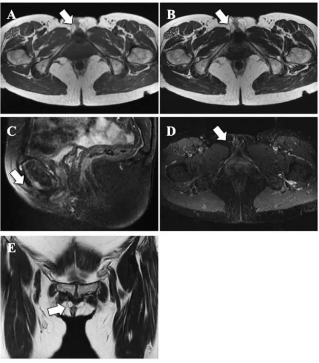
Treatment for subpubic cyst is observation or resection. Almost all cases that presented with symptoms, such as urinary retention, underwent tumor resection ; observation was chosen in almost all asymptomatic cases. Cyst aspiration and instillation of local Figure 1 MRI images show a 1.5
!
1.0!
1.0 cm mass anterior to the middle pubic symphysis (white arrow). A : Axial T1 - WI shows low - intensity attenuation ; B : axial T2 - WI and C : sagittal STIR images show high - intensity attenuation ; D : sagittal contrast - enhanced MRI showing no gad-olinium enhancement of the tumor except for peripheral lesion ; E. coronal T2 - WI.MRI, magnetic resonance imaging ; WI, weighted imaging ; STIR, short T1 inversion recovery
steroids resulted in only a temporary reduction of the mass and not in complete regression (7). Our patient had only dull pain and a pelvic brace may be effective because the pain and mass was dis-appeared after she wore the brace, so we opted for MRI follow - up of the lump.
In previous reports, watchful waiting was undertaken in 4 cases of subpubic cyst (2) ; cyst size did not change in 2 cases that were followed - up at 48 months or in one case that was followed - up at 9 months. Only one case showed slight reduction in size and im-proved symptoms on follow - up at 4 years. In comparison, the sub-pubic cyst in our case was relatively small, at 1.5
!
1.0!
1.0 cm. We speculate that size may influence spontaneous resolution of such benign cysts.In conclusion, subpubic cartilaginous cyst may spontaneously regress. Although surgery would be necessary for severe sympto-matic cases, in the case of asymptosympto-matic or small subpubic cyst, observation and follow - up alone may be sufficient.
CONFLICTS OF INTEREST
The authors have no conflicts of interest to declare.
No funds were received in support of this work and there were no relevant financial activities outside the submitted work. Figure 2 Follow - up MRI at 48 months
A : Axial T1 - WI, B : axial T2 - WI, C : sagittal fat - suppressed T2 - weighted image, D : axial STIR, and E : coronal T2 - WI images show the mass had disappeared completely
MRI, magnetic resonance imaging ; WI, weighted imaging ; STIR, short T1 inversion recovery
ACKNOWLEDGEMENTS
noneREFERENCES
1. Alguacil - Garcia A, Littman CD : Subpubic cartilaginous cyst : report of two cases. Am J Surg Pathol 20 : 975 - 979, 1996 2. Ergun T, Lakadamyali H, Aydin O : Subpubic cartilaginous
cyst : incidental finding detected by abdominopelvic computed tomography. Radiat Med 26 : 46 - 49, 2008
3. Tan TJ, Wong SK, Foo LS : A parasymphyseal pubic cartilagi-nous cyst masquerading as a chondrosarcoma. Clin Radiol 67 : 508 - 510, 2012
4. Wylie KR, Griffiths J, Pye J, Salim F, Inman R : A subpubic car-tilaginous cyst causing neurological and sexual symptoms in a 69 - year - old man. J Sex Med 11 : 2601 - 2604, 2014 5. Kim CE, Beasley HS : MRI diagnosis of subpubic cartilaginous
cyst. AJR Am J Roentgenol 182 : 144 - 146, 2004
6. Bullock RW, Soares DP, Shah S : Subpubic cartilaginous cyst : an unusual cause of a vulval mass. BMJ Case Rep 2009 : 2009 7. Hoogendoorn R, Kayser H, Weening J, van Geloven A :
Sub-pubic cartilaginous cystic lesion presenting as a vulvar mass :
a case report. J Med Case Rep 3 : 7294, 2009
8. Judson PL, Ivy JJ, Zwolak P, Manivel JC, Clohisy DR : A sus-picious vulvar mass diagnosed as a subpubic cartilaginous cyst. Arch Gynecol Obstet 280 : 107 - 109, 2009
9. Sava MR, Rubin B, Sundaram M : Subpubic cyst. Subpubic degenerative cyst arising from the fibrocartilage of the pubic symphysis. Skeletal Radiol 41 : 851 - 852, 2012
10. Farag F, van der Geest I, Hulsbergen- van de Kaa C, Heesakkers J : Subpubic cartilaginous pseudocyst : orthopedic feature with urological consequences. Case Rep Urol 2014 : 176089, 2014
11. Outwater EK, Marchetto BE, Wagner BJ, Siegelman ES : Ag-gressive angiomyxoma : findings on CT and MR imaging. AJR Am J Roentgenol 172 : 435 - 438, 1999
12. Kier R : Nonovarian gynecologic cysts : MR imaging findings. AJR Am J Roentgenol 158 : 1265 - 1269, 1992
13. Dwarkasing RS, Dinkelaar W, Hop WC, Steensma AB, Dohle GR, Krestin GP : MRI evaluation of urethral diverticula and differential diagnosis in symptomatic women. AJR Am J Roent-genol 197 : 676 - 682, 2011
14. Sohaib SA, Richards PS, Ind T, Jeyarajah AR, Shepherd JH, Jacobs IJ, Reznek RH : MR imaging of carcinoma of the vulva. AJR Am J Roentgenol 178 : 373 - 377, 2002
Table. 1 Clinical data for subpubic cartilaginous cyst
Author (Year) Case Sex Age Gravida (G)
and Para (P) Symptoms
Size in MRI (mm) Treatment Follw - up result of observation Complication Alguacil -Garciaet al. (1996) (1) 1 Female Postmenopausal woman NA Pain and urinary disfunction NA Operation NA NA 2 Female Postmenopausal woman NA Painless lump NA Operation NA NA Kimet al. (2004) (5) 3 Female 70 G3P3 Painless lump 30 Operation NA NA Ergunet al. (2008) (2) 4 Female 54 G3 Pain NA Observation after needle biopsy No change after 48 months NA Bullocket al. (2009) (6) 5 Female Postmenopausal woman NA Painless lump 38 Observation No change after 48 months NA Hoogendoornet al.
(2009) (7) 6 Female 55 G4P3 Pain 37
!
36!
35 Operation NAPain after operation Judsonet al.
(2009) (8) 7 Female 62 G2 Pain 18