Long-term Follow-up of a Feline Solitary Osteochondroma
on the Left Mandibular Ramus and Temporomandibular
Joint with Surrounding Tissue Treated by Palliative Surgery
左下顎枝、顎関節およびその周囲に発生した孤立性骨軟骨腫に対し
緩和的外科的切除を実施し長期的経過観察を行った猫の一例
Jun MATSUMOTO1, 2)*, Ayako OKUDA3), Yoshimi UCHIDA1), Naoko OGI1) and Etsuko OGI1) 松本 淳1, 2)* 奥田 綾子3) 内田 佳美1) 小儀 直子1) 小儀 悦子1)
1) Ogi Animal Hospital, 17-3 Takashiro-cho, Suita-shi, Osaka 564-0024, Japan
2) Ibarakimotomachi Animal Hospital, 6-30 Motomachi, Ibaraki-shi, Osaka 567-0882, Japan
3) Vettec Dentistry, 3-20-7 Higashimukojima, Sumida, Tokyo 131-0032, Japan
Summary: A 5 years old, spayed domestic short-hair cat presented because of difficulty opening the mouth and loss of appetite. A firm-swelling was found on her left temporal area. The cat was FeLV negative. Surgical exploration was performed for biopsy and volume reduction. The pathological diagnosis was osteochondroma. The tumor could not be removed completely at two additional surgeries. Fifty-three months after the first surgery, remaining tumor growth decelerated, which was accompanied by the appearance of radio-transparent areas inside of the tumor. The long axis of the tumor increased about 1.3 mm during the final 59 months. The tumor remained in her left temporal area for more than 10 years; the cat continued to be able to eat normally. She died of unknown causes at 16 years of age, with no obvious metastasis and/or acute growth.
Key words: feline, solitary osteochondroma, palliative surgery
要約:5歳、日本猫、避妊済みの雌猫が食欲不振と開口障害を主訴に来院した。一般身体検査で、左側頭部に硬性の腫瘤性 病変を認め、FeLV検査は陰性であった。その腫瘤性病変に対し、診断と減容積を目的とする切除生検を行い、骨軟骨腫と 病理組織診断された。その後、2回の外科的切除を実施したが、完全切除には至らず、開口障害の改善にとどまった。切除 生検後53ヶ月目から腫瘍病変の増大速度の低下とレントゲン透過性の亢進像が認められ、最後の59ヶ月間では1.3 mmの 増大にとどまった。16歳時に骨軟骨腫とは別の要因により死亡した。約10年間の長期的経過観察中、腫瘍は存在したもの の、転移はなく、開口障害もなく、通常の摂食行動が可能であった。 キーワード:猫、孤立性骨軟骨腫、緩和的外科的切除
Jpn. J. Vet. Anesth. Surg. 51(3&4): 52–57, 2020.
Introduction
Osteochondromas are cartilage-capped benign bony tumors arising from the external bone surface,
continuous with that of the underlying bone9, 10).
Osteochondromas can be solitary (monostotic) or multiple (polyostotic). Although they have rarely been reported converting to malignancy, especially if polyostotic and presenting at a relatively older age1, 2, 4, 9, 10), prognosis of the polyostotic type in all
species reported is generally poor9).
Osteochondro-mas develops commonly at the endochondral areas of long bones in humans, dogs, pigs and horses early in life, but the osteochondromas in cats develops long bone as well as skull bones continuously later
age in life9, 10, 12). In domestic cats, the tumor,
com-monly called feline osteochondomatosis, presents
1)小儀動物病院 (〒564-0024 大阪府吹田市高城町17-3)
2)茨木元町どうぶつ病院(〒567-0882 大阪府茨木市元町6-30)
3)Vettec Dentistry(〒131-0032 東京都墨田区向島3丁目20-7)
*Corresponding author: Jun MATSUMOTO, E-mail: [email protected]
53
Long-term Follow-up of a Feline Solitary Osteochondroma Treated by Palliative Surgery
as multiple progressive lesions at 2–3 years of age, and has been linked with feline leukemia virus
(FeLV)7–9, 11, 12).
Case Report
A 5 years old, spayed domestic short-hair cat presented with difficulty opening the mouth and loss of appetite. A firm-swelling was palpated on the left temporal area. While conscious, the mouth could not be opened fully. On physical examination, the cat was normal except for the temporal mass and in-ability to examine the mouth. Complete blood count (CBC) and serum chemistry tests were within normal limits except for high alkaline phosphatase level (290 IU/l). Feline immunodeficiency virus (FIV) and FeLV tests were negative. Serum thyroid hormone level was within the normal range. Chest radiographs were normal.
Under sedation [by isofluren (Isoflurane Inhala-tion SoluInhala-tion, Pfizer, Tokyo, Japan) with inducInhala-tion chamber], the mouth could be opened no further than when the cat was not sedated. On radiographs of the head, a large highly opaque lesion was found affecting the coronoid process of the left mandibular ramus, spreading to the zygomatic arch (Fig. 1); the lesion was unevenly opaque. The limited jaw
move-ment was caused by the coronoid process lesion. Under general anesthesia by isofluren with intuba-tion after premedicated by 0.01 mg/kg glycopyrrolate (Robinul, Aspen, Sydney, Australia) and 0.2 mg/kg butorphanol (Vetorphale, Meiji Seika Pharma, Tokyo, Japan) and with isofluren in an induction chamber, surgical exploration was performed for biopsy and volume reduction. The nodular lesion was removed progressively until the jaw was able to be opened fully and was submitted for pathological examination (Fig. 2). Following surgery, the cat could open her mouth, and had a normal appetite. Mandibular shift to the left was seen 2 weeks after the 1st surgery.
The histopathological diagnosis (Hist Vet. Inc, Kanagawa, Japan) was osteochondroma with prolif-eration of bone and cartilage tissues; there was no evidence of malignancy or obvious mitosis (Fig. 3).
The zygomatic arch, mandibular ramus, condylar process and caudal part of the horizontal ramus of the left mandible were resected palliatively one month after the 1st surgery for maintaining quality of life (QOL). The lesion could not be completely removed because of its extension medially (Fig. 4). Complete crawn amputation without endodontic treatment and covered by gingiva was performed on mandibular canine teeth by preventing occlusal trauma caused by jaw shift and stage 3 of type II resorptive lesions.
Fig. 1. Preoperative radiographs. (A) Head DV image shows calcified mass on the left mandibular ramus extending laterally and distally. The long axis of the calcified mass measured 35 mm. (B) Head slightly oblique lateral image.
Fig. 2. Head DV radiograph immediately after the first surgery for biopsy and volume reduction. Some calcified lesions (white arrows) remain dorsal to the left mandibular ramus and around the left TMJ.
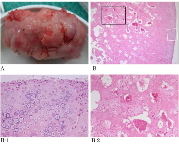
Fig. 3. Part of mass removed at the first surgery. (A) The mass had a hard nodular surface.
(B) Histopathology of the mass surface at low-magnified field (HE×40). B-1 (white square in B): Surface of the mass covered by cartilage that is growing (HE×200). B-2 (black square in B): The center of the mass consisted by bony tissue with growing cartilage undergoing growth. These histological images are similar to those seen in endochondral ossification (HE×200).
Fig. 4. Head DV radiograph after second surgery, at which the center of the zygomatic arch, the mandibular ramus, the condylar process and the caudal part of the mandibular body were removed. Some lesional tissue (white arrow) remains.
55
Long-term Follow-up of a Feline Solitary Osteochondroma Treated by Palliative Surgery
The histopathology was the same as the first sample; mature bone was arranged in an irregular proliferative palisade formation or as islands of bone and cartilage tissue; no mitosis was observed on the sample slides.
Three months after the first surgery, the lesional tissue left in the temporal area had gradually extended, though the mouth was still able to open normally. Eighteen months after the first surgery, growth of the mass had displaced the left eyeball rostrally. Palliative surgery was again performed to reduce the volume of the mass palliatively (Fig. 5). Further coronal
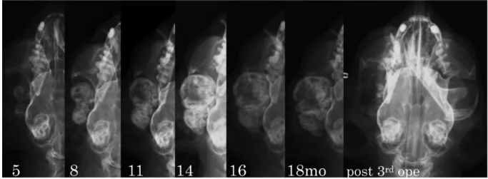
Fig. 5. The radiological changes of the lesion size over time from 5 months after first surgery to the third surgery. (The numbers on the radiographs are months after the first surgery.)
The lesion gradually extended during the first 13 months. At 18 months, the long axis of the lesion was about 30 mm and was displacing the eyeball. Further palliative surgery f was performed, but the lesion was not completely removed.
Fig. 6. The radiological changes of the lesion size over time from 19 months after first surgery to 112 months. (The numbers on the radiographs are months after the first surgery.)
The growth rate of the lesion was slow from the time of the third surgery and a transparent zone is seen in the center of the lesion 45 months after the first surgery. The transparent zone grew larger over time.
reduction was performed to relief soft tissue pain from occlusal trauma. The cat ate normally and was playful. No further treatment was provided because of the owner’s request. The maxillofacial area was palpated and radiographed every 6 months. The tumor continued to enlarge slowly; it was 12 mm on its long axis, with radiological transparency at the center of the tumor 45 months after the first surgery. The radiologically transparent zone inside the lesion gradually enlarged, and growth of the tumor deceler-ated (Fig. 6); the lesion at the final measurement,
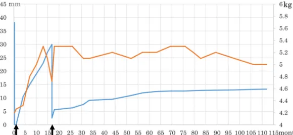
Fig. 7. The long axis of the lesion (blue line) left over time with her body weight (red line). (The numbers on the horizontal axis are months after the first surgery with black arrows of 2nd and 3rd surgery respectively. The numbers on the left vertical axis are the length of the longitudinal axis in mm while the numbers on the right vertical axis are body weight in kg.) The size of lesion increased rapidly from 5 months to 18 months, then growth slowed after the third surgery and was much slower after 55 months. The body weight had kept constantly after the third surgery.
112 months after the first surgery, was 13.3 mm radiographically; it had grown only 1.3 mm during the final 59 months. During observation of her health condition with tumor size, her body weight was kept constantly after palliative surgeries (Fig. 7).
Her health status had been in normal except for cystitis, with no evident renal disease until the cat’s death from unknown causes at 16 years of age (136 months after the first surgery). When her last visit was 123 months after the first surgery, blood tests (CBC and general biochemical tests) and chest radio-graphs were in all normal range. The owner reported the cat’s death by phone; the owner noted that there were no visible changes in the maxillofacial area since the previous examination.
Discussion
Biological behaviors, growth, and metastasis of osteochondoroma in all animal species are
non-specific9). The histopathological features of solitary
and multiple osteochondromas indistinguishable. Feline osteochondromatosis, a multiple osteochon-droma, differs from the osteochondroma in dogs and horses in its clinical setting, skeletal distribution,
histopathological features and prognosis9). The feline
disorder may develop either from endochondral ossification beneath a cap of cartilage, as in dogs and
horses, or directly from the overlying periosteum1, 7, 9).
A solitary osteochondroma was reported in a FeLV
positive cat8); although benign, the lesion
progres-sively expanded9). A possible association with FeLV
has been reported. The condition has been reported
in two FeLV negative cats3, 6); however; the reported
histopathological description appeared more similar to progressive fibrodysplasia ossificans than
osteo-chondoroma9).
The case presented here had FeLV negative serol-ogy, a solitary lesion in the area of the zygomatic arch and mandibular ramus which form by membranous
ossification5), and had no metastasis. The
transpar-ency changes seen on the later radiographs were not examined histopathologically.
Previous reviews reported that osteochondoroma in cats shows progressive enlargement after skeletal maturity, but this case was observed slow
enlarge-ment with aging9, 10, 12). It is suggested that several
palliative surgeries helped the cat live longer with maintaining QOL.
57
Long-term Follow-up of a Feline Solitary Osteochondroma Treated by Palliative Surgery
Acknowledgment
We appreciate the critical reading and helpful comments from Colin E Harvey, BVSc, FRCVS, DipACVS, DipAVDC.
References
1) Doige, C. E. (1987): Multiple Osteochondromas with evidence of malignant transformation in a cat. Vet. Pathol. 24: 457–459. 2) Green, E. M., Adams, W. M., and Steinberg, H. (1999):
Malig-nant transformation of solitary spinal osteochondroma in two mature dogs. Vet. Radiol. Ultrasound. 40: 634–637.
3) Levitin, B., Arloch, I., Aizenberg, I., Foreman, O., and Shmir, M. (2003): Linear osteochondromtosis in a cat. Vet. Radiol. Ultra-sound. 44: 660–664.
4) Murphey, M. D., Choi, J. J., Kransdorf, M. J., Flemming, D. J., and Gannon, F. H. (2000): Imaging of osteochondroma: variants and complications with radiologic and pathologic correlation. Radiographics. 20: 1407–1434.
5) Noden, D. M., and De Lahunta, A. (1985): Craniofacial
skeleto-genesis. In: The Embryology of Domestic Animals, pp. 181– 186, Williams & Wilkins, Baltimore.
6) Nolff, M. F., Puff, C., Langer, B., and Fehr, M. (2014): Feline osteochondromatosis in a FELV-negative European shorthair cat. Tierarztl. Prax. 42(1): 55–59.
7) Pool, R. R., and Carrig, C. B. (1972): Multiple cartilaginous ex-ostoses in a cat. Vet. Path. 9: 350–359.
8) Rosa, C., and Kirberger, R. M. (2012): Extraskeletal osteochon-droma on a cats elbow. J. S. Afr. Vet. Med. Assoc. 83: 1–4. 9) Thompson, K. G., and Dittmer, K. E. (2017): Benign tumors of
bones. In Tumors in Domestic Animals, 5th ed, by Meuten DJ, pp. 359–371, Wiley Blackwell, Ames.
10) Toner, M., and van Heerden WFP (2017): Osteochondroma, be-nign maxillofacial bone and cartilage tumours in WHO classifi-cation of head and neck tumors, (El-Naggar, A. K., Chan, J. K. C., Grandis, J. R., Takata, T., Slootweg, P. J., ed.) p. 255, WHO.
11) Turrel, J. M., and Pool, R. R. (1982): Primary bone tumors in the cat. A retrospective study of 15 cats and literature review. Vet. Radiol. 23: 152–166.
12) Voss, K. (2009): Disease of bone. In: Feline Orthopedic Surgery and Musculoskeletal Disease (Montavon, P. M., Voss, K., and Langley-Hobbs, S. J. ed.) pp. 60–62. Elsevier, Philadelphia.