Case Report TWMUJ 4: 98-103, 2020
Robot-Assisted Thoracoscopic Tumor Resection of Lymph Node Metastasis of
an Unknown Primary Site: A Case Report
Tamami Isaka,1 Shota Mitsuboshi,1 Hiroaki Shidei,1 Hiroe Aoshima,1 Takako Matsumoto,1 Tatsuo Sawada,2 Yoji Nagashima,3
and Masato Kanzaki1
1
Department of Thoracic Surgery, Tokyo Women’s Medical University, Tokyo, Japan
2Division of Pathological Neuroscience, Department of Pathology, Tokyo Women’s Medical University School of Medicine, Tokyo, Japan 3
Department of Surgical Pathology, Tokyo Women’s Medical University, Tokyo, Japan (Accepted August 12, 2020)
(Advance Publication by J-STAGE September 25, 2020)
Cancer of an unknown primary site (CUP) accounts for approximately 3% of all cancer cases. Among these cases, mediasti-nal lymph node cancer is comparatively rare, and there are no established treatment methods or approaches. Excision of CUP is feasible with the best methods. We herein report a case of mediastinal lymph node CUP resection by robot-assisted thora-coscopic surgery (RATS). The patient was a 60-year-old man. A chest computed tomography (CT) scan identified swelling in the mediastinal lymph node (right tracheobronchial lymph node; #4R) with another organ suspected as the site of the pri-mary lesion, but it was not detected through positron emission tomography (PET). The patient was diagnosed with CUP with lymph node metastasis and mediastinal lymph node dissection was performed via RATS. Histopathological examination re-vealed desmoplasia in the lymph nodes. Immunohistochemically, the tumor was positive for CK7, TTF1, and napsin A, but negative for CK20, CK5/6, CDX2, p40, and AFP, indicating lung-derived adenocarcinoma. In the narrow space such as the mediastinum, RATS is a useful procedure for resectable mediastinal tumors.
Key Words: robot-assisted thoracoscopic surgery (RATS), cancer of unknown primary site (CUP)
Introduction
Cancer of an unknown primary site (CUP) accounts for approximately 3% of all the clinical cancer cases, and it is frequently linked with mortality.1,2
Among these cases, mediastinal lymph nodal involvement is comparatively rare, and there are no established therapeutic strategies.3
To determine the primary site and select an appropriate therapeutic regimen, biopsy and/or excision of the CUP
is required. Furthermore, the resected tissue sample can be used for comprehensive cancer genome analysis which will also assist with selecting an appropriate thera-peutic option for CUP.3,4
We herein report a case of CUP metastasizing to the mediastinal lymph node. The lesion was successfully re-sected by robot-assisted thoracoscopic surgery (RATS). Histopathologically, the lung was indicated as the pri-mary site.
Corresponding Author: Masato Kanzaki, Department of Thoracic Surgery, Tokyo Women’s Medical University, 8-1 Kawada-cho, Shinjuku-ku, Tokyo 162-8666, Japan. [email protected]
doi: 10.24488/twmuj.2020006
CopyrightⒸ 2020 Society of Tokyo Women’s Medical University. This is an open access article distributed under the terms of Creative Commons Attribution License (CC BY), which permits unrestricted use, distribution, and reproduction in any medium, provided the original source is properly credited.
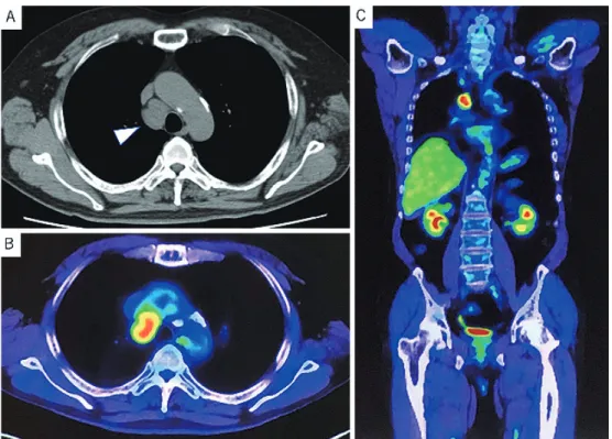
Figure 1 Preoperative imaging findings.
(A) Chest computed tomography scan showing a solitary mediastinal mass with 37 mm in maximum diameter without invasion surrounding tissues.
(B) 18F-fluorodeoxyglucose positron emission tomography (FDG-PET) of the patient showing an ab-normal uptake of FDG with a maximum standardized uptake value of 5.92 in the tumor.
(C) Coronal section of the FDG-PET showing no abnormal FDG accumulation other than the tumor.
Case Presentation
A 60-year-old man presented with a mediastinal tumor by chest computed tomography ( CT ) during medical checkup and was referred to our hospital. His physical findings were normal. He had been smoking 30 cigarettes per day for 37 years, had a comorbidity of hypertension, but had no past history of malignant tumors.
Although blood tests found elevated Sialyl Lewis X (SLX) levels of 61 U/mL, both carcinoembryonic antigen (CEA) and cytokeratin subunit 19 fragment (CYFRA) levels were normal. No other abnormalities were identi-fied in the blood sample.
Chest CT revealed an isolated mediastinal mass meas-uring 37 mm in the maximum diameter without invasion to the adjacent tissues. No abnormal shadows were iden-tified in the lung fields ( Figure 1 A ) . 18 F-fluorodeoxyglucose (FDG) accumulation was identified in the tumor using FDG positron emission tomography (PET), and the maximum standardized uptake value was 5.92. No abnormal FDG accumulation was found in
other organs (Figure 1B and C).
Endobronchial ultrasound transbronchial needle aspi-ration (EBUS-TBNA) was performed and he was diag-nosed with adenocarcinoma. Since no primary site organ could be identified, the diagnosis was mediastinal lymph node CUP, and robot-assisted thoracoscopic surgery (RATS) was performed.
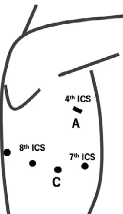
The patient was intubated with a double-lumen en-dotracheal tube for one-lung ventilation under general anesthesia, and placed in a left lateral decubitus position. RATS excision of the mediastinal tumor was performed with four port incisions and a 4-cm utility thoracotomy as an assistant port. The da Vinci Surgical System (Model Xi; Intuitive Surgical, Inc, Sunnyvale, CA, USA ) was used. RATS began with a total of four 8-mm ports in-serted in the 7th intercostal space (ICS) at the anterior side as the 1st arm, in the 8th ICS mid-axillary line as the 2nd arm, in the 8th posterior axillary line as the 3rd arm, and in the 8th posterior side as the 4th arm, plus an assis-tant port in the 4th ICS (Figure 2). A robotic arm was mounted to the port in the 8th ICS, with Cadiere grasping
Figure 2 Schematic illustration of four port incisions and a 4-cm utility thoracotomy as an assistant port.
A total of four 8-mm ports (circle) were inserted in anterior to 7th intercostal space (ICS), 8th ICS mid-axillary line, posterior axil-lary line, and posterior. An assistant port (line) was made at 4th ICS.
ICS, intercostal space; A, assistant port; C, camera.
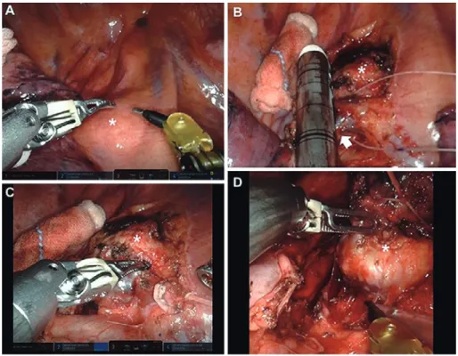
forceps on the 1st arm, bipolar fenestrated grasping for-ceps on the 2nd arm, and monopolar spatula on the 4th arm. The camera scope was inserted into the 8 th ICS mid-axillary line port and was used for 30° oblique view-ing. No adhesions were found within the right thoracic cavity, and there were no abnormalities in the lungs. The tumor was located in the right lower paratracheal lymph nodes (4R) on the cranial side of the azygos vein (Fig-ure 3A). After the pleura was incised in a cephalad direc-tion, the azygos vein and tumor were exposed. For ampu-tating the azygos vein, an EndoWrist stapler was used through the 4th arm port (Figure 3B). The mediastinal fat pad including the tumor was dissected off the superior vena cava and anterolateral tracheal surface ( Fig-ure 3 C and D ) . The resected mediastinal tumor was placed in a bag and removed through the assistant port, followed by the insertion of a 21-Fr silicon tube through the camera port. The tumor was measured 4.5× 3.5 × 2.5 cm (Figure 4). Macroscopic finding of the tumor was yellow-whitish surface.
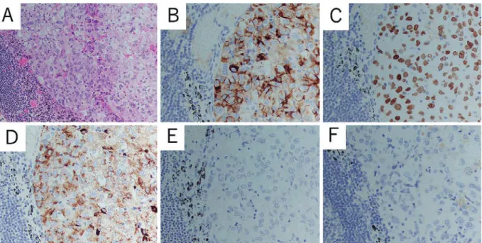
Histopathologically, the resected lymph node was oc-cupied by tumor cells with a solid architecture. The
tu-mor cells were large in size and polygonal in shape. Their nuclei were large with a vesicular appearance and prominent nucleoli. The cytoplasm was pale. Based on these findings, a germ cell tumor, lymphoma, and malig-nant melanoma were considered as differential diagnostic candidates along with metastatic carcinoma (Figure 5A). Immunohistologically, the tumor cells were positive for cytokeratin (CK) 7, thyroid transcription factor-1 (TTF-1), and napsin A, while negative for CK20, CK5/6, cau-dal related homeobox gene type 2(CDX2, p40, and α-fetoprotein (AFP) (Figure 5B-F). Based on these find-ings, he was diagnosed with metastatic adenocarcinoma, the primary site of which was considered to be the lung. Genetic tests failed to reveal any mutation of epidermal growth factor receptor (EGFR) and translocation involv-ing anaplastic lymphoma kinase (ALK) and c-ros onco-gene 1 (ROS1). The postoperative course was uneventful. The chest tube was removed on the 4th day, and he was discharged on the 7th day after surgery. Postoperative ad-juvant chemotherapy was received according to the treat-ment for lung cancer. The patient showed no evidence of recurrence at the 15-month follow-up.
Discussion
CUP refers to metastasized cancer for which the primary site cannot be identified. This condition is usually seen in the liver, lungs, and bones. In this case, the mediastinal lymph nodes showed isolated metastasis, although it ac-counts for just 1.5% of all CUP involvements.5
Most common histological types of CUP are adenocarcinoma, followed by undifferentiated carcinoma and squamous cell carcinoma.1,6
Sampling procedures of mediastinal lymph node CUP include bronchoscopy, thoracoscopy, mediastinoscopy, and surgical procedures. Differentiating diagnosis be-tween CUP, other tumors originating in the mediastinum, and malignant lymphoma is essential. In addition, since future developments in cancer genome analysis are help-ful for both the diagnosis and treatment selection of CUP,2
sampling of sufficient tumor tissue is important. As a result, biopsy and/or excision of mediastinal tumors are the best methods.
The robotic surgical system provides a 3-dimensional view and dedicated robotic instruments that have
com-Figure 3 The intraoperative screenshot of the robot-assisted thoracoscopic surgery (RATS). (A) The tumor (*) was located in right lower paratracheal lymph nodes (4R) on the cranial side of the azygos vein.
(B) After the pleura was incised in a cephalad direction, azygos vein (white arrow) and the tumor were exposed. The azygos vein was amputated by robotic stapler.
(C) The mediastinal fat pad including the tumor was dissected off superior vena cava.
(D) The mediastinal fat pad including the tumor was dissected off the anterolateral tracheal surface and removed.
Figure 4 The intraoperative view of the tumor.
The intraoperative view of the tumor was extracted from digital video data taken during the operation and was provided on a per-sonal computer after operation.
fortable motions with 7 degrees of freedom in the tho-racic cavity. Although the mediastinum with lymph node is tightly packed with intertwined organs, the robotic sur-gical system allowed for a smooth and natural
manipula-tion when performing surgical dissecmanipula-tion. Especially, compared to the traditional video-assisted thoracic sur-gery (VATS) approach, robot-assisted sursur-gery has been reported to be more effective for mediastinal lymph node dissection.7-9
Pathologically, seminoma, lymphoma, and malignant melanoma were considered as differential diagnostic can-didates along with metastatic carcinoma. To determine the histological type and tumor origin, immunostaining was performed. Combined immunostainings for CK7 and CK20 are frequently employed to determine the primary site, especially of adenocarcinoma. TTF-1, SPA, and napsin A are characteristically positive for lung cancer, especially adenocarcinoma.10,11
In this case, the primary lesion site was in the lungs because the tumor was posi-tive for TTF-1, CK 7, and napsin A but negaposi-tive for CK20.
Although most CUP are considered to signify ad-vanced stage cancers, adjuvant chemotherapy following surgical resection is necessary for CUP. For mediastinal
Figure 5 Microscopic findings of the resected specimen.
(A) Hematoxylin and eosin staining showing desmoplasia in the lymph nodes.
(B-F) Immunohistochemical examination revealing positive stain in (B) cytokeratin (CK) 7, (C) Thy-roid transcription factor-1 (TTF-1), and (D) napsin A, and negative stain in (E) α-fetoprotein (AFP) and (F) CK20.
lymph node CUP, there are various postoperative care regimens. A variety of surgical treatments, such as pul-monary lobectomy and lymph node dissection are used. Observation and other treatments, including adjuvant chemotherapy, radiotherapy, or a combination of both chemotherapy and radiotherapy, are performed after sur-gical resection.3,12,13
The 5-year survival rate for CUP has been reported to range from 2-6%, but there are also re-ports that isolated mediastinal lymph node CUP has a comparatively better prognosis.13
In some reports, careful postsurgical investigation is required for mass shadows that appear in the lungs after the excision of mediastinal lymph node CUP.4,14,15
Conclusion
RATS tumor resection was performed for mediastinal lymph node CUP. In narrow spaces, such as the mediasti-num, RATS is a useful procedure for resectable CUP with mediastinal metastasis for the diagnosis and estab-lishment of a therapeutic strategy.
Conflicts of Interest: The authors confirm no conflicts of
interest.
References
1.Briasoulis E, Pavlidis N : Cancer of unknown primary origin. Oncologist 2 (3): 142―152, 1997
2.Vajdic CM, Perez-Concha O, Rhee JJ et al: Health-related predictors of cancer registry-notified cancer of unknown primary site (CUP). Cancer Epidemiol 61: 1―7, 2019
3.Nakano T, Watanabe H, Takeichi H et al: A case of me-diastinal lymph node carcinoma of unknown primary site. Tokai J Exp Clin Med 43 (4): 148―152, 2018 4.Kawasaki H, Arakaki K, Taira N et al: Lung cancer
de-tected 5 years after resection of cancer of unknown pri-mary in a mediastinal lymph node: A case report and re-view of relevant cases from the literature). Ann Thorac Cardiovasc Surg 22 (2): 116―121, 2016
5.Holmes FF, Fouts TL : Metastatic cancer of unknown primary site. Cancer 26 (4): 816―820, 1970
6.Agca M, Kosif A: Metastatic squamous cell cancer with unknown primary origin in the mediastinal lymph node. Niger J Clin Pract 21 (10): 1384―1386, 2018
7.Kanzaki M : Current status of robot-assisted thora-coscopic surgery for lung cancer. Surg Today 49: 795― 802, 2019
8.Nasir BS, Bryant AS, Minnich DJ et al: Performing ro-botic lobectomy and segmentectomy: cost, profitability, and outcomes. Ann Thorac Surg 98 (1): 203―208, 2014 9.Novellis P, Bottoni E, Voulaz E et al: Robotic surgery,
video-assisted thoracic surgery, and open surgery for early stage lung cancer : comparison of costs and out-comes at a single institute. J Thorac Dis 10 (2): 790―798, 2018
10.Hirano T, Auer G, Maeda M et al: Human tissue distri-bution of TA02, which is homologous with a new type of aspartic proteinase, napsin A. Jpn J Cancer Res 91 (10): 1015―1021, 2000
11.Chhieng DC, Cangiarella JF, Zakowski MF et al: Use of thyroid transcription factor 1, PE-10, and cytokeratins 7 and 20 in discriminating between primary lung carcino-mas and metastatic lesions in fine-needle aspiration bi-opsy specimens. Cancer 93 (5): 330―336, 2001
12.Morio A, Miyamoto H, Izumi H et al: A case report of adenocarcinoma of unknown origin metastatic to the me-diastinal lymph nodes with a review of 21 cases reported in Japan. Jpn J Lung Cancer 41: 73―78, 2001 (in Japa-nese)
13.Riquet M, Badoual C, Le Pimpec BF et al: Metastatic thoracic lymph node carcinoma with unknown primary site. Ann Thorac Surg 75 (1): 244―249, 2003
14.Higuchi T, Nagata A, Hamada T et al: A case of primary lung cancer developing 9 years after surgery for metas-tatic mediastinal lymph node carcinoma. J Jpn Assoc Chest Surg 29: 220―225, 2015
15.Hayashi K : A case report of a long-surviving patient with mediastinal lymph node carcinoma from an un-known primary site, with a review of 31 cases reported in Japan. J Jpn Assoc Chest Surg 21 (4): 624―629, 2007