INTRODUCTION
Photodynamic therapy (PDT) was approved as a treatment for age - related macular degeneration (AMD) in Japan since May, 2004. The number of institutions dealing with PDT and cases actually done have been increasing year by year. PDT has been regarded as the treatment of choice for AMD now. Recently many studies on PDT were reported and some studies mentioned that PDT has shown
more effective than the results of The Japanese AMD Trial, an open - label, multicenter, prospective, and noncontrolled study (1). On the other hand, the number of reports on the complications of PDT has been increasing (2 - 5). Massive subretinal hemor-rhage, one of the side effects after PDT, might lead to grave visual disturbance. However the cause or mechanism of subretinal hemorrhage after PDT is still unclear. We report four cases with massive subretinal hemorrhage after PDT.
PATIENTS AND METHODS
This study consisted of retrospective consecutive series of patients who were treated with PDT for
ORIGINAL
The prognosis of cases with massive subretinal
hemor-rhage after photodynamic therapy
Shingo Matsushita, Takeshi Naito, Masaru Takebayashi, Hiroyuki Sato, and
Hiroshi Shiota
Department of Ophthalmology, Institute of Health Biosciences, the University of Tokushima Graduate School, Tokushima, Japan
Abstract : Purpose : To investigate cases with massive subretinal hemorrhage after pho-todynamic therapy(PDT).
Subjects and Methods : We studied four cases(3 men and 1 woman, mean 80.5 yeas old)
with massive subretinal hemorrhage after PDT about type of disease, spot size, period to the onset of hemorrhage, visual acuity (VA) before and after PDT.
Results : Four cases consisted of one with age-related macula degeneration (AMD) and 3
with polypoidal choroidal vasculopathy (PCV). The average spot size of PDT was 5525
!
m. Two patients underwent the second PDT about 3 months after the initial PDT. Four periods to the onset of hemorrhage were 22, 27, 137, and 208 days respectively. The latter 2 cases underwent PDT twice, and the periods were the days from the initial PDT. Two patients among four underwent vitrectomy. Average VA before PDT was 0.20, and the fi-nal one was 0.02 after PDT.Conclusions : PDT has a risk to cause massive subretinal hemorrhage leading to grave
visual disturbance. Especially PDT for PCV seems to have more risk. Therefore we need to inform patients about the risk before PDT, since treating patients with massive subreti-nal hemorrhage is very difficult J. Med. Invest. 55 : 231-235, August, 2008
Keywords : PDT, AMD, PCV, subretinal hemorrhage, risk
Received for publication May 1, 2008 ; accepted June 10, 2008. Address correspondence and reprint requests to Takeshi Naito, Department of Ophthalmology, Institute of Health Biosciences, the University of Tokushima Graduate School, Kuramoto - cho, Tokushima 770 - 8503, Japan and Fax : +81 - 88 - 631 - 4848.
AMD at our university hospital from October 2004 to September 2006. All patients gave their informed consent to be treated with PDT. A total of 4 cases with massive subretinal hemorrhage out of 149 cases undergone PDT were investigated carefully. All patients underwent pretreatment ophthalmologi-cal examinations including visual acuity (VA), fluo-rescein angiography (FA), indocyanin green angi-ography (IA), and an optical coherence tomograph (OCT, Stratus OCT3000 ; Carl Zeiss, Dublin, CA, USA). We also studied spot size, period to the on-set of hemorrhage, visual acuity before and after PDT. We defined as massive subretinal hemorrhage if the spread is more than the area of vasucular ar-cade of the retina.
RESULTS
Among 149 cases treated with PDT, 103 cases were AMD and 46 cases were polypoidal choroidal vasculopathy (PCV). The four cases consisted of one with AMD and 3 with PCV. The incidence of massive subretinal hemorrhage is 3 (6.5%) of 46 cases with PCV and one (0.9%) of 103 cases with AMD. Therefore, the incidence is much higher in
the patients with PCV than the patients with AMD. The average spot size of PDT was 5525 μm. Two out of four patients underwent the second PDT about 3 months after the initial PDT. The periods to the onset of hemorrhage were 22, 27, 137, and 208 days respectively. The latter 2 cases underwent PDT twice, and the period was calculated from the initial PDT. Two patients underwent vitrectomy. The average VA before PDT was 0.20, and the final was 0.02 after PDT. The profiles of these four cases were shown in Table 1. The following is a typical case.
Case 1
A 78 - year - old woman was diagnosed as macu-lar degeneration in her right eye four months be-fore and followed up by a nearby doctor. Her visual acuity deteriorated gradually, then she was referred to our hospital on September 21, 2005. On exami-nation, her visual acuities were 0.4 in the right and 0.7 in the left eye. Intraocular pressure was normal in both eyes and no abnormal findings were ob-served in the anterior segment and the ocular me-dia. A subretinal hemorrhage, fibrin, hard exudate, and reddish - orange nodules were recognized in the right macula (Fig. 1). Fluorescein angiography (FA) showed leakage and hyperfluorescence at the lesion,
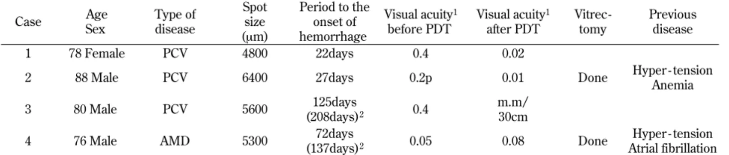
Table 1 The profiles of four cases with massive subretinal hemorrhage after PDT
Case Age Sex Type of disease Spot size (μm) Period to the onset of hemorrhage Visual acuity1 before PDT Visual acuity1 after PDT Vitrec-tomy Previous disease 1 78 Female PCV 4800 22days 0.4 0.02
2 88 Male PCV 6400 27days 0.2p 0.01 Done Hyper - tension
Anemia
3 80 Male PCV 5600 125days
(208days)2 0.4
m.m/ 30cm
4 76 Male AMD 5300 72days
(137days)2 0.05 0.08 Done
Hyper - tension Atrial fibrillation 1)visual acuity is corrected eyesight
2)case3 and 4 underwent PDT twice, and the days after initial the PDT are shown in parentheses
Fig. 1 Clinical photograph before PDT showed a subretinal hemorrhage, fibrin, hard exudate, and reddish - orange nodules were recognized in the right macula
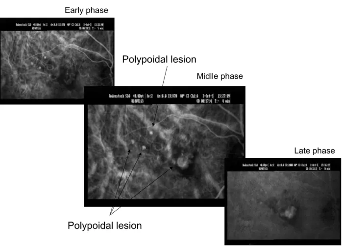
showing classic choroidal neovascularization (CNV) (Fig. 2). Indocyanine green angiography (IA) showed multiple polypoidal choroidal vascular le-sions (Fig. 3). She was diagnosed as PCV from these findings. PDT was performed with a 4800 μm spot size, as guided by the abnormalities shown in the FA on October, 28, 2005. No complications were recognized during the treatment. The patient complained of a rapid decrease in her right vision on 22 days after PDT. We fond the massive
subreti-nal hemorrhage which spread more than the area of vasucular arcade of the retina in her right eye on November 30, 2005 (Fig. 4). Her right visual acu-ity decreased to 0.04. She was followed up and treated with oral use of carbazochrome sodium sul-fonate. Despite the absorption of the hemorrhage, fibrin deposition and organization of the hemorrhage were recognized. The final visual acuity became 0.02.
Fig. 3 Indocyanine green angiography showed multiple polypoidal choroidal vascular lesions.
Fig. 2 Fluorescein angiography before PDT showed leakage and hyperfluorescence at the lesion, showing classic choroidal ne-ovascularization.
DISCUSSION
Although our number is limited, three of 4 cases were PCV. The incidence of massive subretinal hemorrhage is 6.5% in the patients with PCV and almost same as the previous report (11). The inci-dence is much higher in the patients with PCV than the patients with AMD. Therefore, our study alarms that PDT for PCV seems to cause massive subretinal hemorrhage. Spot size of 4 cases was relatively large. Two out of four patients had mas-sive subretinal hemorrhage after the second PDT, so care must be taken even if subretinal hemor-rhage dose not appear after the initial PDT. But it is not clear whether PDT itself causes massive subretinal hemorrhage or not, because the periods to the onset of hemorrhage of thses 2 cases were long. Among Japanese the proportion of PCV among exudative AMD is large (6), and PDT for PCV has shown to be more effective than that for other types of AMD (7 - 10). This leads that we have more chance of PDT for PCV and the num-ber of cases with subretinal hemorrhage after PDT will increase. PDT itself leads to inflammation at the exposure field and ischemia by angiopathic mechanism. The inflammation or ischemia produces vascular endothelial growth factor(VEGF) in the retina, and then angiogenesis or vascular perme-ability occurs. That may trigger subretinal hemor-rhage. Larger spot size is likely to induce wide-spread inflammation and ischemia, and trigger subretinal hemorrhage. Recently, IA is more popu-lar than FA in guiding a suitable laser spot size. This enables to treat by minimal greatest linear di-mension (GLD) and to minimize inflammation and ischemia by PDT. But it is possible that we fail to
cover lesions hiding under hemorrhage when PDT is delivered only as guided by the abnormalities shown by IA. Especially it is necessary to cover le-sions like abnormal network of choroidal neovas-cularization in PDT for PCV, in order to prevent a recurrence or hemorrhage after PDT. From now on, we need comprehensively check lesions and conditions from findings not only of FA and IA, but also of OCT. Then custom - made treatment for each case can be done. We also should consider PDT combined with juxafoveal injection of triamcinolone acetonide or intravitreous injection of anti - VEGF antibodies.
REFERENCES
1. The Japanese Age - related Macular Degeneration Trial (JAT) Study Group : Japanese age -related macular degeneration trial : 1 - year re-sults of photodynamic therapy with vertepor-fin in Japanese patients with subfoveal choroi-dal neovascularization secondary to age-related macular degeneration. Am J Ophthalmol 136 : 1049 - 1061, 2003
2. Wachtlin J, Behme T, Heimann H, Kellner U, Foerster MH : Concentric retinal pigment epi-thelium atrophy after a single photodynamic therapy. Graefes Arch Clin Exp Ophthalmol 241 : 518-521, 2003
3. Schmidt-Erfurth U, Schlotzer-Schrehard U, Cursiefen C, Michels S, Beckendorf A, Naumann GOH : Influence of photodynamic therapy on expression of vascular endothelial growth factor(VEGF), VEGF receptor3, and pigment epithelium - derived factor. Invest Oph-thalmol Vis Sci 44 : 4473 - 4480, 2003
4. Tzekov R, Lin T, Zhang K, Jackson B, Oyejide A, Orilla W, Kulkarni AD, Kuppermann BD, Wheeler L, Burke J : Ocular changes after photodynamic therapy. Invest Ophthalmol Vis Sci 47 : 377 - 385, 2006
5. Lai TY, Chan WM, Lam DS : Transient reduc-tion in retinal funcreduc-tion revealed by multifocal electroretinogram after photodynamic therapy. Am J Ophthalmol 137 : 826 - 833, 2004
6. Sho K, Takahashi K, Yamada H, Wada M, Nagai Y, Otsuji T, Nishikawa M, Mitsuma Y, Yamazaki Y, Matsumura M, Uyama M : Poly-poidal choroidal vasculopathy : incidence, demographic features, and clinical character-istics. Arch Ophthalmol 121 : 1392 - 1396, 2003
Fig. 4 Clinical photograph after PDT showed the massive subretinal hemorrhage which spread more than the area of vasu-cular arcade of retina.
7. Spaide RF, Donsoff I, Lam DL, Yannuzzi LA, Jampol LM, Slakter J, Sorenson J, Freund KB : Treatment of polypoidal choroidal vascu-lopathy with photodynamic therapy. Retina 22 : 529 - 535, 2002
8. Lee SC, Seong YS, Kim SS, Koh HJ, Kwon OW : Photodynamic therapy with verteporfin for polypoidal choroidal vasculopathy of the macula. Ophthalmologica 218 : 193-201, 2004 9. Chan WM, Lam DS, Lai TY, Liu DT, Li KK,
Yao Y, Wong TH : Photodynamic therapy with verteporfin for symptomatic polypoidal choroi-dal vasculopathy one year result of a
prospective case series. Ophthalmology 111 : 1576 -1584, 2004
10. Silva RM, Figueria J, Cachulo ML, Duarte L, Faria de Abreu JR, Cunha-Vaz JG : Polypoidal choroidal vasculopathy and photodynamic therapy with verteporfin. Graefes Arch Clin Exp Ophthalmol 243 : 973 - 979, 2005
11. Hirami Y, Tsujikawa A, Otani A, Yodoi Y, Aikawa H, Mandai M, Yoshimura N : Hemor-rhagic complications after photodynamic ther-apy for polypoidal choroidal vasculopathy. Ret-ina 27 : 335 - 341, 2007