D
efinitive concurrent chemoradiotherapy (CCRT) is one of the standard treatments for unresect-able stage III lung cancer. When administered concur-rently with a cisplatin/docetaxel regimen, it shows a trend toward improved median progression-free timeand 2-year overall survival (OS) compared with second- generation chemotherapy [1]. Patients receiving cispla-tin/docetaxel also tend to show a lower rate of distant metastases at the first recurrence site in comparison with those who receive second-generation chemother-apy. Recent studies have shown that the administration
CopyrightⒸ 2021 by Okayama University Medical School.
http ://escholarship.lib.okayama-u.ac.jp/amo/
Original Article
Volumetric PET Parameters Predict Prognosis after Definitive
Chemoradiotherapy with Cisplatin/Docetaxel for Stage III
Non-Small Cell Lung Cancer
Kuniaki Katsui
a*, Takeshi Ogata
b, Akihiro Tada
c, Soichi Sugiyama
d,
Kotaro Yoshio
d, Masahiro Kuroda
e, Katsuyuki Kiura
f, Yoshinobu Maeda
g,
Shinichi Toyooka
h, Takao Hiraki
i, and Susumu Kanazawa
iDepartments of aProton Beam Therapy, gHematology, Oncology and Respiratory Medicine,
hGeneral Thoracic Surgery and Breast and Endocrinological Surgery, iRadiology,
Okayama University Graduate School of Medicine, Dentistry and Pharmaceutical Sciences, Departments of dRadiology,
fAllergy and Respiratory Medicine, Okayama University Hospital, eDepartment of Radiological Technology,
Graduate School of Health Sciences, Okayama University, Okayama 700-8558, Japan,
bDepartment of Radiology, Iwakuni Clinical Center, Iwakuni, Yamaguchi 740-8510, Japan,
cDepartment of Radiology, Okayama Diagnostic Imaging Center, Okayama 700-0913, Japan
The aim of this study was to investigate whether volumetric positron emission tomography (PET) parameters are prognostic predictors in stage III non-small cell lung cancer patients receiving definitive concurrent chemo-radiotherapy (CCRT) with cisplatin/docetaxel. Cases involving definitive CCRT were reviewed retrospectively, and the maximum standardized uptake value, metabolic tumor volume (MTV) and total lesion glycolysis (TLG) were calculated. The relationships between these PET parameters and prognosis were analyzed. MTV and TLG were significant predictors of distant metastasis-free survival (DMFS) (p=0.0003 and 0.0005, respectively) and progression-free survival (PFS) (p=0.001 and 0.0007, respectively). The three-year DMFS rates in patients with low and high MTV were 13.3% and 64.6%, respectively, and the corresponding values in those with low and high TLG were 13.3% and 65.2%, respectively. The three-year PFS rates in patients with low and high MTV were 13.3% and 57.8%, respectively, and the corresponding values in patients with low and high TLG were 13.3% and 57.8%, respectively. However, MTV and TLG were not predictors of local control or overall sur-vival. We demonstrated that volumetric PET parameters were predictors of patients receiving definitive CCRT. Our findings contradict the findings of previous reports and warrant further research to validate them.
Key words: volumetric positron emission tomography parameters, distant metastasis-free survival,
chemoradio-therapy, cisplatin/docetaxel, non-small cell lung cancer
Received May 20, 2020 ; accepted September 23, 2020.
*Corresponding author. Phone : +81-86-235-7313; Fax : +81-86-235-7316
lesions was 33.8% in the CCRT-alone group and 22.5% in the durvalumab group, which was probably attribut-able to the improvement in PFS as a result of the sup-pression of distant metastases. Since the appearance of distant metastases suggests that curing the disease has generally become more difficult, biomarkers for pre-dicting distant metastases are desired.
18F-Fluorodeoxyglucose (18F-FDG) positron emis-sion tomography/computed tomography (PET/CT) is useful for the staging and prognosis of malignant tumors. Although the maximum standard uptake value (SUVmax) has been adopted as a PET parameter [2], volumetric evaluations can facilitate assessments of overall tumor activity in addition to single-voxel assess-ments such as SUVmax [3,4]. Volumetric PET param-eters for lung cancer have been reported to be useful in assessing responses to CCRT [5,6] and chemotherapy [7]. Although some studies have examined the correla-tion between volumetric PET parameters and OS or PFS in patients receiving chemoradiotherapy, only a few studies have examined the distant metastasis-free sur-vival rate (DMFS). Moreover, even fewer reports have included cases in which a cisplatin/docetaxel regimen was concurrently used [8].
Thus, the purpose of this study is to investigate whether volumetric PET parameters can serve as pre-dictors of DMFS for non-small cell lung cancer (NSCLC) patients who received CCRT with cisplatin/ docetaxel.
Materials and Methods
Patients. The study was approved by the institu-tional ethics committee (No. 1809-018). Patients who received CCRT between April 2006 and December 2017 at the authors’ hospital were reviewed retrospectively. Staging was performed according to the 7th edition of the TNM Classification of Malignant Tumors. Lymph nodes with a short diameter of 1 cm or more were defined as metastases. Lymph nodes of less than 1 cm were defined as metastases if accumulations were observed on PET/CT as judged by a board-certified radiologist. If a biopsy or resection was required in addition to imaging, at the discretion of the respiratory physicians, lymph node metastasis was diagnosed pathologically. The eligibility criteria were as follows:
chemotherapy with cisplatin/docetaxel [1], and no administration of neoadjuvant therapy before the start of CCRT. After treatment, follow-up CT and magnetic resonance imaging were performed periodically, with intervals determined by the individual respiratory phy-sician. All procedures were in compliance with the ethical standards of the 1964 Declaration of Helsinki and subsequent modifications. Written informed con-sent was obtained before CCRT. In accordance with the rules of the ethics committee, opportunities to opt out of the study were provided before this research was started.
Treatment. Indications for definitive CCRT for all cases were discussed at the respiratory conference and finally determined by a board-certified chest medi-cal oncologist and a radiation oncologist. All patients received three-dimensional conformal radiotherapy with an isocenter prescription. CT scans for the radia-tion treatment plan and irradiaradia-tion were performed at 2-10 mm intervals. The gross tumor volume included the primary tumor and a clinically diagnosed metastatic lymph node. The clinical target volume margin and planning target volume margin were both 5-10 mm. Non-metastatic subcarinal and ipsilateral hilar nodal stations were included in cases of elective nodal irradia-tion [9,10]. The definitive radiairradia-tion dose was delivered by a linear accelerator (Mevatron, ONCOR, or Primus; Canon Medical Systems, Tochigi, Japan). The chemo-therapy regimens consisted of cisplatin/docetaxel based on a prospective study of Okayama Lung Cancer Study Group Trial 0007 [1]. Durvalumab was not used because this drug was not launched in Japan within the survey period.
PET/CT protocol. All patients fasted for at least 6 hours before the imaging assessments. The PET/CT scans were acquired using a Biograph 16 PET/CT scan-ner (Siemens Healthcare, Erlangen, Germany) at 90 min after the intravenous administration of FDG (3.7 MBq/kg). For photon attenuation correction and anatomical localization, a low-dose CT scan was ini-tially obtained from the level of the head to the mid-thigh with a tube voltage of 120 kV, an automatic tube current modulation using a reference tube current of 50 mAs, and a section thickness of 3 mm. Subsequently, PET imaging was performed in three-dimensional mode for 3 min per bed position. PET data were
recon-structed using an ordered subsets expectation maximi-zation algorithm and the following parameters: 5 itera-tions, 4 subsets, 168×168 matrix, and a section thickness of 3 mm.
PET/CT image analysis. Measurements of
SUVmax, metabolic tumor volume (MTV), and total lesion glycolysis (TLG) were performed using special-ized software (Syngo. via Siemens Healthcare, Erlangen, Germany). The volume of interest (VOI) was manually drawn at the primary tumor and lymph node metastasis on PET/CT images, and the software automatically delineated the contour of the target lesion (primary tumor and lymph node metastases) inside the VOI using an isocontour threshold method. We set the absolute threshold for SUVmax at 2.5 to define the MTV based on a meta-analysis showing that fixed abso-lute thresholds were suitable for evaluating the prog-nostic value of MTV [4], and the cutoff for the fixed value was an SUV of 2.5 [3]. The delineation of the target lesion was visually checked by a board-certified radiologist who did not have access to the patients’ prognoses. The SUVmax, MTV, and TLG values of the delineated target lesion were automatically calculated. The MTV was defined as the metabolic volume of the target lesion with an SUV exceeding the defined thresh-old of 2.5. TLG values were calculated by multiplying the mean SUV of the target lesion by the MTV.
Statistical analysis. Survival curves were esti-mated using the Kaplan-Meier method. The medians of the volumetric PET parameters were used as the cutoff values. Variables were grouped into 2 categories for statistical analyses, and the medians were adopted as cutoff values for continuous variables. The relation-ships between the PET/CT parameters and the local control rate (LC), DMFS rate, PFS rate, and OS rate were analyzed using the log-rank test for univariate analysis and the Cox proportional hazard model for multivariate analysis. The comparison between the two groups was performed using Student’s t-test. We calcu-lated the mean follow-up interval of the CT, magnetic resonance imaging, and PET/CT examinations until the last day of follow-up for patients without recurrence and until the day of recurrence for patients with recur-rence. A p value <0.05 (two-sided) was considered to be statistically significant. R software, version 3.5.1 (R Foundation for Statistical Computing) was used for all statistical analyses.
Results
The median follow-up period from the start of treat-ment was 25.1 (range: 5.9-118.1) months. Thirty patients met the eligibility criteria, and Table 1 shows their characteristics. The total dose of definitive radio-therapy was 60 Gy/30 fractions in all patients. Cisplatin/ docetaxel chemotherapy was used for all patients, and cisplatin/vinorelbine was used as the second course in one patient because of a drug allergy. For mediastinal tumors, 11 patients underwent endobronchial ultra-sound-guided transbronchial needle aspiration biopsy, 1 underwent endoscopic ultrasound-guided fine-needle aspiration biopsy, 1 underwent endobronchial ultra-sound-guided transbronchial needle aspiration biopsy and open chest biopsy, and 1 underwent a biopsy by mediastinoscopy. For supraclavicular lymph nodes, 3 patients underwent a biopsy. Among recurrent patients, the longest time to recurrence was 40 months; there-fore, the examination intervals were measured up to 40 months for patients without recurrence. The average examination intervals in the high and low TLG groups were 10.98 weeks and 8.00 weeks (95% confidence interval of difference: −0.51 to +6.47), respectively, and the difference was not significant (p=0.09052). The median SUVmax, MTV, and TLG were 16.04 (range: 5.90-35.83), 78.82 cm3 (range: 15.09-547.00 cm3), and 588.07 (range: 47.09-4160.73), respectively. There were no significant differences in the average values of SUVmax, MTV2.5, or TLG2.5 between the adenocar-cinoma and squamous cell caradenocar-cinoma groups (p=0.0656, 0.196 and 0.103, respectively).
Table 2 shows the results of the univariate analysis of factors related to DMFS and PFS. In the univariate analysis, MTV and TLG were significant predictors of DMFS (p=0.0003 and 0.0005, respectively), and SUVmax was not (p=0.2). MTV and TLG were signifi-cant predictors of PFS (p=0.001 and 0.0007, respec-tively), and SUVmax was not (p=0.08). There were no significant factors in the multivariate analysis.
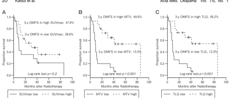
Figures 1 and 2 show the Kaplan-Meier curves of DMFS and PFS divided into 2 groups by the median values of the SUVmax, MTV, and TLG. The three-year DMFS rate was 28.6% in patients with a low SUVmax and 47.6% in those with a high SUVmax, 13.3% in patients with a low MTV and 64.6% in those with a high MTV, and 13.3% in patients with a low TLG and 65.2% in those with a high TLG (Fig.1). The three-year PFS
rate was 28.6% in patients with a low SUVmax and 41.2% in those with a high SUVmax, 13.3% in patients with a low MTV and 57.8% in those with a high MTV, and 13.3% in patients with a low TLG and 57.8% in those with a high TLG (Fig.2).
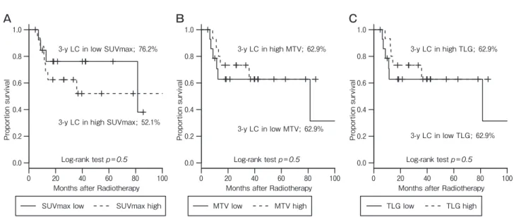
SUVmax, MTV, and TLG were not predictors for LC (p=0.5, 0.5, and 0.5, respectively) (Fig.3). Lower age, histology of adenocarcinoma, and two cycles of concurrent chemotherapy were associated with a higher LC in the univariate analysis (p=0.03, 0.03, and 0.005, respectively). There were no significant factors in the multivariate analysis. SUVmax, MTV, and TLG were not predictors for OS (p=0.7, 0. 5 and 0. 6,
respec-tively) (Fig.4). The histology of the adenocarcinoma was associated with a higher OS on univariate analysis (p=0.0008). Elective nodal irradiation was performed in 7 patients and was not found to be correlated with LC (p=0.5), DMFS (p=0.8), PFS (p=0.3), or OS (p=0.8).
Discussion
In this study, we demonstrated that volumetric PET parameters were predictors of which patients had received definitive CCRT.
The outcome of chemoradiotherapy for stage III lung cancer has been shown to be significantly improved by
Age (years) Median (range) 64 (36-84)
-Sex Male 26 87 Female 4 13 T stage 1 6 20 2 6 20 3 1 3 4 14 47 x 3 10 N stage 0 1 3 1 5 17 2 10 33 3 14 47
Clinical stage IIIA 7 23
IIIB 23 77
Histology Adenocarcinoma 11 37
Squamous cell carcinoma 17 57
Non-small cell carcinoma 2 7
Lobea Upper 21 70
Lower 6 20
Lateralitya Right 17 57
Left 11 33
Smoking History Never 1 3
Former 15 50
Current 14 47
FEV1 (l)a Median (range) 2.17 (1.12-4.11)
-ECOG-PS 0 12 40
1 18 60
Cycles of concurrent chemotherapy 1 1 3
2 29 97
Cycles of adjuvant chemotherapy 0 28 93
1 2 7
SUVmax Median (range) 16.04 (5.90-35.83)
-MTV (cm3) Median (range) 78.82 (15.09-547.00)
-TLG Median (range) 588.07 (47.09-4160.73)
-ECOG-PS, Eastern Cooperative Oncology Group performance status; FEV1, forced expiratory volume in 1 sec; SUVmax, maximum stan-dardized uptake value; MTV, metabolic tumor volume of SUVmax more than 2.5; TLG, total lesion glycolysis of SUVmax more than 2.5. aThese factors have missing values.
the use of durvalumab [11,12]. The improvement in PFS and OS could be attributed to the reduction of dis-tant metastases. However, radiation pneumonitis is one of the serious adverse events of radiotherapy, and the rate of grade 5 adverse events has been reported to be 2.5% [13] and 4.4% [10]. In addition, one of the major adverse events associated with immune checkpoint inhibitors is pneumonitis [14]. Thus, these drugs can be used only after considering the balance between their benefits and the risk of pneumonitis; therefore, predic-tive biomarkers of distant metastases prior to CCRT are desirable, as they can assist the attending physician in
determining whether to administer durvalumab. PET is a useful imaging technique for prediction in addition to diagnosis, staging, and radiotherapy plan-ning in the case of malignancy [15,16]. Although SUVmax was initially adopted as a predictor for patients who received radiotherapy [2], volumetric PET param-eters have recently been adopted as prognostic factors. In our study, SUVmax was not a predictor of LC, DMFS, PFS, or OS, but the volumetric PET parame-ters MTV and TLG were predictors of DMFS and PFS. Because SUVmax is a single-voxel value representing the most intense FDG uptake in the mass [4], it does Table 2 Univariate analyses of factors associated with distant metastasis-free survival and progression-free survival
Factor DMFS PFS
event/total p-value event/total p-value
Age (years) <64 10/15 0.5 10/15 0.6 ≥64 11/15 11/15 Sex Male 19/26 0.8 19/26 0.6 Female 2/4 2/4 T stagea 1-2 10/12 0.7 10/12 0.8 3-4 10/15 10/15 N stage 0-1 4/6 0.8 4/6 0.9 2-3 17/24 17/24
Clinical stage IIIA 4/7 0.5 4/7 0.9
IIIB 17/23 17/23
Histology Adenocarcinoma 8/11 0.05 8/11 0.2
others 13/19 13/19
Lobea Lower lobe 5/6 0.8 5/6 0.6
Upper lobe 15/21 15/21
Lateralitya Right 12/17 0.3 12/17 0.6
Left 8/10 8/10
Smoking history Never/Former 10/16 0.2 10/16 0.3
Current 11/14 11/14
FEV1 (l)a <2.2 11/15 0.8 11/15 0.9
≥2.2 10/13 10/13
ECOG-PS 0 8/12 0.7 8/12 0.9
1 14/18 13/18
Cycles of concurrent chemotherapy 1 1/1 0.7 1/1 0.4
2 20/29 20/29
Cycles of adjuvant chemotherapy 0 19/28 0.2 19/28 0.2
1 2/2 2/2 SUVmax <16 12/14 0.2 12/14 0.09 ≥16 9/16 9/16 MTV (cm3) <79 14/15 0.0003 14/15 0.0005 ≥79 7/15 7/15 TLG <590 14/15 0.001 14/15 0.0007 ≥590 7/15 7/15
DMFS, distant metastases free survival; ECOG-PS, Eastern Cooperative Oncology Group performance status; FEV1, forced expiratory volume in 1 sec; SUVmax, maximum standardized uptake value; MTV, metabolic tumor volume of SUVmax more than 2.5; PFS, pro-gression-free survival; TLG, total lesion glycolysis of SUVmax more than 2.5.
not always represent the volume or burden of the highly active lesions in malignant tumors. In addition, the tumor composition is heterogeneous, and tumors are composed of non-active components such as necrotic tissue [17] or fibrotic scars, which may cause inaccura-cies in assessments based on SUVmax values. SUVmax is also highly sensitive to image noise [18]. In contrast to SUVmax, the MTV and TLG of volumetric PET rep-resent both the three-dimensional tumor volume and metabolic activity. In studies involving surgical resec-tion for early stage disease [19], CCRT for stage II-III disease [5,20], preoperative CCRT followed by surgery
for stage IIIA-N2 disease [21], and chemotherapy for stage III-IV disease [7], researchers concluded that vol-umetric PET parameters are more useful as predictors than SUVmax. Consistent with these reports, volumet-ric PET parameters were more useful than SUVmax as prognostic factors in our study.
In our study, higher MTV and TLG resulted in higher DMFS and PFS rates, which contradicted the results of a previous report [8]. To the best of our knowledge, no papers have obtained the same results as our study. The treatment options for stage III lung can-cer are wide-ranging and include surgery, preoperative
0 20 40 60 80 100 0.0 0.2 0.4 0.6 0.8
Months after Radiotherapy
Propo
rtion su
rvi
val
SUVmax low SUVmax high 3-y DMFS in low SUVmax; 28.6%
Log-rank test p=0.2 0 20 40 60 80 100 0.0 0.2 0.4 0.6 0.8
Months after Radiotherapy
Propo rtion su rvi val MTV low MTV high 3-y DMFS in low MTV; 13.3% Log-rank test p<0.001 0 20 40 60 80 100 0.0 0.2 0.4 0.6 0.8
Months after Radiotherapy
Propo rtion su rvi val TLG low TLG high 3-y DMFS in low TLG; 13.3% Log-rank test p<0.001
Fig. 1 Subgroup analysis of distant metastasis-free survival; (A) SUVmax; (B) MTV; (C) TLG.
0 20 40 60 80 100 0.0 0.2 0.4 0.6 0.8 1.0
Months after Radiotherapy
Propo
rtion su
rvi
val
A
SUVmax low SUVmax high 3-y PFS in high SUVmax; 41.0%
3-y PFS in low SUVmax; 28.6% Log-rank test p=0.09 0 20 40 60 80 100 0.0 0.2 0.4 0.6 0.8 1.0
Months after Radiotherapy
Propo rtion su rvi val B MTV low MTV high 3-y PFS in high MTV; 57.8% 3-y PFS in low MTV; 13.3% Log-rank test p=0.001 0 20 40 60 80 100 0.0 0.2 0.4 0.6 0.8 1.0
Months after Radiotherapy
Propo rtion su rvi val C TLG low TLG high 3-y PFS in high TLG; 57.8% 3-y PFS in low TLG; 13.3% Log-rank test p<0.001
radiotherapy followed by surgery, chemoradiotherapy, and chemotherapy; therefore, indications for definitive radiotherapy may vary greatly among institutions. While our study used standardized regimens of chemo-therapy and radiation doses, many previous reports have mixed different regimens and radiation doses [8], and this may have been responsible for the variation in results. Other possible explanations for the discrepant results are described below. PET accumulation is higher in poorly differentiated tumors [22]. Radiation therapy is highly effective for poorly differentiated tumors, which may explain the successful results in the high
MTV and TLG groups. Docetaxel leads to a G2-M-phase cell cycle arrest of tumor cells and has a synergis-tic effect with radiotherapy [23]. There may have been more tumor cells with higher activity and shorter dou-bling times in the group with higher PET parameters, which may have resulted in a greater effect of CCRT in these patients. Ganeshan et al. found that the computed tomographic texture in NSCLC is associated with tumor hypoxia and angiogenesis [24]. Cook et al. reported that prognosis can be predicted by texture analysis of intra-tumoral image heterogeneity with PET images of NCSLC patients who received chemoradiotherapy [25].
0 20 40 60 80 100 0.0 0.2 0.4 0.6 0.8 1.0
Months after Radiotherapy
Propo
rtion su
rvi
val
A
SUVmax low SUVmax high 3-y LC in high SUVmax; 52.1% 3-y LC in low SUVmax; 76.2%
Log-rank test p=0.5 0 20 40 60 80 100 0.0 0.2 0.4 0.6 0.8 1.0
Months after Radiotherapy
Propo rtion su rvi val B MTV low MTV high 3-y LC in high MTV; 62.9% 3-y LC in low MTV; 62.9% Log-rank test p=0.5 0 20 40 60 80 100 0.0 0.2 0.4 0.6 0.8 1.0
Months after Radiotherapy
Propo rtion su rvi val C TLG low TLG high 3-y LC in high TLG; 62.9% 3-y LC in low TLG; 62.9% Log-rank test p=0.5
Fig. 3 Subgroup analysis of local control rate; (A) SUVmax; (B) MTV; (C) TLG.
0 20 40 60 80 100 0.0 0.2 0.4 0.6 0.8 1.0
Months after Radiotherapy
Propo
rtion su
rvi
val
A
SUVmax low SUVmax high 3-y OS in high SUVmax; 57.4%
3-y OS in low SUVmax; 57.1% Log-rank test p=0.7 0 20 40 60 80 100 0.0 0.2 0.4 0.6 0.8 1.0
Months after Radiotherapy
Propo rtion su rvi val B MTV low MTV high 3-y OS in high MTV; 63.8% 3-y OS in low MTV; 51.3% Log-rank test p=0.5 0 20 40 60 80 100 0.0 0.2 0.4 0.6 0.8 1.0
Months after Radiotherapy
Propo rtion su rvi val C TLG low TLG high 3-y OS in high TLG; 64.2% 3-y OS in low TLG; 50.9% Log-rank test p=0.6
indicate the influence of these more complicated factors. Data from additional studies and more research are warranted to validate these points.
Volumetric PET parameters were not prognostic factors for OS despite being prognostic factors for PFS. This difference in the predictive utility of PET for OS can be attributed to post-recurrence treatments such as molecular target drugs and immune checkpoint inhibi-tors [26]. The pathology of adenocarcinoma was asso-ciated with a high OS rate. Pemetrexed is not recom-mended because of its lower benefit in patients with squamous cell carcinoma compared to patients with adenocarcinoma [27], while bevacizumab is not rec-ommended because squamous cell carcinoma is a risk factor for serious adverse events [28]. It has been spec-ulated that adenocarcinomatous pathology was a favor-able prognostic factor in OS because of differences in drug indications and efficacy after recurrence.
We believe that the results of our study can help determine treatment strategies and the frequency of follow-up examinations after definitive CCRT. The addition of durvalumab after definitive CCRT improves the prognosis and should be prescribed by physicians as much as possible. Due to concerns regarding radiation pneumonitis, cases with a V20 of 35% or more were excluded from the PACIFIC study [11,12], and clinical use in such cases requires great caution. Since the pro-ton beam can produce a better dose distribution than the X-ray, the V20 value can be reduced [29]. Thus, the option of proton beam therapy may also be considered for patients with high V20 values who are likely to develop distant metastases as a result of volumetric PET parameters. The prognosis is poor for those patients having both spinal metastases and rapidly progressing symptoms. The 6-month survival rate for the good prognosis group with slower symptom progression is 92.0%, whereas that for the poor prognosis group with rapid symptom progression is 11.3% [30]. Therefore, early identification of distant metastases is as important as the treatment protocol. It may be possible to improve a patient’s prognosis by assessing metastases at a short interval after CCRT in a group in which distant metas-tasis is likely to occur based on volumetric PET param-eters.
There are some limitations to our study. First, this study was a retrospective analysis with a small sample
ing physicians in the authors’ institutions tended to prioritize surgery after preoperative CCRT over defini-tive CCRT in stage III NCSLC. Third, Cook et al. reported that PET texture analysis of intra-tumoral image heterogeneity can predict prognosis [25], but we did not perform texture analysis. Fourth, since there were only 6 patients with a known pathological differ-entiation grade, we could not perform a statistical anal-ysis of the effect of the degree of differentiation on the PET value. Fifth, the interval at which the examina-tions were performed was not standardized, which could have affected DMFS and PFS. Despite these lim-itations, we showed that the group with high volumet-ric PET parameters had a high DMFS rate.
In conclusion, we demonstrated that volumetric PET parameters could serve as predictors in stage III NSCLC patients who received definitive CCRT with cisplatin/docetaxel. Volumetric PET parameters may help physicians determine the best treatment modality and the most effective timing of follow-up examina-tions. Our findings are contrary to those of previous reports, and further research is warranted to validate these results.
Acknowledgments. This study was supported by a donation from Tsuyama Chuo Hospital.
References
1. Segawa Y, Kiura K, Takigawa N, Kamei H, Harita S, Hiraki S, Watanabe Y, Sugimoto K, Shibayama T, Yonei T, Ueoka H, Takemoto M, Kanazawa S, Takata I, Nogami N, Hotta K, Hiraki A, Tabata M, Matsuo K and Tanimoto M: Phase III trial comparing docetaxel and cis-platin combination chemotherapy with mitomycin, vindesine, and cispla-tin combination chemotherapy with concurrent thoracic radiotherapy in locally advanced non-small-cell lung cancer: OLCSG 0007. J Clin Oncol (2010) 28: 3299-3306.
2. Na F, Wang J, Li C, Deng L, Xue J and Lu Y: Primary tumor standard-ized uptake value measured on F18-Fluorodeoxyglucose positron emission tomography is of prediction value for survival and local control in non-small-cell lung cancer receiving radiotherapy: meta-analysis. J Thorac Oncol (2014) 9: 834-842.
3. Im HJ, Pak K, Cheon GJ, Kang KW, Kim SJ, Kim IJ, Chung JK, Kim EE and Lee DS: Prognostic value of volumetric parameters of (18)F-FDG PET in non-small-cell lung cancer: a meta-analysis. Eur J Nucl Med Mol Imaging (2015) 42: 241-251.
4. Im HJ, Bradshaw T, Solaiyappan M and Cho SY: Current Methods to Define Metabolic Tumor Volume in Positron Emission Tomography: Which One is Better? Nucl Med Mol Imaging (2018) 52: 5-15.
5. Grootjans W, Usmanij EA, Oyen WJ, van der Heijden EH, Visser EP, Visvikis D, Hatt M, Bussink J and de Geus-Oei LF: Perfor mance of auto-matic image segmentation algorithms for calculating total lesion
glycoly-sis for early response monitoring in non-small cell lung cancer patients during concomitant chemoradiotherapy. Radiother Oncol (2016) 119: 473-479.
6. Salavati A, Duan F, Snyder BS, Wei B, Houshmand S, Khiewvan B, Opanowski A, Simone CB, 2nd, Siegel BA, Machtay M and Alavi A: Optimal FDG PET/CT volumetric parameters for risk stratification in patients with locally advanced non-small cell lung cancer: results from the ACRIN 6668/RTOG 0235 trial. Eur J Nucl Med Mol Imaging (2017) 44: 1969-1983.
7. Zaizen Y, Azuma K, Kurata S, Sadashima E, Hattori S, Sasada T, Imamura Y, Kaida H, Kawahara A, Kinoshita T, Ishibashi M and Hoshino T: Prognostic significance of total lesion glycolysis in patients with advanced non-small cell lung cancer receiving chemotherapy. Eur J Radiol (2012) 81: 4179-4184.
8. Cremonesi M, Gilardi L, Ferrari ME, Piperno G, Travaini LL, Timmerman R, Botta F, Baroni G, Grana CM, Ronchi S, Ciardo D, Jereczek-Fossa BA, Garibaldi C and Orecchia R: Role of interim (18)F-FDG-PET/CT for the early prediction of clinical outcomes of Non-Small Cell Lung Cancer (NSCLC) during radiotherapy or chemo-radiotherapy. A systematic review. Eur J Nucl Med Mol Imaging (2017) 44: 1915-1927.
9. Ogata T, Katsui K, Yoshio K, Ihara H, Katayama N, Soh J, Kuroda M, Kiura K, Maeda Y, Toyooka S and Kanazawa S: Dose-Volume Parameters Predict Radiation Pneumonitis after Surgery with Induction Concurrent Chemoradiotherapy for Non-small Cell Lung Cancer. Acta Med Okayama (2018) 72: 507-513.
10. Katsui K, Ogata T, Watanabe K, Katayama N, Kuroda M, Kiura K, Hiraki T, Maeda Y, Toyooka S and Kanazawa S: Radiation pneumonitis after definitive concurrent chemoradiotherapy with cisplatin/docetaxel for non-small cell lung cancer: Analysis of dose-volume parameters. Cancer Medicine (2020) (Epub ahead of print).
11. Antonia SJ, Villegas A, Daniel D, Vicente D, Murakami S, Hui R, Yokoi T, Chiappori A, Lee KH, de Wit M, Cho BC, Bourhaba M, Quantin X, Tokito T, Mekhail T, Planchard D, Kim YC, Karapetis CS, Hiret S, Ostoros G, Kubota K, Gray JE, Paz-Ares L, de Castro Carpeno J, Wadsworth C, Melillo G, Jiang H, Huang Y, Dennis PA, Ozguroglu M and Investigators P: Durvalumab after Chemoradiotherapy in Stage III Non-Small-Cell Lung Cancer. N Engl J Med (2017) 377: 1919-1929. 12. Antonia SJ, Villegas A, Daniel D, Vicente D, Murakami S, Hui R,
Kurata T, Chiappori A, Lee KH, de Wit M, Cho BC, Bourhaba M, Quantin X, Tokito T, Mekhail T, Planchard D, Kim YC, Karapetis CS, Hiret S, Ostoros G, Kubota K, Gray JE, Paz-Ares L, de Castro Carpeno J, Faivre-Finn C, Reck M, Vansteenkiste J, Spigel DR, Wadsworth C, Melillo G, Taboada M, Dennis PA, Ozguroglu M and Investigators P: Overall Survival with Durvalumab after Chemoradiotherapy in Stage III NSCLC. N Engl J Med (2018) 379: 2342-2350.
13. Tsujino K, Hashimoto T, Shimada T, Yoden E, Fujii O, Ota Y, Satouchi M, Negoro S, Adachi S and Soejima T: Combined analysis of V20, VS5, pulmonary fibrosis score on baseline computed tomography, and patient age improves prediction of severe radiation pneumonitis after concurrent chemoradiotherapy for locally advanced non-small-cell lung cancer. J Thorac Oncol (2014) 9: 983-990.
14. Khunger M, Rakshit S, Pasupuleti V, Hernandez AV, Mazzone P, Stevenson J, Pennell NA and Velcheti V: Incidence of Pneumonitis With Use of Programmed Death 1 and Programmed Death-Ligand 1 Inhibitors in Non-Small Cell Lung Cancer: A Systematic Review and Meta-Analysis of Trials. Chest (2017) 152: 271-281.
15. Ung YC, Maziak DE, Vanderveen JA, Smith CA, Gulenchyn K, Lacchetti C and Evans WK: 18Fluorodeoxyglucose positron emission tomography in the diagnosis and staging of lung cancer: a systematic review. J Natl Cancer Inst (2007) 99: 1753-1767.
16. Konert T, Vogel W, MacManus MP, Nestle U, Belderbos J, Gregoire V, Thorwarth D, Fidarova E, Paez D, Chiti A and Hanna GG: PET/CT imaging for target volume delineation in curative intent radiotherapy of non-small cell lung cancer: IAEA consensus report 2014. Radiother
Oncol (2015) 116: 27-34.
17. Henson JW, Ulmer S and Harris GJ: Brain tumor imaging in clinical tri-als. AJNR Am J Neuroradiol (2008) 29: 419-424.
18. Boellaard R, Krak NC, Hoekstra OS and Lammertsma AA: Effects of noise, image resolution, and ROI definition on the accuracy of standard uptake values: a simulation study. J Nucl Med (2004) 45: 1519-1527. 19. Hyun SH, Choi JY, Kim K, Kim J, Shim YM, Um SW, Kim H, Lee KH
and Kim BT: Volume-based parameters of (18)F-fluorodeoxyglucose posi-tron emission tomography/computed tomography improve outcome pre-diction in early-stage non-small cell lung cancer after surgical resection. Ann Surg (2013) 257: 364-370.
20. Huang W, Fan M, Liu B, Fu Z, Zhou T, Zhang Z, Gong H and Li B: Value of metabolic tumor volume on repeated 18F-FDG PET/CT for early prediction of survival in locally advanced non-small cell lung cancer treated with concurrent chemoradiotherapy. J Nucl Med (2014) 55: 1584-1590.
21. Hyun SH, Ahn HK, Ahn MJ, Ahn YC, Kim J, Shim YM and Choi JY: Volume-Based Assessment With 18F-FDG PET/CT Improves Outcome Prediction for Patients With Stage IIIA-N2 Non-Small Cell Lung Cancer. AJR Am J Roentgenol (2015) 205: 623-628.
22. Higashi K, Matsunari I, Ueda Y, Ikeda R, Guo J, Oguchi M, Tonami H and Yamamoto I: Value of whole-body FDG PET in management of lung cancer. Ann Nucl Med (2003) 17: 1-14.
23. Mason KA, Kishi K, Hunter N, Buchmiller L, Akimoto T, Komaki R and Milas L: Effect of docetaxel on the therapeutic ratio of fractionated radio-therapy in vivo. Clin Cancer Res (1999) 5: 4191-4198.
24. Ganeshan B, Goh V, Mandeville HC, Ng QS, Hoskin PJ and Miles KA: Non-small cell lung cancer: histopathologic correlates for texture parame-ters at CT. Radiology (2013) 266: 326-336.
25. Cook GJ, Yip C, Siddique M, Goh V, Chicklore S, Roy A, Marsden P, Ahmad S and Landau D: Are pretreatment 18F-FDG PET tumor textural features in non-small cell lung cancer associated with response and sur-vival after chemoradiotherapy? J Nucl Med (2013) 54: 19-26.
26. Borghaei H, Paz-Ares L, Horn L, Spigel DR, Steins M, Ready NE, Chow LQ, Vokes EE, Felip E, Holgado E, Barlesi F, Kohlhaufl M, Arrieta O, Burgio MA, Fayette J, Lena H, Poddubskaya E, Gerber DE, Gettinger SN, Rudin CM, Rizvi N, Crino L, Blumenschein GR, Jr., Antonia SJ, Dorange C, Harbison CT, Graf Finckenstein F and Brahmer JR: Nivolumab versus Docetaxel in Advanced Nonsquamous Non-Small-Cell Lung Cancer. N Engl J Med (2015) 373: 1627-1639.
27. Scagliotti GV, Parikh P, von Pawel J, Biesma B, Vansteenkiste J, Manegold C, Serwatowski P, Gatzemeier U, Digumarti R, Zukin M, Lee JS, Mellemgaard A, Park K, Patil S, Rolski J, Goksel T, de Marinis F, Simms L, Sugarman KP and Gandara D: Phase III study comparing cis-platin plus gemcitabine with ciscis-platin plus pemetrexed in chemothera-py-naive patients with advanced-stage non-small-cell lung cancer. J Clin Oncol (2008) 26: 3543-3551.
28. Sandler AB, Schiller JH, Gray R, Dimery I, Brahmer J, Samant M, Wang LI and Johnson DH: Retrospective evaluation of the clinical and radiographic risk factors associated with severe pulmonary hemorrhage in first-line advanced, unresectable non-small-cell lung cancer treated with Carboplatin and Paclitaxel plus bevacizumab. J Clin Oncol (2009) 27: 1405-1412.
29. Zhang X, Li Y, Pan X, Xiaoqiang L, Mohan R, Komaki R, Cox JD and Chang JY: Intensity-modulated proton therapy reduces the dose to normal tissue compared with intensity-modulated radiation therapy or passive scattering proton therapy and enables individualized radical radiotherapy for extensive stage IIIB non-small-cell lung cancer: a virtual clinical study. Int J Radiat Oncol Biol Phys (2010) 77: 357-366.
30. Rades D, Douglas S, Huttenlocher S, Rudat V, Veninga T, Stalpers LJ, Basic H, Karstens JH, Hoskin PJ, Adamietz IA and Schild SE: Validation of a score predicting post-treatment ambulatory status after radiotherapy for metastatic spinal cord compression. Int J Radiat Oncol Biol Phys (2011) 79: 1503-1506.