(Original) Matsumoto Shigaku 19: 10--16, 1993
key words : calcified bodies histopathology ultrastructure electron probe microanalysis postoperatiye maxillary cyst
Observations on Calcified Bodies in the Cyst Wall
of a Postoperative Maxillary Cyst
TOSHIYUKI KAWAKAMI NORIYUKI TAKEI HIDEYO UJI
MOTOYOSHI ANTOH HIROMASA HASEGAWA and SHIGEO EDA
DePartment of Oral Pathology, Matsumoto Dental College(Chief .' Prof S. Eda)
Summary
Light and scanning electron microscopic observations and electron probe
mi-croanalysis were carried out on granular calcified bodies that appeared in the wall of a postoperative maxillary cyst in a 59-year-old man. The granular bodies, stained slightly with hematoxylin, were scattered in the cyst wall. They reacted positively to von Kossa's stain indicating calcification. Based on scanning electron microscopic observation, secon-dary electron images revealed them to be compact bodies, and they appeared as light spots in composition images. Electron probe microanalysis revealed that the bodies were ccomposed mainly of calcium and phosphorus. Some of the large bodies had a membranous calcification core, which was observed especially in von Kossa's stained specimens. Therefore, we believe that these calcification core must be generated cell debris, cell membrane, nuclear membrane or subcellular organelles.Introduction
Pathological calcium deposition in various tissues mainly occurs in connection with necrotic andlor degenerative changes'•2}. Therefore, these calcifications are generally characterized as deposits that the needle like or amorphous in shape depending upon the underlying structures. Although granular calcified bodies commonly occur in meningiomas and adenocarcinomas, some papers have also described microcalcifications, especially microcalculi in salivary glands. In oral
neoplasms, this type of microcalcification appears mainly in salivary gland tumors3-"), particularly in pleomorphic adenomas.
During our histopathological survey of surgically removed tissues at the Clinical Division of Matsumoto Dental College Hospital, we discovered a case of postoperative maxillary cyst having granular calcified bodies scattered in the cyst wall. We describe herein the histopathological and ultrastructural features of these bodies.
Presented in part at the 35th meeting of the Matsumoto Dental College Society held on November 7, 1992. (accepted
zaJ!Ntwnt]}=4 19(1) 1993 11
Materials and Methods
Materials examined in this study were obtained from a postoperative maxillary cyst in a 59-year-old man (MDC 082-91). The specimens were drawn from the patient during surgery , and for histopathological analysis, selected portions were fixed in 10% formalin solution, dehydrated through a graded ethanol series, and then embedded in paraffin. After sectioning, the specimens were stained with hematoxylin-eosin (H-E) and von Kossa's stain and then observed under a light mlcroscope.
For scanning electron microscopy (SEM) and electron probe microanalysis (EPMA), sectioned specimens were mounted on a carbon block and deparaffinized with a xylene solution. They were then processed by critical-point drying, coated with carbon by the cathodic sputtering method, and then examined with a JEOL JCXA 733 super probe.
Results
Histopathological findings :
Light microscopic observation revealed that the cyst wall consisted mainly of collagen bundles having limited lining epithelium (Figs. 1, 2), and in part of the wall signs of reactive bone formation were evident (Fig. 1). The cyst wall connective tissue was partially hyalinized, which showed poor cellular areas, and in these areas some granular and irregular sized bodies stained slightly with hematoxylin were present (Figs. 3, 4). The large masses were globular in shape, and the small ones were sand-like granules. These bodies seemed to have no relationship with the above mentioned reactive bone formation.
These globular masses and sand-like granules reacted strongly with von Kossa's stain, indica-ting the occurrence of calcification (Figs. 5, 6). Regarding the large globular bodies, their positively stained portion appeared to be laminated, and the area around (Fig. 5 arrows), the periphery (Fig. 6 arrows) and the central area of the bodies were strongly positive. In contrast, the entire small sand-like granule was stained uniformly strong. No positive stainings were found in the surrounding hyalinized tissue.
Scanning electron microscopic findings :
Examination by scanning electron microscopy under low magnification showed several light spots in the composition images which corresponded to the von Kossa's-positive material and their distribution in various parts of the hyalinized collagen bundles (Figs. 7, 8); The secondary electron images revealed that the sectioned surface of the cyst wall was relatively solid, and globular-shaped bodies were scattered in the cyst wall (Figs. 9, 10). These globular structures were approximately 1 to 20 ptm in diameter (Figs. 9, 10).
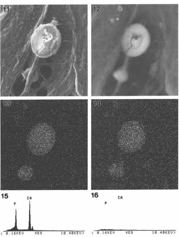
Electron probe microanalytic findings :
Analytically, these materials were calcified (Figs. 11, 12) as judged from their composition determined from radiographic (Ca-Ka and P) images (Figs. 13, 14). EPMA demonstrated that the calcified masses consisted mainly of calcium and phosphorus (Fig. 15), indicating calcium phos-phate ; in contrast, the surrounding tissues showed no trace of these elements (Fig. 16).
Discussion
According to the literature, pathological calcification, which is composed mainly of hydrox-yapatite crystals, is often associated with membranous cellular debris, and these crystals are
12 Kawakami, et al. : Observations on Calcified Bodies in the Cyst Wall of a Postoperative Maxillary Cyst .i,,--.#t=t2.#.I:.=]-.pttz-'.'.6ivÅre=!-ig-ex#yss.N
3
,sk,.a. Kx xg•i-, tl'•si •-ij -;•• .k -...'-trt'k}.5,i, ,g!yef
Sigis".,},.ym. S,iajiIlliili'
'"'` i tS/H . Fig. 1 fig. 2. Fig. 3. Fig. 4. Fig, 5. Fig. 6, 1ec#
.kF - .S!A
,,i! ttl ''i t"••
ei.://"""1 s.i''z
. }l/1 'v{ "- ' 1,y' : ts ,//fi 1 :..., '2f'1.," ,--xXL. ' 1" tt'I' -" t .. :.ti •l •-;tt"' " '-'' ' 'iÅék X' '"'
hsl!N. .. 1/kMl(of'tlsw's/liesiillllglts/wLfo.nt.i""\`/iMi4f,.,eg{g, tk r-;".X. ,tit., ,k •'. ' ,6
f t"' ' .-.. .-`".Iles
-t Ig. .g1.+. . ,,
' .1'' iS' "'•i-,,i,, -E,Å~40). -E, Å~ 150). Xh l.l'3,l,J':•I:'.}'tiJ:;'•""ti'seT3,;//,.i/r.titlt-•.i•,:l:l","•Fi';"i.'t/
' t ' -1.. .,.. .. .,.t)tr -. .
t.
,s
',N ";•tt
"tt"').IJ. " . ,,;
'
tt
.t
rl s' ,ofk ' s
}ge." ' '
" '-;)tiIE,'l•.,., ,g,..,si' '''"/'i"'1.is }i"l "',L "l' .rk"\'l,tS,,"1 ,"3s. yk l')
Histopathological view of the cyst wall (H Lining epithelium of the cyst wall (H
Collagenous tissue of the cyst wall (H-E,Å~480). Globular bodies and sand-like materials in the tissue (H Arrows showing the von
t' .. ;g.-.J,,, . 'e, •-v-gis•l.Pi. r- E,Å~480)- t-- .
:
V"' L. , r. Sl,tl.::e:!;l •l'.I-ag
J " -, :". '.".)':' 't-'i:t l
l't
/!"ie
.; .;pp •vl ' ;'t`j' . Jt.;A} i- •.1 .i'
-.
lr'
---bt
. ., . ,. F: -l;t.j/t jvt'
S,, S .ttt:tdi4' ,. },,,i,.l ,}S}!• .'a
'
t- t tt/ttll/. .J/
'
Kossa's-positive stainings around the large body (Å~600).
Positive stainings existing in the periphery (arrows) and the center of the large bodies (von Kossa's, Å~600).
i![}Jztsiig':']tL'" 19(1) 1993 13
Fig. 7. Fig. 8. Fig. 9. Fig. 10,
Composition image showing numerous light spots in the tissue (Å~600). Composition image showing light spots in the tissue (Å~360).
Enlarged photograph of the area shown in Fig. 8 (Secondary image,Å~1,200). Composition image of the same area of Fig. 9.
14 Kawakami, et aL : Observations on Calcified Bodies
15
c fiin the Cyst Wall of a Postoperative Maxillary Cyst
16
p
eA
Åq e
Åq e,16KEU
XES
te,4eKEuÅr
Fig. 11. Fig. 12. Fig. 13. Fig. 14. Fig. 15. Fig. 16.
Enlarged graph of Fig. 9 demonstrating large globular body Composition irnage of same calcified body seen in Fig. 11. Ca-Ka image of the same area shown in Fig. 11. Phosphorus image of the same area shown in Fig. 11. Result of EPMA of calcified bodies.
Result of EPMA of surrounding tissues.
iflrJ!XcaRt]i!.4 19(1) 1993 15
thought to have started to form on these membranous structuresi). Regarding a case of calcinosis universalis, Kawakami et al. (1986)2) reported that the calcification site was closely related to foci of fibrinoid degeneration ; they proposed that the globular and/or membranous structures seen were derived from degenerating cells. They suggested, therefore, that these globular and/or membranous structures might be involved in the initial calcification in this case. Furthermore, Kawakami et al.8•9) described membranous structures in the pathologic calcification sites elicited by calcium hydroxide -containing dental material, and discussed the relationship between the membranous structure and initial calcification. We believe that some of these structures have a matrix vesicle-Iike function, although we think that the calcified bodies are caused mainly by dystrophic changes because of their appearanceiO). Our previous papers described the ultrastructure of these calcifications in salivary gland tumor cases3"'}, and we suggested that the calcium binding capacity of these materials may be closely related to these microcalcifications. Furthermore, in a case of central neurinoma these microcalcifications were also observedii).
In our present case, the relationship between the calcium depositions and the vesicular membranes was not evident, and we were unable to find obvious membranous structures in the present case despite careful observation. However, we believe that the microcalcifications are closely related to cellular degeneration. In general, the cyst wall is invaded by inflammatory cells at the early stage of its development but these cells disappear with the passage of time. At the time of inflammatory cell disappearance, some of these cells may degenerate. Thus, we speculate that the initial calcification will occur around these degenerating structures. In fact, we found core
struc-tures on the periphery of large calcified bodies by light microscopic observation of von
Kossa's-stained specimens and by scanning electron microscopic examination. We believe these membranous calcification cores to be that of generated cell debris, cell membrane or nuclear membrane, as judged from their diameter. However, there were no core structures in the center of the small bodies, suggesting that the microcalcification in them occurred in close relation with the degeneration of subcellular organelles, i. e. mitochondria, rough and smoothed surfaced-endoplas-mic reticulum, Golgi apparatus, and so on. According to the results of electron probe surfaced-endoplas-microanalysis, the elemental composition of the calcified bodies was similar to that in other cases already describedii•i2).
Acknowledgement
The authors wish to express their thanks to Professor M. Yamaoka, Department of Oral and Maxillofacial Surgery II, Matsumoto Dental College, for supplying the surgical material from the patient. Chief Technologist S. Akahane, Laboratory of Electron Microscope, Matsumoto Dental College, is acknowledged for his technical assistance in electron microscopy.
References
1) Kim, K. M. and Huang, S.(1971) UItrastructural study of calcification of human aortic valve. Lab. Invest. 25 : 357-366.
2) Kawakami, T., Nakamura, C., Hasegawa, H., Eda, S., Akahane, S., Yamazaki, T. and Takasu, N. (1986) Ultrastructural study of calcinosis universalis with dermatomyositis. J. Cutan. Pathol. 13 : 135
143.
3 ) Akahane, S., Kawakami, T., Nakamura, C., Hasegawa, H., Eda, S., Komatsu, M., Furusawa, K. and Ideguchi, H. (1984) Electron microscopic studies on mucoepidermoid carcinoma, II calcified materials appeared in the stroma. Matsumoto Shigaku, 10 : 29-41.
16 Kawakami, et al. : Observations on Calcified Bodies in the Cyst Wall of a Postoperative Maxillary Cyst 4 ) Kawakami, T., Nakamura, C, Hasegawa, H., Eda, S., Komatsu, M., Furusawa, K. and Akahane, S.
(1986) Ultrastructure of stromal calcification in mucoepidermoid carcinoma. Jpn. J. Oral Biol. 28 : 217
-222.
5 ) Yamazaki, T., Kotani, A. and Kawakami, T. (1987) Basal cell adenoma of the sublingual gland. J. Oral Maxillofac. Surg. 45 : 270-273.
6 ) Hasegawa, H., Kawakami, T., Nakamura, C, Eda, S., Furusawa, K. and Kiga, M. (1987) Ultrastructural study of varied calcified materials in the pleomorphic adenoma occurring in the soft palate. Matsumoto Shigaku, 13 : 115-121.
7 ) Nakamura, C, Kawakami, T., Hasegawa, H., Eda, S., Akahane, S. and Yamazaki, T. (1987) Light and electron microscopic studies of microcalcifications appearing in monomorphic adanomas. Matsumoto Shigaku, 13 : 329-336.
8) Kawakami, T., Nakamura, C, Hayashi, T., Eda, S. and Akahane, S. (1979) Studies on the tissue reactions to the paste of calcium hydroxide added iodoform (root canal filling material : Vitapex ®), second report an electron-microscopic study. Matsumoto Shigaku, 5 : 161-170.
9 ) Kawakami, T. (1984) An experimental study on tissue reactions to a paste made of calcium hydroxide and iodoform with an addition of silicone oil -with special reference to absorption of and calcification by the paste-. Shikwa Gakuho, 84 : 1563-1593.
10) Kawakami, T., Nakamura, C, Hasegawa, H., Akahane, S. and Eda, S. (1987) Ultrastructural study of initial calcification in the rat subcutaneous tissues. Oral Surg. 63 : 360-365.
ll) Kawakami, T., Hasegawa, H., Nakamura, C, Eda, S., Yagasaki, T., Kitamura, Y., Chino, T. and Akahane, S. (1988) An electron microscopic observation of psammoma body-type microcalcifications in a case of intraosseous neurinoma. J. Clin. Electron Microsc, 21 : 167-171.
12) Kawakami, T., Nakamura, C, Hasegawa, H., Eda, S. and Akahane, S. (1988) Analytical electron microscopic study of mineral deposits in a case of calcinosis universalis. Matsumoto Shigaku, 14 : 41