Correlation between Serum Dehydroepiandrosterone Sulfate
and Coronary Artery Disease in M iddle-Aged and Elderly M en
Hiroyuki Sumino , Shuichi Ichikawa , Shu Kasama , Akiko Ichikawa , Yoshihiro Araki ,
Hiroyoshi Kanai , Toshio Itoh , Toshiya Iwasaki , Yoshie Sawada , Takashi Takahashi ,
Hisao Kumakura , Yoshiaki Takayama , Kazutomo Minami , Masahiko Kurabayashi
and Masami Murakami
1 Department of Internal Medicine, Cardiovascular Hospital of Central Japan (Kitakanto Junkanki Hospital), 740 Shimohakoda, Hokkitsu-machi, Shibukawa, Gunma 377-0061, Japan
2 Department of Clinical Laboratory Medicine,Gunma University Graduate School of Medicine,3-39-22 Showa-machi,Maebashi,Gunma 371-8511, Japan
3 Laboratory of Infectious Diseases, Graduate School of Infection Control Sciences, Kitasato University, 5-9-1 Shirokane, Minato-ku, Tokyo 108-8641, Japan
4 Department of Medicine and Biological Science, Gunma University Graduate School of Medicine, 3-39-22 Showa-machi, Maebashi, Gunma 371-8511, Japan
Abstract
Background & Aims:The correlation between low serum dehydroepiandrosterone sulfate (DHEA-S) levels and coronary artery disease (CAD) is yet unclear. Therefore, we investigated the correlation between DHEA-S levels and the presence or severity of CAD in middle-aged and elderly men.
M ethods:Fifty-six consecutive middle-aged and elderly men who underwent selective coronary angiography were divided into the CAD group (n=41) and control group (without CAD; n=15). CAD was defined as 75% occlusion in 1 major coronary arteries. Serum DHEA-S,lipid,and plasma glucose levels were determined after a 12-h fast. Coronary artery stenosis severity was estimated using the Gensini score.
Results:DHEA-S levels were significantly lower in the CAD group than in the control group;however, other clinical parameters showed no difference between the groups. An age-adjusted multiple regression analysis revealed that DHEA-S-dependent determinants were significantly correlated with the Gensini score (β=−0.291, p<0.05), but not with other clinical parameters.
Conclusions:Middle-aged and elderly men with CAD may have lower serum DHEA-S levels than age-matched men without CAD,and serum DHEA-S levels may negatively correlate with coronary artery stenosis severity in all men, suggesting a cardioprotective effect of DHEA-S.
Introduction
The secretion and circulating levels of the adrenal steroid dehydroepiandrosterone (DHEA) and its metabolite, DHEA sulfate (DHEA-S), decrease with aging; however, the biological significance of this decrease is unclear. DHEA is known to exert direct effects on cells through the activation of known mecha-nisms such as peroxisome proliferator-activated receptor-α and/or an unidentified membrane rece-ptor. DHEA has been correlated with bone physiol-ogy, body composition, insulin sensitivity, immune functions, and vascular physiology.
With regard to the correlation between serum or plasma DHEA-S levels and atherosclerotic disease, including cardiovascular disease (CVD) and coronary artery disease (CAD), in men, several studies have demonstrated a correlation between low serum or plasma DHEA-S levels and increased CVD-associated morbidity or mortality. In contrast, other studies reported no significant correlation between these fac-tors. Furthermore, one clinical study reported an inverse correlation between plasma DHEA-S levels
Article Information Key words:
coronary angiography, coronary artery disease, dehydroepiandrosterone sulfate Publication history: Received: January 25, 2016 Accepted: March 10, 2016 Corresponding author: Hiroyuki Sumino
Department of Clinical Laboratory Medicine, Gunma University Graduate School of Medicine, 3-39-22 Showa-machi, Maebashi, Gunma 371-8511, Japan
Tel: +81-27-220-8576
E-mail: suminoh@gunma-u.ac.jp Original
and the number of diseased coronary arteries deter-mined by angiography(any stenosis 50%)in men. Therefore, consensus regarding this correlation is yet unclear.
The Gensini score is often used to assess the severity of coronary artery stenosis in patients rather than the number of significant coronary artery stenoses. This score is computed by assigning a severity score to each coronary stenosis according to the degree of luminal narrowing and geographic importance. Although the Gensini score is superior to the number of significant coronary artery stenoses when estimating the severity of coronary stenosis in patients, no study has reported a correlation between circulating DHEA-S levels and Gensini score-estimated coronary artery stenosis severity in men. Therefore,the clinical significance of DHEA-S for preventing CVD and CAD in men has not been completely elucidated.
Therefore,we aimed to investigate the correlation between endogenous serum DHEA-S levels and the presence or severity of CAD in men. Furthermore,we aimed to determine whether serum DHEA-S levels varied in middle-aged and elderly men with and without CAD who underwent selective coronary an-giography and whether serum DHEA-S levels correlat-ed with coronary artery stenosis severity in this popula-tion.
Patients and methods
Study population
One hundred and thirty-five consecutive middle-aged and elderly Japanese male patients (mean age, 65.9±9.7[range:37-86]years)who underwent selective coronary angiography at the Cardiovascular Hospital of Central Japan (Kitakanto Junkanki Hospital) par-ticipated in this study. Patients were referred for coronary angiography because of chest pain,a positive exertion test, positive myocardial perfusion scan, and history of angina pectoris or myocardial infarction. Individuals with 75% occlusion of at least one major coronary artery were defined as having CAD; those without CAD comprised the control group. Physical examination,chest roentgenogram,laboratory tests, electrocardiography, and echocardiography were performed to exclude structural heart defects.
Subjects with acute, recent, or past myocardial infarction; unstable or vasospastic angina pectoris; heart failure; malignancy; renal or liver dysfunction; autoimmune disease;infection were excluded;further-more,those who were treated with medications known to affect sex hormone levels including androgens,ster-oid hormones,or antiandrogen treatments for prostate carcinoma were excluded. Finally, a total of 56 male patients (mean age, 65.4±9.4[37-86]years) were enrolled in this study and divided into two groups:the CAD (n=41) group and the control group (n=15). Written informed consent was obtained from each participant prior to admission to the study. The study protocol was approved by the Ethics Committee of the
Cardiovascular Hospital of Central Japan (Kitakanto Junkanki Hospital) and conducted according to the ethical guidelines outlined in the Declaration of Hel-sinki.
Physical examination
Measurements of height, weight, and blood pres-sure were performed in the morning after a 12-h over-night fast. Body mass index (BMI) was calculated as weight (kg)/height (m). Blood pressure was measured twice using a sphygmomanometer on the right arm after the subject had rested in a seated position for 10 min.
Assays
Blood samples were drawn from the antecubital vein between 7:00 AM and 9:00 AM after a 12-h overnight fast. Within 1-h of collection, the fasting blood samples were centrifuged for 15 min at 4℃ and 3,000g, placed in tightly sealed containers, and stored at −80℃ for a maximum of 3 months before measure-ments. The serum concentrations of DHEA-S were measured by radioimmunoassay at a commercial labo-ratory(LSI Medience,Co.,Tokyo,Japan). The serum total cholesterol, high-density lipoprotein cholesterol, and triglyceride concentrations were determined using standard laboratory techniques (Health Sciences Research Institute East Japan CO, LTD, Konosu, Japan). The concentrations of low-density lipoprotein cholesterol were calculated using the Friedwald for-mula. Plasma glucose concentrations were assayed using the glucose oxidase method, and hemoglobin A1c (HbA1c) levels were measured via high-performance liquid chromatography (Health Sciences Research Institute East Japan CO, LTD). The intra-and inter-assay coefficients of variation were< 5% for DHEA-S,< 6% for lipids,< 6% for plasma glucose, and < 5% for HbA1c.
Determination of coronary artery stenosis severity Coronary artery stenosis severity was estimated using the Gensini score. The Gensini system scores coronary artery luminal narrowing as follows: 1= 1%-25% narrowing; 2=26%-50% narrowing; 4= 51%-75% narrowing; 8=76%-90% narrowing; 16= 91%-99% narrowing;and 32= total occlusion. This score is then multiplied by a factor that incorporates the importance of the lesion position in the coronary arterial tree as follows:left main coronary artery,×5; proximal left anterior descending or left circumflex coronary artery,×2.5;mid-segment of the left anterior descending artery,×1.5;distal left anterior descending, right coronary,or mid-distal left circumflex artery,×1; and any other artery,×0.5.
Statistical analysis
Data are expressed as means±standard deviations (SD). The chi-square test and Fishers exact test were used to compare categorical data. Regarding inter-group comparisons, the Mann-Whitney U test was
sis of covariance (ANCOVA) adjusted for age, BMI, and the use of nitrates and antiplatelet agents was used to compare laboratory data. A Pearson s correlation coefficient analysis was used to examine correlations between DHEA-S concentrations and clinical parame-ters,including age,BMI,blood pressure,Gensini score, and blood chemistry. An age-adjusted multiple regres-sion analysis was then used to examine the correlation between the DHEA-S concentration and Gensini score. All probability values were two-tailed,and a p value<0.05 was considered significant. All statistical analyses were performed using IBM SPSS software (v17.0;Chicago, IL, USA).
Results
Clinical characteristics, complications,and medications Table 1 details the clinical characteristics,compli-cations, and medications of patients in the CAD and control groups. Age, BMI, smoking status, blood pressure, and medical complications did not differ between the two groups. However, the CAD group had a significantly higher Gensini score relative to the control group (p<0.001).In addition,a larger
propor-antiplatelet agents, compared with the control group (p<0.05 for both factors). However, there were no significant differences between the groups in the pro-portions of patients using other medications.
Blood chemistry and DHEA-S
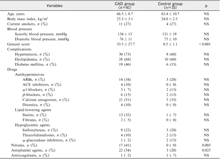
Table 2 details the blood chemistry and DHEA-S concentrations of patients in the CAD and control groups. There were no significant differences in lipid, fasting plasma glucose,and HbA1c levels between the groups. However, DHEA-S levels were significantly lower in the CAD group than in the control group (p< 0.05), and this difference remained after adjusting for age,BMI,and the use of nitrates and antiplatelet agents (52.8±39.2μg/dl vs.81.3±42.0μg/dl, p<0.05;Figure 1).
Correlations between DHEA-S levels and clinical parameters, including age, BMI, blood pressure, Gensini score, and blood chemistry
Table 3 shows correlations between DHEA-S levels and clinical parameters, including age, BMI, blood pressure, Gensini score, and blood chemistry. The univariate regression analysis revealed a
signifi-Table 1 Clinical characteristics, complications, and medications of patients in the CAD and control groups
Variables CAD group(n=41) Control group(n=15) p
Age, years 66.5± 8.7 62.4± 10.7 NS
Body mass index, kg/m 25.3± 3.1 24.0± 2.5 NS
Current smokers, n (%) 11 (27) 4 (27) NS
Blood pressure
Systolic blood pressure, mmHg 134± 13 131± 19 NS
Diastolic blood pressure, mmHg 76± 11 75± 10 NS
Gensini score 33.5± 27.7 0.5± 1.1 <0.001 Complications Hypertension, n (%) 30 (73) 9 (60) NS Dyslipidemia, n (%) 28 (68) 10 (60) NS Diabetes mellitus, n (%) 19 (46) 6 (33) NS Drugs Antihypertensives ARBs, n (%) 14 (34) 3 (20) NS ACE inhibitors, n (%) 4 (10) 0 ( 0) NS α1-blockers, n (%) 3 ( 7) 2 (13) NS β-blockers, n (%) 6 (15) 2 (13) NS Calcium antagonists, n (%) 21 (51) 5 (33) NS Diuretics, n (%) 4 (10) 0 ( 0) NS Lipid-lowering agents Statins, n (%) 13 (32) 1 ( 7) NS Fibrates, n (%) 2 ( 5) 0 ( 0) NS Hypoglycemic agents Sulfonylureas, n (%) 9 (22) 3 (20) NS Thiazolidinediones, n (%) 4 (10) 2 (13) NS α-glucosidase inhibitors, n (%) 1 ( 2) 2 (13) NS Nitrates, n (%) 17 (41) 0 ( 0) 0.003 Antiplatelet agents, n (%) 22 (54) 3 (20) 0.025 Anticoagulants, n (%) 1 ( 2) 1 ( 7) NS
Data are presented as means± standard deviations unless otherwise noted.
CAD=coronary artery disease;ARBs=angiotensin II type 1 receptor blockers;ACE=angiotensin-converting enzyme; NS=not significant.
Table 2 Blood chemistry and DHEA-S levels in patients of the CAD and control groups
Variables CAD group(n=41) Control group(n=15) p
Total cholesterol, mg/dl 185.3± 36.0 188.4± 30.7 NS
HDL cholesterol, mg/dl 47.4± 12.3 53.6± 17.1 NS
Triglyceride, mg/dl 147.8± 74.1 137.8± 74.9 NS
LDL cholesterol, mg/dl 108.2± 34.1 110.7± 33.7 NS
Fasting plasma glucose, mg/dl 118.7± 24.8 112.4± 24.9 NS
Hemoglobin A1c, % 6.6± 1.0 6.3± 1.6 NS
Uric acid, mg/dl 6.2± 1.1 6.1± 1.1 NS
Alanine aminotransferase, U/l 28.7± 17.9 22.5± 10.3 NS
Aspartate aminotransferase, U/l 26.2± 11.1 29.5± 29.8 NS
Gamma-glutamyl transferase, U/l 51.3± 38.6 42.4± 36.8 NS
Creatinine, mg/dl 0.92± 0.19 0.86± 0.16 NS
DHEA-S, μg/dl 52.4± 22.3 82.3± 60.2 0.04
Data are presented as means± standard deviations.
CAD=coronary artery disease;DHEA-S=dehydroepiandrosterone sulfate;HDL=high-density lipoprotein; LDL=low-density lipoprotein ; NS=not significant.
Table 3 Correlation between dehydroepiandrosterone-sulfate levels and clinical parameters, including age, body mass index, blood pressure, Gensini score, and blood chemistry
Variables r p
Age −0.120 NS
Body mass index −0.074 NS
Systolic blood pressure 0.149 NS
Diastolic blood pressure 0.107 NS
Gensini score −0.303 <0.05
Total cholesterol, mg/dl 0.196 NS
HDL cholesterol, mg/dl 0.099 NS
Triglyceride, mg/dl 0.050 NS
LDL cholesterol, mg/dl 0.161 NS
Fasting plasma glucose, mg/dl 0.209 NS
Hemoglobin A1c, % 0.053 NS
Uric acid, mg/dl 0.106 NS
Alanine aminotransferase, U/l 0.026 NS
Aspartate aminotransferase, U/l −0.059 NS
Gamma−glutamyl transferase, U/l −0.163 NS
Creatinine, mg/dl −0.170 NS
DHEA-S=dehydroepiandrosterone sulfate;HDL=high-density lipoprotein;LDL=low−density lipoprotein;NS=not significant.
Fig. 1 Comparison of dehydroepiandrosterone sulfate (DHEA-S) levels between the coronary artery disease(CAD) and control groups after adjusting for age, body mass index, and the use of nitrates and antiplatelet agents. DHEA-S levels were significantly lower in the CAD group than in the control group ( p<0.05 versus control group).
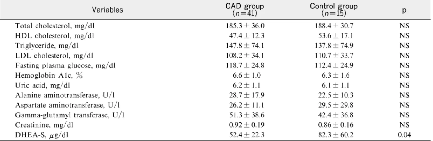
Fig. 2 Correlation between dehydroepiandrosterone sulfate (DHEA-S) levels and coronary angiographic Gensini scores in patients with and without coronary artery dis-ease (CAD). The univariate regression analysis revealed a significant negative correlation between DHEA-S levels and Gensini scores (r=−0.303, p<0.05)
cant negative correlation between the DHEA-S level and Gensini score (r=−0.303, p<0.05;Figure 2), but found no significant correlations between DHEA-S levels and other clinical parameters. The results of an age-adjusted multiple regression analysis revealed that DHEA-S-dependent determinants remained signifi-cantly correlated with the Gensini score (β=−0.291, p<0.05;data not shown).
Discussion
Our study identified lower serum levels of DHEA-S in middle-aged and elderly men with CAD than in age-matched men without CAD and a negative correla-tion between DHEA-S levels and the Gensini score,an indicator of coronary artery stenosis severity. These findings support the results of previous studies that reported correlations between low DHEA-S levels and CVD-associated morbidity or mortality in men. For example, Barrett-Connor et al. reported a strong correlation between low DHEA-S levels and 12-year CVD-associated mortality in 1,029 middle-aged and elderly men from the Rancho Bernardo study. Trivedi et al. demonstrated a correlation between increased mortality risk and low DHEA-S levels in 963 older men from the Cambridge General Practice Health Study. Ohlsson et al. demonstrated that low serum levels of DHEA-S were predictive of death from all causes, CVD, and ischemic heart disease during a 4.5-year follow-up study of 2,644 elderly Swedish men. Feldman et al. reported that in 1,167 middle-aged men from the Massachusetts Male Aging Study, patients with low DHEA or DHEA-S levels were significantly more likely to develop ischemic heart disease during a 9-year follow-up period. Tivesten et al. also reported that low serum levels of DHEA or DHEA-S predicted an increased risk of coronary heart disease events during a 5-year follow-up study of 2,416 elderly Swedish men. Additionally,Herrington et al. demonstrated significantly lower DHEA-S levels in men with at least one stenosis 50%, compared to those without any such lesions.
In addition,several clinical studies reported nega-tive correlation between DHEA-S levels and clinical atherosclerosis severity in men. Herrington et al. reported that in men, plasma levels of DHEA-S were inversely related to the number of diseased coronary arteries (any stenosis 50%) as determined by angio-graphy. Regarding the use of a surrogate marker of clinical atherosclerosis, Yoshida et al. demonstrated that low DHEA-S levels were correlated with increased carotid intimal-medial thickness in middle-aged men. Moreover, Kawano et al. found that DHEA sup-plementation for 12 weeks improved flow-mediated vasodilation of the brachial artery in men. The results of these previous studies support the findings of the present study,in which DHEA-S levels were negatively correlated with the Gensini score in middle-aged and elderly men who underwent selective coronary angio-graphy.
Although the mechanisms by which DHEA and DHEA-S exert antiatherogenic effects in men are not yet clearly understood, we might speculate on several possible explanations. DHEA inhibits human vascu-lar smooth muscle cell proliferation and plaque for-mation, and decreases platelet aggregation and plasminogen activator inhibitor levels. Furthermore, DHEA reduces the levels of atherogenic cytokines such as interleukin-6 and tumor necrosis factor-α. Collectively, these findings suggest a protective effect of DHEA and DHEA-S on the cardiovascular system. We must note several study limitations. First,the number of subjects in this study was relatively small; accordingly,a large cohort clinical study is required to confirm our findings. Second, this was a cross-sectional study, and a causal correlation between en-dogenous DHEA-S levels and the presence of CAD could not be determined. Longitudinal studies of these subjects might contribute more information.
In conclusion, middle-aged and elderly men with CAD in the present study had lower serum DHEA-S levels than in age-matched men without CAD, and serum DHEA-S levels correlated negatively with coro-nary artery stenosis severity in this population,suggest-ing a protective effect of DHEA-S on the cardiovas-cular system. This study may provide mechanistic insight into the role of endogenous DHEA-S in CVD development in middle-aged and elderly men. Further large-scale studies are needed to explore the correlation between endogenous DHEA-S levels and CAD in this population.
Disclosure Statement
No conflicts of interest have affected the results of this study. The study protocol was approved by the Ethics Committee of the Cardiovascular Hospital of Central Japan (Kitakanto Junkanki Hospital).
Funding
This work was financially supported in part by Grants-in-Aid for Scientific Research (C)(No.19590699 and No.23590661) from the Ministry of Education, Culture, Sports, Science, and Technology of Japan.
References
1. Orentreich N, Brind JL, Rizer RL, et al. Age changes and sex differences in serum dehydroepiandrosterone sulfate concentrations throughout adulthood. J Clin Endocrinol Metab 1984;59:551-555.
2. Orentreich N, Brind JL, Vogelman JH, et al. Long-term longitudinal measurements of plasma dehydroepiandroster-one sulfate in normal men. J Clin Endocrinol Metab 1992; 75:1002-1004.
3. Webb SJ,Geoghegan TE,Prough RA,et al. The biological actions of dehydroepiandrosterone involves multiple rece-ptors. Drug Metab Rev 2006;38:89-116.
4. Tchernof A, Labrie F. Dehydroepiandrosterone, obesity and cardiovascular disease risk:a review of human studies.
Eur J Endocrinol 2004;151:1-14.
5. Bhagra S,Nippoldt TB,Nair KS. Dehydroepiandrosterone in adrenal insufficiency and ageing. Curr Opin Endocrinol Diabetes Obes 2008;15:239-243.
6. Arlt W, Hewison M. Hormones and immune function: implications of aging. Aging Cell 2004;3:209-216. 7. Dillon JS. Dehydroepiandrosterone,
dehydroepiandroster-one sulfate and related steroids:their role in inflammatory, allergic and immunological disorders. Curr Drug Targets Inflamm Allergy 2005;4:377-385.
8. Chen CC, Parker Jr CR. Adrenal androgens and the immune system. Semin Reprod Med 2004;22:369-377. 9. Barrett-Connor E,Khaw KT,Yen SS. A prospective study
of dehydroepiandrosterone sulfate,mortality,and cardiovas-cular disease. N Engl J Med 1986;315:1519-1524. 10. Trivedi DP, Khaw KT. Dehydroepiandrosterone sulfate
and mortality in elderly men and women. J Clin Endo-crinol Metab 2001;86:4171-4177.
11. Ohlsson C, Labrie F, Barrett-Connor E, et al. Low serum levels of dehydroepiandrosterone sulfate predict all-cause and cardiovascular mortality in elderly Swedish men. J Clin Endocrinol Metab 2010;95:4406-4414.
12. Feldman HA, Johannes CB, Araujo AB, et al. Low dehy-droepiandrosterone and ischemic heart disease in middle-aged men:prospective results from the Massachusetts Male Aging Study. Am J Epidemiol 2001;153:79-89.
13. Tivesten A,Vandenput L,Carlzon D,et al. Dehydroepian-drosterone and its sulfate predict the 5-year risk of coronary heart disease events in elderly men. J Am Coll Cardiol 2014;64:1801-1810.
14. Barrett-Connor E, Goodman-Gruen D. The epidemiology of DHEAS and cardiovascular disease. Ann NY Acad Sci 1995;774:259-270.
15. Tilvis RS,Kahonen M,Harkonen M. Dehydroepiandroster-one sulfate,diseases and mortality in a general aged popula-tion. Aging (Milano) 1999;11:30-34.
16. Herrington DM, Gordon GB, Achuff SC, et al. Plasma
dehydroepiandrosterone and dehydroepiandrosterone sul-fate in patients undergoing diagnostic coronary angiogra-phy. J Am Coll Cardiol 1990;16:862-870.
17. Gensini GG. A more meaningful scoring system for deter-mining the severity of coronary heart disease. Am J Cardiol 1983;51:606.
18. Yoshida S, Aihara K,Azuma H,et al. Dehydroepiandros-terone sulfate is inversely associated with sex-dependent diverse carotid atherosclerosis regardless of endothelial func-tion. Atherosclerosis 2010;212:310-315.
19. Kawano H, Yasue H, Kitagawa A, et al. Dehydroepian-drosterone supplementation improves endothelial function and insulin sensitivity in men. J Clin Endocrinol Metab 2003;88:3190-3195.
20. Williams MR,Ling S,Dawood T,et al. Dehydroepiandros-terone inhibits human vascular smooth muscle cell prolifera-tion independent of ARs and ERs. J Clin Endocrinol Metab 2002;87:176-181.
21. Arad Y,Badimon JJ,Badimon L,et al. Dehydroepiandros-terone feeding prevents aortic fatty streak formation and cholesterol accumulation in cholesterol-fed rabbits. Arterio-sclerosis 1989;9:159-166.
22. Jesse RL,Loesser K,Eich DM,et al. Dehydroepiandroster-one inhibits human platelet aggregation in vitro and in vivo. Ann N Y Acad Sci 1995;774:281-290.
23. Straub RH, Konecna L, Hrach S, et al. Serum dehydroe-piandrosterone (DHEA) and DHEA sulfate are negatively correlated with serum interleukin-6 (IL-6), and DHEA inhibits IL-6 secretion from mononuclear cells in man in vitro: possible link between endocrinosenescence and im-munosenescence. J Clin Endocrinol Metab 1998;83:2012-2017.
24. Kimura M,Tanaka S,Yamada Y,et al. Dehydroepiandros-terone decreases serum tumor necrosis factor-alpha and restores insulin sensitivity: independent effect from secon-dary weight reduction in genetically obese Zucker fatty rats. Endocrinology 1998;139:3249-3253.