1. Introduction
The incidence of diabetes is increasing worldwide. Saquib et al. (2012) reported that the number of diabetes patients in Bangladesh has increased from 3.8% in 1995 to 9% in 2010. The prevalence of diabetes is increasing in Bangladesh due to inadequate healthcare facilities in rural areas, lack of health consciousness, poor economic conditions, lifestyle changes caused by rapid urbanization, and changes in food habits. The Global Change Makers Program also indicated that the poor people in Bangladesh are suffering lifestyle-related diseases such as diabetes (Global Change
Makers Program, 2013).
Although the numbers of diabetic patients are rapidly increasing, countermeasures have not been developed in Bangladesh. There are several reasons for the difficulty in coping with diabetes in Bangladesh, e.g., poverty, limitations of health care facilities, low capability of spending on health care expenses, lack of understanding of health condition, lack of education, lack of information about diabetes, inadequate opportunities for annual health checks and lack of knowledge prevent people in this country from taking modern medicines. Especially, facilities for diabetes treatment are not available in health centers, and nurse practitioners do not have sufficient
Inhibitory effects of Bangladeshi medicinal plant leaf extracts
on α-glucosidase activity
Mst Tamanna Niger
*1, Kazuhiro Ohtani
1, Bhuiyan Feroze Ahamed
2Graduate School of Kuroshio Science, Kochi University, 200 Otsu Monobe Nankoku-shi, Kochi 783-8502, Japan
1Laboratory of Human Health and Medical Science, Graduate School of Kuroshio Science, Kochi
University, 200 Otsu Monobe, Nankoku-shi, Kochi 783-8502, Japan
2 Research Institute of Molecular Genetics, Kochi University, 200 Otsu Monobe, Nankoku-shi,
Kochi 783-8502, Japan
Abstract
One of the best strategies in treatment of diabetes mellitus management involves control of postprandial hyperglycemia through enzymatic inhibition of starch degradation. Emblica officinalis, Terminalia bellirica, and Terminalia chebula are used as remedies in Ayurvedic medicine. In this study, methanol extracts of the leaves of these three plant species were screened for their α -glucosidase inhibitory potential activities in vitro. Methanol extract of T. chebula showed maximum inhibitory activity against yeast α-glucosidase with a half maximal inhibitory concentration (IC50) of
15μg/ml, followed by T. bellirica with IC50value of 34μg/ml and E. officinalis with a value of 50
μg/ml, compared with the standard drug, acarbose (IC50value: 13 mg/ml). Rat intestinal sucrase
inhibitory activity was also investigated. The methanol extracts of these three plants at 1 mg/ml showed considerable rat intestinal sucrase inhibitory activity. The hexane extract, ethyl acetate extract, butanol extract, and water extract from the three plants were screened for yeast α -glucosidase and rat intestinal sucrase inhibitory activity. Among the three plants, butanol extract exhibited potent α-glucosidase inhibitory activity. Methanol and butanol extracts of the three plants also showed excellent inhibitory activity against rat intestinal sucrase. E. officinalis, T. bellirica, and T. chebula are potential plant sources of for α-glucosidase inhibitors that can be used to treat diabetes. Key words: α-glucosidase inhibitor, diabetes, Emblica officinalis, Terminalia bellirica, Terminalia chebula
Received April 28, 2016; Accepted August 3, 2016 *Corresponding author: e-mail : [email protected], Tel: + 81-80 3925-9900
training for early identification of prediabetes in Bangladesh (Islam et al., 2013).
Traditional medicines, which are commonly used in rural areas, represent one solution to these problems. Traditional medicine has a long history of use in Bangladesh, and has the advantage of reduced medical expenses. Rahman (2013) reported that large numbers of people use traditional medicines for various diseases, such as skin diseases, joint pain, diarrhea, cough, stomach problems, and diabetes. Ayurvedic traditional medicine has been used since ancient times in Bangladesh (Yoshida et al., 2016). Traditional medicine has been reported to show fewer side effects than allopathic medicines, and Vipula (2014) reported a good response to Ayurvedic treatment compared to allopathic treatment, with no side effects.
Traditional medicinal plants are also used to provide affordable treatments for diabetes mellitus with advances in many herbal medicinal health care systems in Bangladesh. Various types of medicinal plants and plant parts are used in traditional medicine for diabetes. In addition to traditional practitioners reporting good responses, their treatments can be validated scientifically based on the antidiabetic properties of components of their medicinal plants. Grover et al. (2002) showed that antihyperglycemic agents are present in various plant extracts that have been used as traditional medicine.
In this study, we focused on three plants, Emblica officinalis, Terminalia bellirica, and Terminalia chebula (Fig. 1), which are widely distributed southern India, Pakistan, Bangladesh and other South-East Asian countries and have been extensively used in traditional medicine in Bangladesh and the Indian subcontinent. The traditional herbal formulation consisting of a combination of fruits of these three plants called “triphala” is very popular in traditional Ayurvedic medicine, and has been used for thousands of years against various types of disease. Mukherjee et al. (2006)
reported that “triphala and triphala mixture” follows a standardized ayurvedic formula, which has been clinically tested and shown to exhibit good results against constipation and appetite problems.
These three plants are well known as individual herbal remedies. E. officinalis has been reported to possess antiinflammatory (Golechha et al., 2014), antioxidant (Scartezzini et al., 2006), hepatoprotective (Jose et al., 2000), antipyretic and analgesic activities (Perianayagam et al., 2004). T. chebula has been reported to show anticancer (Ahuja et al., 2013), antiviral (Kim et al., 2001), antibacterial (Kannan et al., 2009), antioxidant (Cheng et al., 2003), and wound healing activities (Singh et al., 2009). T. bellirica has been studied for its antimicrobial (Devi et al., 2014), anti-HIV-1, antimalarial, antifungal (Valsaraj et al., 1997), analgesic, and antipyretic activities (Sharma et al., 2010).
Moreover, Sabu and Kuttan (2002) reported that oral administration of an extract of E. officinalis, T. bellirica, and T. chebula fruits effectively decreased the serum glucose level in diabetic rats. T. chebula seed extracts showed a remarkable decrease in blood glucose level by increasing insulin secretion in both short and long term studies (Rao and Nammi, 2006). The therapeutic potentials of various parts of the above three plants from different regions have been evaluated in previous studies. However, the antidiabetic potential of the leaves collected from Bangladesh has not been reported. While the fruits and seeds of E. officinalis, T. chebula, and T. bellirica have been reported to show antihyperglycemic effects, there have been no studies regarding the α -glucosidase inhibitory activity of leaves of these three plants collected from Bangladesh. The present study was designed to evaluate the antidiabetic potentials of different extracts of these three plant leaves through monitoring of the inhibitory effects on yeast and rat intestinal α-glucosidase.
Alfa-glucosidases, which include maltase,
maltase-Fig.1. Plant leaves used in this study.
(A) E. officinalis belongs to the family Euphorbiaceae. (B) T. chebula belongs to the family Combretaceae. (C) T. bellirica belongs to the family Combretaceae.
glucoamylase, etc., are the key enzymes. The α-glucosidase from the yeast, Saccharomyces cerevisiae is commonly used to search for biologically active compounds with inhibitory effects on the enzyme in vitro. Hogan et al. (2010) used yeast α-glucosidase as a model for isolation and identification of inhibitory substances from natural products. The p-nitrophenyl-α-D-glucopyranoside was used as a synthetic substrate in the assay of yeast α- glucosidase, which catalyzes glucose and nitrophenol. Yeast α-glucosidase and p-nitrophenyl-α-D-glucopyranoside substrates were developed for analysis of maltase inhibitory activity. Starch is the most important carbohydrate in the human body, and is catalyzed into maltose by α-amylase, which in turn is catalyzed into glucose by maltase in the brush-border surface membrane of the small intestine.
Sucrose is the most commonly used sugar all over the world, and is degraded into glucose and fructose by the α-glucosidase, sucrase. Rat intestinal acetone powder has been used as a source of sucrase for in vitro assays with sucrose as the substrate. Ohta et al. (2002) reported that the rat intestinal acetone powder α-glucosidase closely mimics that in the mammalian system in vivo and may be a superior model for use in studies to develop methods for the control of postprandial blood glucose. Enzymes from different sources catalyze different substrates. Therefore, we used two types of α-glucosidase, i.e., yeast glucosidase and rat intestinal α-glucosidase, in this study.
Diabetes mellitus is a group of serious metabolic disorders associated with high blood glucose levels due to a failure of the body to produce insulin properly. At present, three main types of diabetes are known, i.e., type I insulin-dependent diabetes, type II non-insulin-insulin-dependent diabetes, and gestational diabetes (International Diabetes Federation, 2013). Type II has the highest prevalence rate, and the control of postprandial blood glucose level is important in its treatment. The inhibition of α-glucosidase is essential to delay carbohydrate digestion and absorption, reduce blood glucose levels, and finally control postprandial hyperglycemia. Acarbose, miglitol, and voglibose are well- known clinically approved inhibitors of α-glucosidase activity (Van de Laar, 2008). However, these α-glucosidase inhibitors have a number of undesirable side effects, including gastrointestinal problems (Cheng et al., 2005). Therefore, natural α-glucosidase inhibitors that are safe, effective, and have no or only minor side effects are required.
2. Materials and Methods
2.1) Plant collection and extraction
The leaves of the three plant species were collected from
the Bangladesh Agricultural University, packed in the newspaper, and dried in the sun. The dried samples were crushed in a blender, and then boiled in methanol at 60℃ -70℃ three times. Boiled leaf extracts were dried under reduced pressure using a rotary evaporator. These dried methanol extracts were dissolved in 30% hexane/70% methanol solution and successively partitioned with the same volume of hexane, ethyl acetate, butanol, and water by adding each solution as shown in Fig. 2. The solvents were again evaporated and the remaining substances were used as the test materials. The methanol extracts and their partitioned fractions were monitored for inhibitory effects on α-glucosidase activity.
Generally, plant bioactive compounds consist of various classes of chemicals, such as alkaloids, glycosides, lignins, tannins, terpenoids, etc. The methanol extracts of the plants were used because many molecules, including fatty acids, water-soluble materials, and other bioactive compounds, are soluble in methanol. The major biological compound, bartogenic acid, was identified from the methanol extract of Barringtonia racemosa seed (Gowri et al., 2007). Therefore, methanol is important in screening for biologically active compounds, and was used in the present study.
2.2) Yeast α-glucosidase inhibitory assay
The inhibitory activity of yeast α-glucosidase (Wako Pure Chemical Industries, Ltd., Osaka, Japan) was determined by the method of Babu et al. (2004) with slightly modifications. Aliquots of 20μl of the test samples dissolved in methanol at 10 mg/ml were serially diluted in 96-well microtiter plates. The plant extracts diluted in10μl of phosphate buffer 0. 1 mol/l (pH 6. 8) and 150μl (5 mmol/ml) of p-nitrophenyl-α-D-glucopyranoside (PNPG) were added to each well. The reaction was started by addition of 20μl (5 μg/ml) of the enzyme to the reaction mixture in the 96-well Fig.2. Separation by partition scheme with several solvents. The upper layers ( ) are hexane, ethyl acetate, and butanol. The lower layers ( ) are water.plates. Individual blanks where the substrate was replaced with 40μl of phosphate buffer were prepared to correct for background absorbance. The controls contained 20μl of phosphate buffer in place of the test sample, while the leaf extract was replaced with acarbose in positive controls. All determinations were performed in triplicate. The changes in absorbance at 405 nm (A405) were recorded at 1-minute intervals for 10 minutes,
and the percentage inhibition was estimated from the slope using the following equation:
Inhibition % = (1- slope of test sample/slope of control) × 100. The concentration that gave the half-maximal response (IC50) was determined from the sample concentration vs.
percentage inhibition rate. α-glucosidase inhibitory activity was expressed as inhibition % and IC50 value, with a lower
IC50value was indicating higher inhibitory activity.
2.3) Rat intestinal α-glucosidase (sucrase) inhibitory
assay
Rat intestinal α-glucosidase inhibitory activity was determined by a modification of the method of Babu et al. (2004). Aliquots of 20μl of plant extract test samples dissolved in methanol at 10 mg/ml were added to each well of 96-well plates. Then, 150μl of 5 mg/ml saccharose was added to each well. Rat intestinal acetone powder (Sigma-Aldrich, St. Louis, MO) was added at 100 mg/ml to phosphate buffer 0.1 mol/l (pH 6.8) and sonicated for 5 minutes. The suspension was centrifuged at 2500 rpm for 5 minutes to remove particulate matter, and the resulting supernatant was used as the enzyme solution. The reaction was initiated by addition of 30μl of the enzyme to the reaction mixture in 96-well plates. The reaction mixture was incubated at 37 C for 30 minutes followed by 70 C for 3 minutes on a heating block to stop the reaction. The reaction mixture was cooled to room temperature for 10 minutes and aliquots of 20μl were transferred to another plate. The reaction was started by addition of 150 μl of reagent (Glucose C2; Wako) and cooled to 25 C for 15 minutes. Individual blanks where the substrate was replaced with 50μl of phosphate buffer were prepared to correct for background absorbance. The controls contained 20μl of phosphate buffer in place of the test sample, while the leaf extract was replaced with acarbose in positive controls. All determinations were performed in triplicate. The changes in absorbance at 492 nm (A492) were recorded at 1-minute. The percentage inhibition
was estimated using the following equation:
Inhibition % = (1 - (absorbance of test sample-absorbance of blank)/(absorbance of control-absorbance of blank)) × 100.
2.4) Statistical analysis
The effects of crude methanol extracts (Fig. 3) and
fractionated extracts (Fig. 4) of the leaves from three medicinal plants, E. officinalis, T. bellirica, and T. chebula were examined by analysis of variance (ANOVA) followed by Tukey’s multiple comparison tests using SPSS version 16.0 for windows (SPSS Inc., Chicago, IL). In all analyses, p < 0.01 was taken to indicate statistical significance.
3. Results
E. officinalis yielded the highest percentage (18%) of methanol extract from 50 g of plant material (Table 1, with T. chebula and T. bellirica showing methanol extract yields of 15% and 12%, respectively (Table 1). The methanol extracts Fig.3. Effects of methanol extracts of the leaves from three medicinal plants on yeast α-glucosidase activity (12.5 μ g/ml). The same letters above the bars in the graph indicate that the mean did not differ significantly, while different letters indicate significant variation at P < 0. 01. The values are expressed as means ± standard deviation, n = 3.
Fig. 4. Inhibitory effects of fractionated extracts of the leaves from three medicinal plants on yeast α-glucosidase activity (10 μ g/ml).
The same letters above the bars in the graph indicate that the mean did not differ significantly, while different letters indicate significant variation at P < 0. 01. The values are expressed as means ± standard deviation, n = 3.
were screened for their α-glucosidase inhibitory activities. The methanol extracts of the three plants showed significant in vitro α-glucosidase inhibitory activity compared with acarbose (13 mg/ml), with T. chebula showing the highest inhibitory activity (IC50 =15μg/ml), followed by T. bellirica (IC50=
34μg/ml), and E. officinalis (IC50=50μg/ml) (Table 2).
The hexane fraction of T. chebula leaves was inactive, while the ethyl acetate, butanol, and water fractions showed α -glucosidase inhibitory activity. The T. chebula butanol fraction showed a strong inhibitory effect with an IC50value
of 10μg/ml (Table 3). The hexane fraction of E. officinalis leaf extract showed no inhibitory activity against α-glucosidase. The butanol and ethyl acetate fractions of E. officinalis showed almost the same inhibitory activities, with IC50values
of 18μg/ml and 19μg/ml, respectively (Table 3). The hexane fraction of T. bellirica leaves was also less active than the ethyl acetate, butanol, and water fractions, with IC50values for Table 1. Efficiency of methanol extraction from the leaves of three Bangladeshi medicinal plants.
Percentage extract yield (w/w) was calculated as (dry extract weight/dry starting material weight) × 100. Table 2: Effects of methanol extracts from the leaves of
three medicinal plants against yeast α-glucosidase.
Acarbose was used as a positive control.
IC50: Concentration of the antagonist that inhibited the
enzyme reaction by 50%.
Table 3. Inhibitory effects of fractionated extracts of three medicinal plants on yeast α-glucosidase activity.
Acarbose was used as a positive inhibitory molecule.
IC50: Concentration of the antagonist that inhibited the enzyme reaction by 50%.
the butanol and ethyl acetate fractions of 12μg/ml and 18 μg/ml, respectively (Table 3).
The inhibitory activities of the methanol extracts (12.5 μg/ml) and fractionated extracts (10μg/ml) from the three plants against yeast α-glucosidase were analyzed and the results are shown in Figures 3 and 4, respectively. There were significant differences in the effects of the methanol extracts between the plant species, with that of T. chebula showing the maximum inhibitory effect (46%) followed by E. officinalis (32%) and T. bellirica (30%) (Fig. 3). There were no significant differences in the inhibitory effects of ethyl acetate extracts among the three plant species (Fig. 4). The butanol extracts of T. chebula and T. bellirica showed similar inhibitory effects (50% and 46%, respectively), while E. officinalis showed a weaker effect (32%) (Fig. 4). The water extract of T. chebula showed the greatest inhibitory effect (50%), with E. officinalis and T. bellirica are showing lower inhibitory effects (21% and 23%, respectively) (Fig. 4).
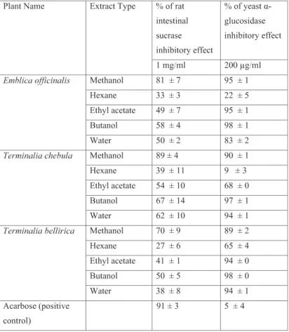
In this study, the inhibitory effects of leaf extracts from the three plant species against sucrase activity were determined and compared with that of acarbose (91%,
Supplemental Data, Table 1). The methanol extracts of E. officinalis, T. chebula, and T. bellirica leaves at 1 mg/ml in the reaction mixtures showed significant inhibitory effects against sucrase enzyme activity (Supplemental Data, Table 1), with that of T. chebula showing the greatest effect (89%). We also investigated the inhibitory effects of hexane, ethyl acetate, butanol, and water extracts of the leaves from these plants against sucrase activity. The butanol extract of E. officinalis and water extract of E. officinalis were shown to inhibit the enzyme activity by 58% and 50%, respectively (Supplemental Data, Table 1). However, the hexane extract of E. officinalis showed only a weak inhibitory effect (33%) on sucrase activity, while acarbose as a positive control showed 91% inhibition of the enzyme activity at a concentration of 1 mg/ml (Supplemental Data, Table 1).
Ethyl acetate, butanol, and water fractions of T. chebula leaves showed the highest sucrase inhibitory effects (54%, 67%, and 62%, respectively), and the weakest effect was observed for the hexane extract of T. chebula (39%, Supplemental Data, Table 1). The butanol extract of T. bellirica leaves showed an inhibitory effect of 50%, while the Supplemental Data.
Table 1. Inhibitory effects of extracts of the leaves from three medicinal plants on rat intestinal sucrase and yeast α-glucosidase.
hexane, ethyl acetate, and water extracts showed lower inhibitory activities of 27%, 41%, and 38%, respectively (Supplemental Data, Table 1).
4. Discussion
Although acarbose is a well-known antidiabetic medication, Oki et al. (1999) reported that acarbose did not inhibit yeast α-glucosidase activity, while it showed inhibitory effects on rat, rabbit, and pig small intestinal α-glucosidase activities. Kim et al. (2004) reported a similar result where pine bark extracts showed strong inhibitory effects against yeast α-glucosidase, while the standard drug, acarbose, showed no inhibitory effect on yeast α-glucosidase activity. These observations were compatible with those of the present study, in which acarbose showed only a weak inhibitory effect against yeast α-glucosidase with an IC50value of 13 mg/ml,
while the three plant extracts showed strong inhibitory effects against the activity of this enzyme (Table 3). In this study, acarbose had an IC50 value of 13mg/ml for yeast
α-glucosidase at 5μg/ml in the reaction mixture, whereas Shai et al. (2011) reported that acarbose had an IC50 value of 1.5
mg/ml for yeast α-glucosidase at 0.5 mg/ml in the reaction mixture. These results indicated that the different inhibitory effect of acarbose on yeast α-glucosidase, which may have been due to the use of different concentration of the enzyme. Shai et al. (2011) also mentioned that the different concentrations of the enzyme are responsible for different IC50
value of acarbose.
The methanol and butanol extracts of the leaves from the three plant species, E. officinalis, T. bellirica, and T. chebula, showed stronger inhibitory effects against yeast α-glucosidase than rat intestinal sucrase (Supplemental Data, table 1). At 200μg/ml, methanol and butanol extracts of T. chebula inhibited yeast α-glucosidase activity by 90% and 97%, respectively, while at 1 mg/ml, methanol and butanol extracts of the effects of that inhibited on rat intestinal sucrase were lower at 89% and 67%, respectively. This result was supported by those of Shai et al. (2011), who found that acetone extracts of different plants had stronger inhibitory effects against yeast glucosidase than mammalian α-glucosidase. Havsteen (1983) classified flavonoids into six groups based on the variation of benzopyran and phenyl groups and their ring and linkage sites, i.e., flavone, flavonol, flavanone, isoflavone, flavan-3-ol, and anthocyanidin. Tadera et al. (2006) reported that the inhibitory effects on yeast α-glucosidase and rat intestinal α-α-glucosidase differed according to the chemical structure of flavonoids based on the presence of absence of the OH group at the C-3 position of the flavone (quercetin > luteolin; kaempferol > apigenin), hydroxyl substitution on the B ring (flavonol: myricetin =
quercetin > kaempferol; flavone: luteolin > apigenin), linkage of the B-3 position (isoflavone and flavone groups: genistein >apigenin), and 1,2- and 3,4-double bonds and lack of 4-CO of cyanidin, which is responsible for significant inhibition of yeast α-glucosidase. On the other hand, the anthocyanidin group and isoflavone group were associated with weak inhibition of rat intestinal α-glucosidase activity.
In the present study, butanol extracts of T. chebula and T. bellirica showed stronger inhibitory effects against yeast α-glucosidase activities with IC50 values of 10μg/ml and 12
μg/ml, respectively (Table 3), than against rat intestinal α-glucosidase (Supplemental Data, table 1). This result was supported by the previous report of Tadera et al. (2006), who also found that yeast α-glucosidase was strongly inhibited by flavonoids with IC50values < 15μM, and that rat small intestinal
α-glucosidase was weakly inhibited by many flavonoids and faintly by the isoflavone and anthocyanidin groups. Sim et al. (2010) reported that the substrate specificity is responsible for different reactions because the differences of active site of enzyme between N-terminal maltase-glucoamylase and N-terminal sucrase-isomaltase where maltose could be hydrolyzed efficiently by both N-terminal sucrase-isomaltase and N-terminal maltase-glucoamylase, because N-terminal sucrase-isomaltase has a narrow + 1 subsite which accommodates both its 1,4 (maltose) and α-1, 6 (isomaltose) substrates, but N-terminal maltase-glucoamylase has a wide + 1 subsite, which is specific for the α-1,4 substrate. The present study results showed different reactions against yeast glucosidase and rat intestinal α-glucosidase using the different extract of three plant leaves, these different reactions might be due to the substrate specificity, which was demonstrated by Sim et al. (2010).
The methanol extracts of T. chebula, T. bellirica, and E. officinalis had IC50 values for yeast α-glucosidase of 15
μg/ml, 34μg/ml, and 50μg/ml, respectively (Table 2). On the other hand, Prihantini et al. (2014) reported that the methanol extract of Distylium racemosum showed a strong inhibitory effect on yeast α-glucosidase with an IC50value of 22.6 ±
1.9μg/ml, followed by Acer mono Maxim and Elaeocarpus sylvestris with IC50 values of 56.5 ± 0.9μg/ml and 74.4 ±
0.9μg/ml, respectively. These results indicated that the α-glucosidase inhibitory activity of methanol extract of T. chebula was much stronger than that of D. racemosum, which has been identified as a α-glucosidase inhibitor. Nampoothiri et al. (2011) reported that T. bellirica fruit extract showed stronger inhibitory activity against α-glucosidase than E. officinalis fruit extract (IC50values 0.75μg/ml and 1.0μg/ml,
respectively). These results indicated higher inhibitory activities than those seen in the present study, which may have been due to the use of different parts of the plants (i.e., fruit vs. leaf extracts, respectively). In addition, Oboh et al. (2014)
reported that different parts of Persea americana (avocado pear) plants showed different α-glucosidase inhibitory activities. These results of the present study indicated that the methanol extract of T. chebula leaves had the highest inhibitory potency against rat intestinal sucrase inhibitory activity (89%), followed by those of E. officinalis (81%) and T. bellirica (70%) (Supplemental Data, table 1), while Sabu and Kuttan (2002) reported that the methanol extract of T. bellirica fruits showed maximum inhibitory activity (52.74%), followed by those of T. chebula (50.98%) and E. officinalis (29. 52%). These discrepancies may have been due to differences in the experimental approach, because our experiments were performed in vitro, whereas Sabu and Kuttan (2002) performed in vivo experiments. In the present study, ethyl acetate extract of T. chebula leaves showed 54% inhibition (1 mg/ml) against rat intestinal sucrase, while Kim et al. (2011) reported that a high dose of the ethyl acetate portion of ethanolic extract of T. chebula fruit (500 mg/kg) reduced the levels of blood glucose. This result showed good agreement with those of the present study.
The results of the present study indicated that the methanol and butanol extracts of T. chebula leaves and butanol extracts of T. bellirica leaves showed maximum inhibitory effects on yeast α-glucosidase activity. Methanol and butanol extracts of the leaves of the three plants, T. chebula, T. bellirica, and E. officinalis, showed the strong inhibitory effects on rat intestinal sucrase activity. Thus, the leaves of these three plant species may contain antihyperglycemic compounds. Further structural elucidation and characterization are required to identify for identifying the biologically active constituents of these extracts, and in vivo studies are also necessary to confirm these observations.
5. Conclusions
The leaf extracts of three Bangladeshi medicinal plants T. chebula, T. bellirica, and E. officinalis, showed significant inhibitory effects against α-glucosidase and sucrase activities. Butanol and methanol extracts of T. chebula leaves and butanol extract of T. bellirica leaves showed significant inhibitory effects on carbohydrate digestive enzymes, which may function in controlling postprandial glucose levels. However, further studies are needed to determine their potential efficacy in the treatment of diabetes.
References
Ahuja, R., Agrawal, N. and Mukerjee, A. 2013. Evaluation of anticancer potential of Terminalia chebula fruits against Ehrlich Ascites Carcinoma induced cancer in mice. J. Sci. Innovative Res., 2: 3; 549-554.
Babu, K.S., Tiwari, A.K., Srinivas, P.V., Ali, A.Z., Raju, B.C. and Rao, J. M. 2004. Yeast and mammalian α-glucosidase inhibitory constituents from Himalayan Rhubarb Rheum emodi Wall, Ex Meisson. Bioorg. Med. Chem. Lett., 14: 384-13845.
Cheng, A.Y.Y. and Fantus, I.G. 2005. Oral anti hyperglycemic therapy for type 2 diabetes mellitus. CMAJ, 18: 172(2); 213-26.
Cheng, H.Y., Lin, T.C., Yu, K.H., Yang, C.M. and Lin, C.C. 2003. Antioxidant and free radical scavenging activities of Terminalia chebula. Biol. Pharm. Bull., 26: 9; 1331-1335.
Devi, P. N., Kaleeswari, S. and Poonkothai, M. 2014. Antimicrobial activity and phytochemical analysis of fruit extracts of Terminalia bellirica. Int. J. Pharm. Pharm. Sci., 6: 5; 639-642.
Global Change Makers Program (GCMP) report. 2013. A report of villagers’ consciousness about health and disease (http://www.gcmp.com/wcontent/uploads /2014/01/report 2013w _be.pdf) p 1-19.
Golechha, M., Sarangal, V., Ojha, S., Bhatia, J. and Aryal, D. S. 2014. Anti-Inflaatory Effect of Emblica officinalis in rodent models of acute and chronic inflammation: involvement of possible mechanisms. Int. J. Inflamm., Volume 2014, Article ID 178408, 6 pages.
Gowri, P.M., Tiwari, A.K., Ali, A.Z. and Rao, J.M. 2007. Inhibition of α-glucosidase and amylase by bartogenic acid isolated from Barringtonia racemosa Roxb. seeds. Phytother. Res., 21: 8; 796-799.
Grover, J.K., Yadav, S. and Vats, V. 2002. Medicinal plants of India with antidiabetic potential. J. Ethnopharmacol., 81: 1; 81-100.
Havsteen, B. 1983. Flavonoids, a class of natural products of high pharmacological potency. Biochem. Pharmacol., 32: 7; 1141-1148.
Hogan, S., Zhang, L., Li, J., Sun, S., Canning, C. and Zohu, K. 2010. Antioxidant rich grape pomace extract suppresses postprandial hyperglycemia in diabetic mice by specifically inhibiting alphaglucosidase. Nutr. Metab. (Lond)., 7: 71; 1- 9.
International Diabetes Federation (IDF). 2013. Diabetes Atlas, Sixth Edition, pp 1-160.
Islam, S.M.S., Lechner, A., Ferrari, U., Froeschl, G., Niessen, L. W., Seissler, J. and Alam, D. S. 2013. Social and economic impact of diabetics in Bangladesh: protocol for a case-control study. BMC Public Health, 13: 1217; 1- 9. Jose, J.K. and Kuttan, R. 2000. Hepatoprotective activity of Emblica officinalis and Chyavanaprash. J. Ethnopharmacol., 72: (1-2); 135-140.
Kannan, P., Ramadevi, S. R. and Hopper, W. 2009. Antibacterial activity of Terminalia chebula fruit extract.
Afr. J. Microbiols. Res., 3: 4; 180-184.
Kim, J.H., Hong, C.O., Koo, Y.C., Kim, S.J. and Lee, K.W. 2011. Oral administration of ethyl acetate-soluble portion of Terminalia chebula conferring protection from streptozotocin-induced diabetic mellitus and its complications. Biol. Pharm. Bull., 34: 11; 1702-1709. Kim, T.G., Kang, S.Y., Jung, K.K., Kang, J.H., Lee, E., Han,
H.M. and Kim, S.H. 2001. Antiviral activities of extracts isolated from Terminalia chebula Retz., Sanguisorba officinalis L., Rubus coreanus Miq. and Rheum palmatum L. against hepatitis B virus. Phytother. Res., 15: 8; 718-720.
Kim, Y.M., Jeong, Y.K.,Wang, M.H., Lee, W.Y. and Rhee, H. I. 2005. Inhibitory effect of pine extract on α -glucosidase activity and postprandial hyperglycemia. Nutrition., 21: 6; 756-761.
Mukherjee, P.K., Rai, S., Bhattacharyya, S., Debnath, P.K., Biswas, T.K., Jana, U., Pandit, S., Saha, B.P. and Paul, P. K. 2006. Clinical study of 'Triphala'-a well known phytomedicine from India. J. Pharmacol. Ther., 5: 1; 51-54.
Nampoothiri, S.V., Prathapan, A., Cherian, O.L., Raghu, K.G., Venugopalan, V.V. and Sundaresan, A. 2011. In vitro antioxidant and inhibitory potential of Terminalia bellerica and Emblica officinalis fruits against LDL oxidation and key enzymes linked to type 2 diabetes. Food Chem. Toxicol., 49: 1; 125-131.
Ohta, T., Sasaki, S., Oohori, T., Yoshikawa, S. and Kurihara, H. 2002. Alpha-glucosidase inhibitory activity of a 70% methanol extract from ezoishige (Pelvetia babingtonii de Toni) and its effect on the elevation of blood glucose level in rats. Biosci. Biotechnol. Biochem., 66: 7; 1552-1554.
Oki, T., Matsui, T. and Osajima, Y. 1999. Inhibitory effect of α-glucosidase inhibitors varies according to its origin. J. Agric. Food. Chem., 47: 2; 550-553.
Oboh, G., Isaac, A.T., Akinyemi, A.J. and Ajani, R.A. 2014. Inhibition of key enzymes linked to type 2 diabetes and sodium nitroprusside induced lipid peroxidation in rats' pancreas by phenolic extracts of avocado pear leaves and fruit. Int. J. Biomed. Sci., 10: 3; 208-216.
Perianayagam, J.B., Sharma, S.K., Joseph, A. and Christina, A. J. M. 2004. Evaluation of anti-pyretic and analgesic activity of Emblica officinalis Gaertn. J. Ethnopharmacol., 95:1; 83-85.
Prihantini, A.I., Tachibana, S. and Itoh, K. 2014. Evaluation of antioxidant and α-glucosidase inhibitory activities of some subtropical plants. PaK. J. Biol. Sci., 17: 10; 1106-1114. Rahman, M.H. 2013. A Study on exploration of ethnobotanical knowledge of rural community in Bangladesh: Basis for biodiversity conservation. ISRN Biodiversity, V 2013:
1-10.
Rao, N.K. and Nammi, S. 2006. Antidiabetic and renoprotective effects of the chloroform extract of Terminalia chebula retz. seeds in streptozotocin-induced diabetic rats. BMC Complementary Altern. Med., 6: 17; 1-6.
Sabu, M. C. and Kuttan, R. 2002. Anti-diabetic activity of medicinal plants and its relationship with their antioxidant property. J. Ethnopharmacol., 81: 2; 155-160.
Saquib, N., Saquib, J., Ahmed, T., Khanam, M.A. and Cullen, M.R. 2012. Cardiovascular diseases and type 2 diabetes in Bangladesh: a systematic review and meta-analysis of studies between 1995 and 2010. BMC Public Health, 12: 434; 1-10.
Scartezzini, P., Antognoni, F., Raggi, M. A., Poli, F. and Sabbioni, C. 2006. Vitamin C content and antioxidant activity of the fruit and of the Ayurvedic preparation of Emblica officinalis Gaertn. J. Ethnopharmacol, 104: 1-2; 113-118.
Shai, L.J., Magano, S.R., Lebelo, S.L. and Mogale, A.M. 2011. Inhibitory effects of five medicinal plants on rat alpha-glucosidase: Comparison with their effects on yeast alpha-glucosidase. J. Med. Plants Res., 5: 13; 2863-2867. Sharma, U.S., Sharma, U.K., Singh, A., Sutar, N. and Singh, P.J. 2010. Screening of Terminalia bellirica fruits extracts for its analgesic and antipyretic activities. J. Biol. Sci., 3: 3; 121-124.
Singh, M.P. and Sharma, C.S. 2009. Wound healing activity of Terminalia chebula in experimentally induced diabetic rats. Int. J. Pharm. Tech. Res., 1: 4; 1267-1270. Sim, L., Willemsma, C., Mohan, S., Naim, H.Y, Pinto, B.M.
and Rose, D. R. 2010. Structural basis for substrate selectivity in human maltase-glucoamylase and sucrase-isomaltase N-terminal domains. J. Biol. Chem., 285: 23; 17763-17770.
Tadera, K., Minami, Y., Takamatsu, K. and Matsuoka, T. 2006. Inhibition of α-glucosidase and α-amylase by flavonoids. J. Nutr. Sci. Vitaminol., 52: 2; 149-153. Valsaraj, R., Pushpangadan, P., Smitt, U. W, Adsersen, A.,
Christensen, S.B., Sittie, A., Nyman, U. and Nielsen, C.E. 1997. New anti-HIV-1, antimalarial, and antifungal compounds from Terminalia bellirica. J. Nat. Prod., 60: 7; 739-742.
Van de Laar, F.A. 2008. Alpha-glucosidase inhibitors in the early treatment type 2 diabetes. J. Vasc. Health Risk Management., 4: 6; 1189-1195.
Vipula. 2014. Ayurveda & allopathy- an integrated approach. Guru Drone J. Pharm. Res., 2: 4; 17-22.
Yoshida, Y., Rashid, M.H.O., Yoshida, Y. and Alim, M.A. 2016. Perceptions of Ayurvedic medicine by citizens in Dhaka, Bangladesh. Nagoya J. Med. Sci., 78: 99-107.