ZOOTAXA
ISSN 1175-5326 (print edition)
ISSN1175-5334(online edition) Copyright © 2011 · Magnolia Press
Zootaxa 3102: 1–26 (2011)
www.mapress.com/zootaxa/
Article
Rove beetles (Coleoptera: Staphylinidae) associated with Aenictus laeviceps
(Hymenoptera: Formicidae) in Sarawak, Malaysia: Strict host specificity,
and first myrmecoid Aleocharini
MUNETOSHI MARUYAMA1,2,4, TAKASHI MATSUMOTO3 & TAKAO ITIOKA3
1Zoology Department, Field Museum of Natural History, 1400 South Lake Shore Drive, Chicago, IL 60605 USA
2The Kyushu University Museum, Hakozaki 6-10-1, Higashi-ku, Fukuoka-shi, Fukuoka, 812-8581 Japan
3Graduate School of Human and Environmental Studies, Kyoto University,Yoshida-nihonmatsu-cho, Sakyo-ku, Kyoto, 606-8501 Japan
4Corresponding author. E-mail: [email protected]
Abstract
The fauna of myrmecophilous rove beetles associated with Aenictus laeviceps (sensu Wilson 1964) at Lambir Hills Na- tional Park, Sarawak, Malaysia was investigated. Eight species belonging to six genera of the subfamily Aleocharinae in- cluding the following new taxa are recorded and/or described: Myrmecosticta exceptionalis Maruyama gen. et sp. nov., Aenictocleptis hirsutoides Maruyama sp. nov., Aenictocleptis lambirensis Maruyama sp. nov., Mimaenictus matsumotoi Maruyama sp. nov., Procantonnetia opacithorax Maruyama sp. nov., Weissflogia pubescens Maruyama sp. nov. Three morphotypes (L1, L2 and S) are recognized in A. laeviceps, and strict host specificity by the rove beetles for the morpho- types was observed. Myrmecosticta exceptionalis is the first known myrmecoid species of the tribe Aleocharini and be- longs to the Tetrasticta generic group.
Key words: Aleocharinae, Aenictinae, Lambir Hills National Park, Borneo, new genus, new species, myrmecophily
Introduction
Many species of ant colony guests, so-called “myrmecophiles”, are known to be associated with army ants of the subfamilies Dorylinae, Aenictinae and Ecitoninae (Gottwald, 1995; Hölldobler & Wilson, 1990; Kistner, 1979; Rettenmeyer, 1962; Seevers, 1965). In the Asian tropics, ants of the genus Aenictus Schuckard, 1840 (Aenictinae) are the most prominent hosts and harbour numerous myrmecophile species, the majority of which are rove beetles (Staphylinidae) of the subfamily Aleocharinae. About 40 Aenictus-associated aleocharine rove beetle species have been described from the Asian tropics, but these taxa constitute a scattered group in material collected from several areas of South East Asia (see, Wheeler, 1932; Kistner & Jacobson, 1975; Kistner, 1993; Kistner, et al. 1997). Recently, one of us (T. Matsumoto) collected a large series of myrmecophilous insects from Lambir Hills National Park, Sarawak, Malaysia, whilst conducting an ecological study of Aenictus ants. Here, we document and describe the aleocharine rove beetles associated with the Aenictus species most commonly encountered at the study site, and indeed throughout Southeast Asia: Aenictus laeviceps (F. Smith, 1858). Furthermore, we present evidence for a strict host specificity of these myrmecophiles: whereas some myrmecophilous taxa are associated with single or a few ant species (e.g. Dinarda rove beetles (Wasmann, 1896; Zerche, 1989), Maculinea butterflies (Akino et al. 1999), Microdon hoverflies (Elmes et al. 1999; Schönrogge et al. 2002; Maruyama & Hironaga, 2006) and Myrme- cophilus crickets (Maruyama, 2004; Komatsu et al. 2008) we find that these rove beetles associate with specific morphotypes of Aenictus laeviceps, distinguishable by body surface pilosity (Wilson, 1964).
Material and methods
Collecting rove beetles. The field research was conducted at the Lambir Hills National Park (Taman Negara Bukit Lambir), northern Sarawak, Borneo, Malaysia, during several collecting events in 2004-2005 by T. Matsumoto and in 2008 by M. Maruyama. We focused on the army ant species identified as Aenictus laeviceps by the key of Wil- son (1964), which is the commonest species of Aenictus found in the park (where at least eight other Aenictus spe- cies have been noted: Yamane, personal communication).
Columns of army ants are classified into: (1) raiding column, with which ants find and carry their prey; and (2) emigration column, a moving colony (Gottwald, 1995; Hölldobler & Wilson, 1990; Schneirla, 1971). The rove beetles associated with Aenictus ants are mostly found in the emigration columns of the ants, though some are also collected from bivouacs, or temporary nests of army ants (Maruyama et al. 2009). The rove beetles were mostly collected from emigration columns using an aspirator. Rove beetles then were put in a 50 ml killing tube with some drops of ethyl acetate, and 3–4 hours later, transferred to 1.5 ml tube with 80 % ethanol for preservation.
All rove beetles dealt with in this paper were collected exclusively from ant colonies and no ant aggression to beetles was observed. On these grounds, the beetles are considered to be fully integrated into these ant societies and therefore myrmecophiles. Our hypothesis of beetle myrmecophily is also supported by morphological features which are almost exclusively observed in myrmecophilous species, e.g., a myrmecoid body shape, a combination of flattened body and transverse pronotum, and development of gland reservoirs probably serving to store defen- sive or appeasement substances. Myrmecophily is also supported by the behavioral data known for some of the congeners (Kistner & Jacobson, 1975; Maruyama et al. 2009). All materials were collected by T. M., except for a few specimens collected by M. M.
M. M. repeatedly conducted field research in 2003-2011 at Ulu Gombak, Selangor, Peninsular Malaysia, where all the congeners of rove beetles dealt with in this paper (except for a new genus) were found. Comparative notes on the rove beetles in the diagnosis and discussion for each species are based on the material from Ulu Gombak.
Systematics. The systematic positions of the Aenictus-associated rove beetles are still being clarified and many taxonomic changes made by Hlaváč et al. 2011) are followed for the tribal affiliations of each genus.
Procedures for the dissection, drawing and morphological terminology we use are adopted from Maruyama (2006). Micrographs were prepared using the Microptics Digital Imaging System and the Zeiss LEO scanning elec- tron microscope (SEM) at the Field Museum of Natural History, Chicago (FMNH).
All measurements in the text are given in millimetres, and the following abbreviations are used: antennal length (AL); body length, approximate whole length (BL); eye length, longest axis (EL); elytral width (ELW); fore body length, from apex of clypeus to posterior margin of elytra (FBL); length of hind tibia (HTL); head width (HW); pronotal length (PL); pronotal width (PW). Numbers of setae and macrosetae are indicated for one body side only.
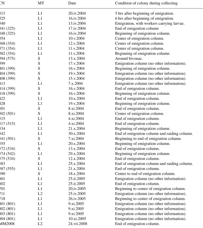
Detailed collecting data are compiled in Table 1. Label data are written out for holotypes, and collecting date, number of specimens and collecting number (inside of brackets) is indicated for paratypes and other specimens.
Holotypes and some paratypes of the new species are deposited in the Sarawak Forestry Department, Sarawak, Malaysia. Additional paratypes and other identified specimens are deposited in FMNH (M. K. Thayer) and the Kyushu University Museum (M. Maruyama).
Results
Morphotypes of ants and host specificities of rove beetles
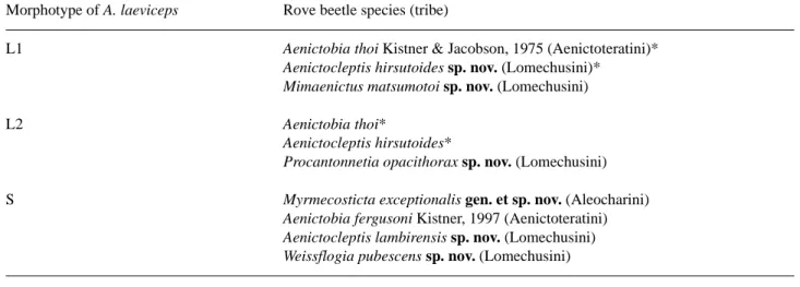
About 350 specimens of rove beetles were collected from approximately 20 colonies of Aenictus laeviceps (sensu Wilson, 1964). The rove beetles were classified into eight species belonging to six genera. We noticed that each ant nest harboured particular rove beetle species (3 or 4 species), and there were three patterns of rove beetle species combinations (Table 2). After a morphological study of the host ants, we found that the ants can be classified into three morphotypes that correspond to the three combinations of rove beetle species (Table 2). Hamaguti et al. (2007) designed microsatellite markers for A. laeviceps and treated these morphotypes based on this information as morphotypes L1, L2 and S.
Host specificities by the rove beetles were very strict except for Aenictobia thoi and Aenictocleptis hirsutoides which were observed with both morphotypes L1 and L2. The composition of the rove beetle species associated with Morphotype S was clearly separated from those of morphotypes L1 and L2.
Morphotypes are diagnosed and discussed in the following paragraphs.
TABLE 1. Collecting data. CN: collecting number (that of the same colony is in brackets); MT: host morphotype of Aenictus laeviceps (sensu Wilson 1964).
CN MT Date Condition of colony during collecting
313 L1 20.iv.2004 5 hrs after beginning of emigration.
325 L1 16.iv.2004 6 hrs after beginning of emigration.
340 L1 13.iv.2004 Emigration, with workers carrying larvae.
341 (325) L1 17.iv.2004 End of emigration column
348 (325) L1 16.iv.2004 Beginning of emigration column.
354 L1 10.v.2004 Center of emigration column.
368 (354) L1 12.v.2004 Center of emigration column.
371 (354) L1 11.v.2004 Center of emigration column.
382 (354) L1 11.v.2004 Beginning of emigration column.
394 (575) S 13.x.2004 Around bivouac.
399 S 17.v.2004 Emigration column (no other information).
401 (399) S 16.v.2004 Beginning of emigration column.
404 (399) S 19.v.2004 Emigration column (no other information).
408 (399) S 15.v.2004 Emigration column (no other information).
413 L1 7.x.2004 Emigration column (no other information).
414 (399) S 16.v.2004 End of emigration column.
418 (399) S 16.v.2004 Beginning of emigration column.
422 L1 10.x.2004 End of emigration column.
428 L1 19.v.2004 Beginning of emigration column.
501 S 8.xi.2004 End of emigration column.
502 (501) S 8.xi.2004 Center of emigration column.
515 L1 4.xi.2004 End of emigration column.
517 (515) L1 4.xi.2004 End of emigration column.
534 L1 21.x.2004 Beginning of emigration column.
542 L1 30.x.2004 End of emigration column and raiding column.
541 (501) S 7.xi.2004 Beginning to end of emigration column.
555 L1 20.x.2004 Beginning of emigration column.
572 (534) S 13.x.2004 End of emigration column.
574 (542) L1 29.x.2004 Beginning of emigration column.
575 (534) S 12.x.2004 End of emigration column.
583 L1 25.x.2004 End of emigration column and raiding column.
587 (555) L1 21.x.2004 End of emigration column.
590 S 18.x.2004 Center to end of emigration column.
601 L1 25.ii.2005 Emigration column (no other information).
602 L1 25.ii.2005 End of emigration column.
701 L1 20.iv.2005 Beginning to center of emigration column.
711 L2 25.iv.2005 Emigration column (no other information).
718 L1 26.iv.2005 Beginning to center of emigration column.
801 (801) L1 9.xi.2005 Emigration column (no other information).
802 (801) L1 9.xi.2005 Emigration column (no other information).
803 (801) L1 9.xi.2005 Emigration column (no other information).
804 (801) L1 10.xi.2005 Emigration column (no other information).
MM2008 L2 24.vii.2008 End of emigration column.
TABLE 2. Aenictus laeviceps host morphotypes and their corresponding myrmecophilous rove beetle species as recorded in the Lambir Hills National Park. Asterisks indicate rove beetle species common to both morphotypes L1 and L2.
Morphotype of A. laeviceps Rove beetle species (tribe)
L1 Aenictobia thoi Kistner & Jacobson, 1975 (Aenictoteratini)* Aenictocleptis hirsutoides sp. nov. (Lomechusini)*
Mimaenictus matsumotoi sp. nov. (Lomechusini)
L2 Aenictobia thoi*
Aenictocleptis hirsutoides*
Procantonnetia opacithorax sp. nov. (Lomechusini)
S Myrmecosticta exceptionalis gen. et sp. nov. (Aleocharini)
Aenictobia fergusoni Kistner, 1997 (Aenictoteratini) Aenictocleptis lambirensis sp. nov. (Lomechusini) Weissflogia pubescens sp. nov. (Lomechusini)
Morphotype L1 of Aenictus laeviceps (Figs. 1–3)
Diagnosis. Antennae slender; alitrunk (Figs. 1, 2) without setae; dorsal tip of petiole (Fig. 3) with 1 or 2 setae; ven- tral process of petiole (Fig. 3) acutely prominent and toward postero-ventrally; postopetiole (Fig. 3) short and high, higher than long; setae on sternite of postopetiole (Fig. 3) irregular in length, those on posterior part longer than those on anterior part.
Comments. With reference to the figure of Wilson (1964), the syntypes of A. laeviceps from Sarawak agree well with Morphotype L1. This morphotype is extremely similar to Morphotype L2, but can be distinguished from it by the presence of setae on the dorsal tip of the petiole, although some individuals are occasionally devoid of this seta.
Morphotype L2 of Aenictus laeviceps (Figs. 4–6)
Diagnosis. Antennae slender; alitrunk (Figs. 4, 5) without setae; dorsal tip of petiole (Fig. 6) without setae; ventral process of petiole (Fig. 6) acutely prominent, longer than in Morphotype L1; postopetiole (Fig. 6) moderate in length and height, almost as high as long; setae on sternite of postopetiole (Fig. 6) almost uniformly short.
Comments. Very similar to Morphotype L1 but clearly distinguished from it by the absence of setae on the pet- iole. Only two colonies of this morphotype were found.
Morphotype S of Aenictus laeviceps (Figs. 7–9)
Diagnosis. Antennae thick and slightly short; alitrunk (Figs. 7, 8) with setae; dorsal tip of petiole (Fig. 9) with 4–6 setae; ventral process of petiole (Fig. 9) acutely prominent but much shorter than in morphotypes L1 and L2; setae on sternite of postopetiole (Fig. 9) irregular in length, with long setae distributed both on anterior and posterior parts.
Comments. This morphotype is distinguished from morphotypes L1 and L2 by several character states, though all morphotypes are generally very similar in general appearance. The same morphotype was also found at Ulu Gombak, which was erroneously identified as “Aenictus fergusoni” by Kistner et al. (1997) (see Comments for Aenictobia fergusoni).
FIGURES 1–9. Three morphotypes of Aenictus laeviceps (sensu Wilson, 1964). 1–3, Morphotype L1; 4–6, Morphotype L2; 7– 9, Morphotype S (all SEM photographs; 1, 4, 7, habitus, laeral view; 2, 5, 8, alitrunk, lateral view; 3, 6, 9, petiole and postpeti- ole, lateral view).
Taxonomy of the rove beetles Tribe Aleocharini Fleming
Aleocharidae Fleming, 1821: 49 (type genus: Aleochara Gravenhorst, 1802).
Myrmecosticta Maruyama, gen. nov.
Type species. Myrmecosticta exceptionalis sp. nov., by present designation.
Etymology. Combination of the Greek myrmex (= ant) and stictos (= spotted), derived from the myrmecophily and the presence of a pair of deep spots (hollows) on the pronotum. The latter word also alludes to Tetrastica, a rep- resentative of the Tetrastica genus group to which Myrmecosticta belongs. Gender feminine.
Distribution. Malaysia (Borneo).
Description. Body (Figs. 10, 11) myrmecoid, shining. Head (Figs. 12–14) oval, depressed dorso-ventrally, with pair of deep hollows at side of gula (base of submentum), strongly constricted at base to form a “neck.” Antenna gently widened apically; segment I long, as long as II and III combined; segments IV–XI sparsely covered with pubescence. Labrum (Fig. 19) much wider than long; anterior margin widely emarginate; surface sparsely covered with pseudopores. Mandibles asymmetric, strongly curved, pointed apically; right mandible (Fig. 20) with a small tooth at middle of inner margin, without serration on inner side of each mandible. Maxilla (Fig. 21) small,
with galea and lacinia shortened; cardo slender, constricted near base, without pore; stipes small, shortened, with- out pore; palpifer without pore. Mentum (Fig. 22) sub-pentangular with anterior margins concave; surface with 3 setae, 1 pore around middle, and with some pores around postero-medial margin. Prementum (Fig. 23) with 2 real pores antero-laterally, 1 setal pore around middle, in total 4 or 5 pseudopores medially, and with a pore postero-lat- erally. Ligula (Fig. 23) slightly bilobed; each lobe with 4 long setulae apically. Lateral lobe of ligula (Fig. 23) gen- eralized; inner margin almost straight. Labial palpus (Fig. 23) with segment I short, slightly longer than wide, as long as II. Pronotum (Fig. 15) oval, with a pair of depressions around middle; proventrite fully visible in lateral view. Mesoventrite (Fig. 16) with anterior part constricted to connect to pronotal base. Metaventrite (Fig. 16) broad; anterior intercoxal process truncate apically; mesocoxal cavity not margined. Elytra (Fig. 10) together about as long as wide; posterior margins slightly convex. Legs (Figs. 10, 11) long; femora and tibiae moderately covered with long setae; mid and hind tibiae curved; hind coxa (Fig. 16) long, with a deep sulcus from near base to apex; hind trochanter (Fig. 16) large, prominent laterally. Abdomen (Figs. 10–11, 17–18) oval, strongly curved dorsally; anterior margins of tergites IV and V broadly emarginate. Male: median lobe of aedeagus (Fig. 24) thin laterally; copulatory piece of inner sac with flagellum. Female: spermatheca (Fig. 25) with a membranous area between api- cal and basal parts.
Diagnosis. This genus can easily be distinguished from other genera of Aleocharini by the myrmecoid body shape. The reasons behind the systematic placement of the genus in Aleocharini are discussed below.
Myrmecosticta exceptionalis Maruyama, sp. nov. (Figs. 10–25)
Holotype, ♂ “MALAYSIA: Sarawak, Lambir Hills National Park, 18 X 2004, Matsumoto T. / ATTM 590 / HOLO- TYPE Myrmecosticta exceptionalis Maruyama” (no. 590). Paratypes: 1, same data as holotype but 17 V 2004 (no. 399); 2, ditto, 16 V 2004 (no. 418); 1, ditto, 8 XI 2004 (no. 501); 2, ditto, 8 XI 2004 (no. 502); 6, ditto, 7 XI 2004 (no. 541); 5, ditto, 13 X 2004 (no. 572); 17, ditto, 12 X 2004 (no. 575); 22, ditto, 18 X 2004 (no. 590). See, Table 1 for detailed collecting data.
Symbiotic host. Morphotype S of the Aenictus laeviceps. Distribution. Malaysia (Borneo).
Etymology. The Latin exceptionalis means exceptional, for the exceptionally myrmecoid body shape within the tribe Aleocharini. The body shapes of other Aleocharini members are of the standard staphylinid-type, similar to most other species of Aleocharinae, and no myrmecoid forms have been described previously.
Description. Body surface smooth, shining. Almost uniformly reddish brown except for mouthparts and tarsi somewhat paler. Head (Figs. 12–14) oval, much longer than wide in dorsal view, widest at eyes, with a v-shaped prominence between antennal insertions in dorsal view; clypeus with a small projection medially; surface sparsely covered with setae; eyes large, about half as long as head. Antennae short, evidently shorter than combined length of head and pronotum; segment I slender, gently widened apically; segment II small, conical; segment III short, slightly longer than II; segments IV–X slightly longer than wide; segment XI parallel-sided around base, rounded at apex. Pronotum (Fig. 15) much longer than wide (length/width=1.43–1.50), strongly convex; surface sparsely and finely punctate, with 4 macrosetae along midline and 1 macroseta antero-laterally. Elytra slightly widened pos- teriorly, wider than long; surface with dense but fine microsculpture, and moderately covered with short setae, without macrosetae. Metaventrite (Fig. 16) shining, with 2 large setae at middle and 3–4 small setae around poste- rior area. Abdomen (Figs. 17–18) with segments III–VIII sparsely covered with setae; number of macrosetae from tergites III–X: 2, 2, 2, 3, 4, 7, 3, 3; with tergite VIII posterior margin rounded; sternite VIII with 10–12 macrosetae. Male: median lobe of aedeagus (Fig. 24) with flagellum coiled 7.5 times; apical lobe of paramere elongate and slightly curved. Female: spermatheca (Fig. 25) with basal part constricted, apical half parallel-sided; apical part somewhat oval.
Measurements. BL, ≈ 3.1–3.4; FBL, ≈ 1.7–1.9; HW, 0.57–0.61; EL, 0.313–0.337; AL, 1.49–1.69; PL, 0.66– 0.75; PW, 0.44–0.53; ELW, 0.66–0.78; HTL, 1.31–1.35. N=5.
Diagnosis. This species can easily be distinguished from any members of the tribe Aleocharini by the myrme- coid body shape. This species can be distinguished from the other myrmecoid species of Aleocharinae by charac- ters which define the tribe Aleocharini, e.g., presence of a pseudosegment on the apex of the labial palpus.
FIGURES 10–18. Myrmecosticta exceptionalis gen. et sp. nov. (12–18, SEM photographs). 10, habitus, dorsal view; 11, ditto, lateral view (holotype); 12, head, pronotum and anterior part of elytra, lateral view; 13, head, dorsal view; 14, head, ventral view; 15, pronotum, dorsal view; 16, meso- and metathorax, coxae and trochanters, ventral view; 17, abdomen, dorsal view; 18, ditto, ventral view.
FIGURES 19–25. Myrmecosticta exceptionalis gen. et sp. nov. 19, labrum, dorsal view; 20, right mandible, dorsal view; 21, right maxilla, ventral view; 22, mentum, ventral view; 23, labium, ventral view; 24, median lobe of aedeagus, lateral view (holotype); 25, spermatheca.
Tribe Aenictoteratini Kistner, 1993
Aenictoteratini Kistner, 1993: 242 (original description; type genus: Aenictoteras Wheeler, 1932).
Aenictobia Seevers
Aenictobia Seevers, 1953: 127 (original description; type species: Aenictobia longicornis Seevers 1952, by original designa- tion). Kistner & Jacobson, 1975: 42 (redescription, description of one additional species). Kistner et al. 1997: 175 (rede- scription, description of one additional species, species key, placed in subtribe Aenictobiina of tribe Lomechusini).
Aenictobia thoi Kistner (Figs. 26–28)
Aenictobia thoi Kistner & Jacobson, 1975: 45 (original description). Kistner et al. 1997: 175 (redescription).
Materials examined. 11, Lambir Hills National Park, Sarawak, Malaysia, 16 IV 2004, T. Matsumoto leg. (no. 325); 2, same data but 13 IV 2004 (no. 340); 4, ditto, 16 IV 2004 (no. 348); 3, ditto, 10 V 2004 (no. 354); 1, ditto, 12 V 2004 (no. 368); 2, ditto, 11 V 2004 (no. 371); 1, ditto, 11 V 2004 (no. 382); 6, ditto, 7 X 2004 (no. 413); 3, ditto, 19 V 2004 (no. 428); 1, ditto, 4 XI 2004 (no. 515); 2, ditto, 30 X 2004 (no. 542); 3, ditto, 20 X 2004 (no. 555); 4, ditto, 29 X 2004 (no. 574); 1, ditto, 25 II 2005 (no. 601); 1, ditto, 25 II 2005 (602); 3, ditto, 20 IV 2005 (701); 12, ditto, 25 IV 2005 (711); 20, ditto, 26 IV 2005 (718); 10, ditto, 9 XI 2005 (no. 801); 1, ditto, 9 XI 2005 (no. 803); 5, ditto, 10 XI 2005 (no. 804). See, Table 1 for detailed collecting data.
Symbiotic host. Morphotypes L1 and L2 of Aenictus laeviceps. Distribution. Malaysia (Peninsula, Borneo), Indonesia (Sumatra).
Diagnosis. This species can be distinguished from Aenictobia fergusoni by the temples of head (Figs. 26, 27) being weakly convex, by the more slender antennae (Fig. 26), by the antennal segment I (Fig. 27) being about 2.5 times as long as wide, by the postero-lateral corner of the pronotum (Fig. 27) being not projected, but almost right angled, and by the setae on the pronotum and elytra (Fig. 28) being somewhat sparser and shorter.
Comments. This species was originally described based on specimens from Ulu Gombak, Peninsular Malaysia and recorded from Sumatra and Sabah.
No morphological differences were detected between populations from Lambir Hills National Park and Ulu Gombak.
Aenictobia fergusoni Kistner (Figs. 29–31)
Aenictobia fergusoni Kistner, in Kistner et al. 1997: 181 (original description).
Materials examined. 1, Lambir Hills National Park, Sarawak, Malaysia, 13 X 2004, T. Matsumoto leg. (no. 394); 2, same data but 15 V 2004 (no. 408); 1, ditto, 16 V 2004 (no. 414); 3, ditto, 16 V 2004 (no. 418); 6, ditto, 8 XI 2004 (no. 502); 1, ditto, 7 XI 2004 (no. 541); 6, ditto, 13 X 2004 (no. 572); 1, ditto, 12 X 2004 (no. 575); 5, ditto, 18 V 2004 (no. 590). See, Table 1 for detailed collecting data.
Symbiotic host. Morphotype S of Aenictus laeviceps. Distribution. Malaysia (Peninsula, Borneo).
Diagnosis. This species can be distinguished from Aenictobia thoi by the temples of head (Figs. 29, 30) being strongly convex, by the thicker antennae (Fig. 29), by the antennal segment I (Fig. 30) being about 1.5 times as long as wide, by the postero-lateral corner of pronotum (Fig. 30) being projected laterally, and by the setae on the pronotum and elytra (Fig. 31) being somewhat denser and longer.
Comments. This species was originally described based on specimens from Ulu Gombak, Peninsular Malay- sia. New record from Borneo.
FIGURES 26–31. Aenictobia spp. 26–28, A. thoi; 29–31, A. fergusoni (27, 28, 30, 31, SEM photographs). 26, 29, habitus, dor- sal view; 27, 30, head, pronotum and anterior part of elytra, dorsal view; 28, 31, sculptures on elytra, dorsal view.
The host ant of the type series was reported as “Aenictus fergusoni Forel, 1901” (Kistner et al. 1997), and, therefore, the same specific epithet was used for the beetle. The host species was, however, found to be misidenti- fied. Pfeiffer (2005) illustrated an ant identified as “Aenictus fergusoni” which was the same nest number as the host ant of the type series of Aenictobia fergusoni in Kistner et al. (1997), but it was apparently Morphotype S of Aenictus laeviceps. During the field research by M. Maruyama in Ulu Gombak, the type locality of Aenictobia fer- gusoni, it was specifically collected from Morphotype S of Aenictus laeviceps (N=4). Therefore, the host ant spe- cies of “Aenictobia fergusoni” in Kistner et al. (1997) is actually Morphotype S of Aenictus laeviceps. The true
Aenictus fergusoni may not be distributed in Peninsular Malaysia, but in India to the Indochinese Peninsula (India, Myanmar, Cambodia, Thailand, etc.) (Maruyama, unpublished data).
Tribe Lomechusini Fleming
Lomechusidae Fleming, 1821: 49 (type genus: Lomechusa Gravenhorst, 1806).
Aenictocleptis Kistner & Jacobson
Aenictocleptis Kistner & Jacobson, 1975: 36 (original description; type species: Aenictocleptis hirsutus Kistner & Jacobson, 1975, by original designation). Kistner et al. 1997: 134 (redescription, descriptions of four additional species, species key).
Aenictocleptis hirsutoides Maruyama, sp. nov. (Figs. 32–34, 38–42)
Holotype, ♂ “MALAYSIA: Sarawak, Lambir Hills National Park, 25 IV 2005 / ATTM 711 / HOLOTYPE Aenicto- cleptis hirsutoides Maruyama” (no. 711). Paratypes: 2, same data as holotype but 13 IV 2004 (no. 340); 1, ditto, 17 IV 2004 (no. 341); 3, ditto, 12 V 2004 (no. 368); 3, ditto, 19 V 2004 (no. 428); 1, ditto, 20 X 2004 (no. 555); 1, ditto, 25 X 2004 (no. 583); 2, ditto, 25 IV 2005 (no. 711). See, Table 1 for detailed collecting data.
Symbiotic host. Morphotypes L1 and L2 of Aenictus laeviceps. Distribution. Malaysia (Borneo).
Etymology. In reference to the close resemblance to Aenictocleptis hirsutus.
Description. Body (Fig. 32) slender. Brown, except for mouth parts, legs, abdominal segments III–V and basal half of abdominal segment VI reddish brown. Head (Fig. 32, 33) with temple not developed. Mandibles strongly asymmetric; right mandible thick, outer margin around apical 1/4 convex. Antennae (Figs. 32, 34) slender, long, reaching just before posterior margins of elytra; segment XI slightly longer than IX and X combined: relative lengths of segments IX–XI: 14, 19, 35. Pronotum (Figs. 32, 34) as long as wide (width/length=1.01–1.03), densely covered with stout setae. Elytra (Figs. 32, 33) densely covered with stout setae. Abdominal segments VIII–IX densely covered with short setae; macrosetae on tergite VIII (Fig. 38) readily distinguished from other setae by thickness and color. Male: median lobe of aedeagus (Figs. 39, 40) with apical lobe elongate, gently narrowed api- cally, acutely pointed at apex; apical lobe of paramere (Fig. 41) parallel-sided, rounded at apex. Female: spermath- eca (Fig. 42) with basal part spherically bulbous at base, without lateral projection.
Measurements. BL, ≈ 5.0–6.1; FBL, ≈ 2.4–2.6; HW, 0.99–1.06; EL, 0.46–0.50; AL, ≈ 2.2–2.4; PL, 0.88–1.02; PW, 0.93–1.05; ELW, ≈ 1.3–1.4; HTL, 1.31–1.38. N=5.
Diagnosis. This species is most similar to Aenictocleptis hirsutus in color, shape of mandibles, pronotal pro- portions, and density of setae on pronotum and elytra, but may be easily distinguished by the shorter setae on the abdominal segments VIII–IX, the shorter apical lobe of the aedeagal median lobe, and the absence of lateral projec- tion in the basal part of the spermatheca.
Aenictocleptis lambirensis Maruyama, sp. nov. (Figs. 35–37, 43–47)
Holotype, ♂ “MALAYSIA: Sarawak, Lambir Hills National Park, 16 V 2004 / ATTM 418 / HOLOTYPE Aenicto- cleptis lambirensis Maruyama” (no. 418). Paratypes: 1 ♂, 1 ♀, 1 sex?, same data as holotype (no. 418). See, Table 1 for detailed collecting data.
Symbiotic host. Morphotype S of Aenictus laeviceps. Distribution. Malaysia (Borneo).
Etymology. Named for the type locality.
FIGURES 32–37. Aenictocleptis spp. 32–34, A. hirsutoides sp. nov.; 35–37, A. lambirensis sp. nov. (33, 34, 36, 37, SEM pho- tographs). 32, 35, habitus, dorsal view; 33, 36, head, pronotum and elytra, dorsal view; 34, 37, right antenna, dorsal view.
Description. Body (Fig. 35) rather robust. Reddish brown, except for mouth parts, legs, lateral margins of pro- notum, abdominal segments III–VII other than medial area of tergites paler. Head (Figs. 35, 36) with temple well developed to eye level. Mandibles rather asymmetric; right mandible gently narrowed and curved apically. Anten-
nae (Figs. 35, 37) somewhat thick, clavate, reaching around middle of elytra; segment XI longer than IX and X combined: relative lengths of segments IX–XI: 12, 15, 30. Pronotum (Figs. 35, 36) much wider than long (width/ length=1.19–1.21), sparsely covered with long and stout setae. Elytra (Figs. 35, 36) densely covered with long and stout setae. Abdominal segments VIII–IX moderately covered with long setae; macrosetae on tergite VIII (Fig. 43) poorly distinguished from other setae.
FIGURES 38–42. Aenictocleptis hirsutoides sp. nov. 38, male tergite VIII, dorsal view, holotype; 39, median lobe of aedea- gus, lateral view; 40, ditto, ventral view; 41, apical lobe of paramere; 42, spermatheca.
FIGURES 43–47. Aenictocleptis lambirensis sp. nov. 43, male tergite VIII, dorsal view (holotype); 44, median lobe of aedea- gus, lateral view; 45, ditto, ventral view; 46, apical lobe of paramere; 47, spermatheca.
Male: Median lobe of aedeagus (Figs. 44, 45) with apical lobe rhomboidal, minutely bilobed at apex; apical lobe of paramere (Fig. 46) subtriangular.
Female: Spermatheca (Fig. 47) with basal part bulbous at base, without lateral projection.
Measurements. BL, ≈ 6.0–7.1; FBL, ≈ 2.6–2.9; HW, 1.09–1.15; EL, 0.53–0.58; AL, ≈ 2.4–2.5; PL, 0.89–0.96; PW, 1.13–1.21; ELW, ≈ 1.4–1.5; HTL, 1.25–1.33. N=3.
Diagnosis. This species is easily distinguished from other Aenictocleptis species by the sparse setation on the pronotum and elytra, and the apical lobe of the aedeagal median lobe being rhomboidal.
Mimaenictus Kistner & Jacobson
Mimaenictus Kistner & Jacobson, 1975: 47 (original description; type species: Mimaenictus wilsoni Kistner & Jacobson, 1975, by original designation; placed in tribe Dorylomimini). Kistner, 1993: 272 (redescription, one additional species, species key, transferred to tribe Aenictoteratini).
Mimaenictus matsumotoi Maruyama, sp. nov. (Figs. 48–59)
Holotype, ♂ “MALAYSIA: Sarawak, Lambir Hills National Park, 29 X 2004 / ATTM 574 / HOLOTYPE Mimaen- ictus matsumotoi Maruyama” (no. 574). Paratypes: 3, same data as holotype but 20 IV 2004 (no. 313); 1, ditto, 13 IV 2004 (no. 340); 1, ditto, 16 IV 2004 (no. 348); 2, ditto, 12 V 2004 (no. 368); 2, ditto, 7 X 2004 (no. 413); 3, ditto, 10 X 2004 (no. 422); 1, ditto, 19 V 2004 (no. 428); 1, ditto, 4 XI 2004 (no. 517); 1, ditto, 21 X 2004 (no. 534); 1, ditto, 30 X 2004 (no. 542); 4, ditto, 29 X 2004 (no. 574); 1, ditto, 20 X 2004 (no. 555); 3, ditto, 21 X 2004 (no. 587); 14, ditto, 20 IV 2005 (no. 701); 10, ditto, 9 XI 2005 (no. 801); 1, ditto, 9 XI 2005 (no. 802); 2, ditto, 9 XI 2005 (no. 803); 6, ditto, 10 XI 2005 (no. 804). See, Table 1 for detailed collecting data.
Symbiotic host. Morphotype L1 of Aenictus laeviceps. Distribution. Malaysia (Borneo).
Etymology. Dedicated to Dr. T. Matsumoto, the second author of this paper and collector of the type series. Description. Almost uniformly reddish brown. Head (Figs. 48–51) with dorsal surface completely glabrous except for several setae in front of eyes. Pronotum (Fig. 52) with ratio length/width=1.24–1.25, with 7 or 8 setae along midline, 1 seta antero-laterally and 1 seta postero-laterally. Elytra with 6–10 minute setae. Abdomen (Figs. 53–55) with numbers of macrosetae from tergite II–VIII: 1, 1, 1, 6, 6, 6; gland openings on sternites III–V clear. Male: median lobe of aedeagus (Figs. 57, 58) gently narrowed apically in lateral view, with lateral ridge which is gradually narrowed from middle to apex in parameral view. Female: spermatheca (Fig. 59) with basal part lacking basal projection; apical lobe with lateral ridge small.
Measurements. BL, ≈ 3.5–3.8; FBL, ≈ 1.7–1.9; HW, 0.53–0.56; EL, 0.254–0.263; AL, ≈ 1.7–1.8; PL, 0.56– 0.58; PW, 0.45–0.46; ELW, 0.67–0.69; HTL, 1.10–1.21. N=5.
Diagnosis. This species is most similar to Mimaenictus wilsoni (M. w.) in the presence of lateral ridge in median lobe of aedeagus, but is clearly distinguished from it by the shorter setae on the pronotum (in M. w., the setae are longer and denser in the anterior area and lap over each other) and the sparser setae on the elytra (M. w. has more than 15 setae). The shape of the median lobe of aedeagus and that of the spermatheca are also different. In M. w. the apical lobe of the median lobe of aedeagus (Figs. 60, 61) is slightly constricted near the base and almost parallel-sided in lateral view, and the lateral ridge is almost semi-circular in parameral view; the basal part of sper- matheca (Fig. 62) is with a basal projection, and the apical lobe is with a large lateral ridge.
Procantonnetia Kistner & Jacobson
Procantonnetia Kistner & Jacobson, 1975: 47 (original description; type species: Procantonnetia malayensis Kistner & Jacob- son, 1975, by original designation; placed in tribe Lomechusini). Kistner, 1993: 263 (redescription, one additional species, species key, transferred to tribe Aenictoteratini).
FIGURES 48–56. Mimaenictus matsumotoi sp. nov. (50–56, SEM photographs). 48, Habitus, dorsal view; 49, ditto, lateral view; 50, head, pronotum and anterior part of elytra, lateral view; 51, head, dorsal view; 52, pronotum, dorsal view; 53, abdom- inal tergites III–VI, dorsal view; 54, abdomen, lateral view; 55, abdominal sternite III–VI, ventral view; 56, gland opening on abdominal sternite IV, ventral view.
FIGURES 57–62. Mimaenictus spp.: 57–59, M. matsumotoi sp. nov.; 60–62, M. wilsoni (57, 60, median lobe of aedeagus, lat- eral view; 58, 61, ditto, ventral view; 59, 62, spermatheca. 57, 58, holotype).
Procantonnetia opacithorax Maruyama, sp. nov. (Figs. 63–73)
Holotype, ♂ “MALAYSIA: Sarawak, Lambir Hills National Park, 25 IV 2004 Matsumoto T. leg. / ATTM 711 / HOLOTYPE Procantonnetia opacithorax Maruyama” (no. 711). Paratypes: 19, same data as holotype (no. 711); 16, ditto but 24 VII 2008, M. Maruyama leg. (no. MM2008). See, Table 1 for detailed collecting data.
Symbiotic host. Morphotype L2 of Aenictus laeviceps.
FIGURES 63–70. Procantonnetia opacithorax sp. nov. (65–70, SEM photographs). 63, Habitus, dorsal view; 64, ditto, lateral view; 65, head, pronotum and anterior part of elytra, lateral view; 66, head, dorsal view; 67, pronotum, dorsal view; 68, abdo- men, dorsal view; 69, ditto, lateral view; 70, ditto, ventral view.
Distribution. Malaysia (Borneo).
Etymology. Combination of the Latin opacus (=opaque, matte) and thorax (= thorax), bringing attention to the opaque thorax.
Description. Almost uniformly brown. Head (Figs. 63–66) around “neck” coarsely punctate with microsculp- ture, with 10–15 small setae around temple and 1 seta between eyes. Pronotum (Fig. 67) with ratio length /width
=1.38–1.46, with surface coarsely punctate except for midline which is glabrous, with 1 seta at middle. Elytra with entire surface coarsely punctate, with 15–20 setae from middle to apices. Abdomen (Figs. 68–70) with numbers of macrosetae from tergite II–VIII: 3, 5, 1, 1, 3, 6. Male: median lobe of aedeagus (Figs. 71, 72) slightly dilated near apex in lateral view. Female: spermatheca (Fig. 73) with basal part with a large basal projection; apical lobe small, with inner wall not wrinkled.
Measurements. BL, ≈ 3.0–3.4; FBL, ≈ 1.7–1.9; HW, 0.55–0.63; EL, 0.241–0.262; AL, ≈ 1.7–1.8; PL, 0.78– 0.83; PW, 0.53–0.60; ELW, 0.89–0.96; HTL, 1.26–1.35. N=5.
Diagnosis. This species is most similar to Procantonnetia borneensis Kistner, 1993 in having pronotum and elytra coarsely punctate, but is distinguishable from the latter by the presence of setae on the pronotum and the number of the macrosetae on the abdominal tergites (Kistner, 1993: 269).
FIGURES 71–73. Procantonnetia opacithorax sp. nov. (71, 72, holotype). 71, Median lobe of aedeagus, lateral view; 72, ditto, ventral view; 73, spermatheca.
Weissflogia Kistner
Weissflogia Kistner, in Kistner et al. 1997: 128 (original description; type species: Weissflogia rhopalogaster Kistner, 1997, by original designation; placed in tribe Aenictoteratini).
Weissflogia pubescens Maruyama, sp. nov. (Figs. 74–84)
Holotype, ♂ “MALAYSIA: Sarawak, Lambir Hills National Park, 12 X 2004 Matsumoto T. leg. / ATTM 575 / HOLOTYPE Weissflogia pubescens Maruyama” (no. 575). Paratypes: 1, same data as holotype but 17 V 2004 (no. 399); 1, ditto, 16 V 2004 (no. 401); 3, ditto, 15 V 2004 (no. 408); 1, ditto, 16 V 2004 (no. 414); 31, ditto, 16 V 2004 (no. 418); 2, ditto, 8 XI 2004 (no. 502); 5, ditto, 13 X 2004 (572); 14, ditto, 12 X 2004 (no. 575); 19, ditto, 18 X 2004 (no. 590). See, Table 1 for detailed collecting data.
Symbiotic host. Morphotype S of Aenictus laeviceps. Distribution. Malaysia (Borneo).
FIGURES 74–81. Weissflogia pubescens sp. nov. (76–81, SEM photographs). 74, Habitus, dorsal view; 75, ditto, lateral view; 76, head, pronotum and anterior part of elytra, lateral view; 77, head, dorsal view; 78, pronotum, dorsal view; 79, abdominal tergites III–VI, dorsal view; 80, abdomen, lateral view; 81, abdominal sternite III–V, ventral view.
FIGURES 82–87. Weissflogia spp. 82–84, W. pubescens sp. nov.; 85–87, W. rhopalogaster (82, 85, median lobe of aedeagus, lateral view; 83, 86, ditto, ventral view; 84, 87, spermatheca; 82, 83, holotype).
Etymology. The Latin pubescens means pubescent, bringing attention to the setae on the head and elytra that are longer and denser than those of the congener W. rhopalogaster.
Description. Almost uniformly reddish brown. Head (Figs. 74–77) sparsely covered with setae except for between eyes. Pronotum (Fig. 78) with ratio length /width =1.40–1.44, with a row of 5 or 6 setae along midline, 10–14 small setae laterally and 2 or 3 setae posteriorly. Elytra (Fig. 78) sparsely covered with minute setae except for medial area of each elytron, with a large seta near base of scutellum. Abdomen (Figs. 79–81) with sternite III widest around apical 1/3; paratergites III–V slightly matte; numbers of macrosetae from tergite II–VIII: 1, 1, 0, 2, 2, 6. Male: median lobe of aedeagus (Figs. 82, 83) with apical lobe dilated apically, base of parameral side shal- lowly emarginate in lateral view; apex of apical lobe thin laterally in parameral view. Female: spermatheca (Fig. 84) with basal part short.
Measurements. BL, ≈ 3.7–4.1; FBL, ≈ 2.0–2.2; HW, 0.63–0.67; EL, 0.268–0.280; AL, ≈ 1.9–2.0; PL, 0.81– 0.83; PW, 0.56–0.59; ELW, 0.80–0.83; HTL, 1.27–1.38. N=5.
Diagnosis. This species differs from Weissflogia rhopalogaster (W. r.), by denser and longer setae on the head and elytra (in W. r. the temples are covered with 7–10 scattered small setae, and the setae on the elytral shoulder are sparser and shorter), the sternite III being widest around apical third (in W. r. it is widest around middle) and the paratergites III–V being slightly matt (in W. r. they are entirely shining). The shapes of the median lobe of aedeagus and the spermatheca are also different. In W. r. the base of the parameral side of the apical lobe (Figs. 85, 86) is deeply emarginate in the lateral view; the apex of apical lobe is gently narrowed in the parameral view. Spermath- eca (Fig. 87) with the basal part long, curved in basal 1/6.
Comments. The host ant of Weissflogia rhopalogaster, the generic type species and the only congener to the newly described species, was reported as “Aenictus fergusoni,” but it was found to be misidentification, and it is actually Morphotype S of Aenictus laeviceps. See the Comments under Aenictobia fergusoni, the species which has been collected together with Weissflogia rhopalogaster at Ulu Gombak, the type locality of W. rhopalogaster (Kist- ner, 1997; Maruyama, unpublished data).
Discussion
Strict host specificity
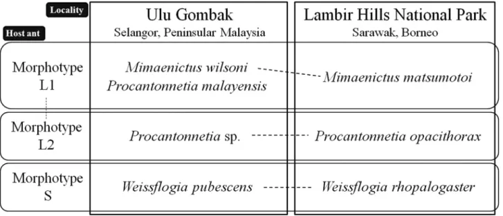
As we have documented above, rove beetles belonging to six genera and eight species were collected from colonies of three distinct Aenictus laeviceps morphotypes (L1, L2 and S). With the exceptions of Aenictobia thoi and Aenic- tocleptis hirsutoides, which were both hosted by L1 and L2, strict host specificity was observed by all beetle spe- cies for a single ant morphotype. Three of these host-specialists belong to three Lomechusini genera: Mimaenictus (L1), Procantonnetia (L2) and Weissflogia (S), and are likely closely related, sharing the synapomorphic presence of large gland reservoirs on abdominal sternites III–V. The association between these genera and Aenictus laevi- ceps morphotypes was also investigated in Ulu Gombak (Gombak), Peninsular Malaysia. The A. laeviceps mor- photypes at Gombak appeared to be the same as were encountered at Lambir, and furthermore, each morphotype harboured a closely related rove beetle congener (Fig. 88). Morphotype L1 again harboured a species of Mimaeni- cus (M. wilsoni), L2 hosted a presently undescribed species of Procantonnetia very similar to P. opacithorax, and Morphotype S hosted Weissflogia rhopalogaster. Hence, the pattern of ant-rove beetle associations is largely the same in Gombak and Lambir. The only notable difference was the presence of an additional species of Procanton- netia, P. malayensis in colonies of the L1 morphotype (Kistner & Jacobson 1975; Maruyama unpublished data) (N=3). Notably however, on morphological grounds P. opacithorax and the undescribed Procantonnetia from Gombak appear to be more closely related to each other than to P. malayensis (see below).
Aenictus ants reproduce only by colony fission because their queens are wingless (e.g., Schneirla 1971). Thus, their geographical expansion is constrained by sea straits. The presence of the same ant morphotypes in these two localities, long-isolated by a sea strait, suggests that the morphotypes arose not recently or locally, but prior to the Malay Peninsula-Borneo split. Given the highly similar patterns of association between myrmecophile congeners with A. laeviceps morphotypes in Gombak and Lambir, it is highly probable these associations likewise predate the separation of the Malay Peninsula and Borneo.
The genera Procantonnetia and Weissflogia form a monophyletic group, strongly supported by the synapomor- phic presence of a pair of gland openings in the abdominal sternites III–V (Figs. 70, 81). Although Mimaenictus also has gland reservoir in the abdominal sternites III–V, their openings are clearly different in shape from those of Procantonnetia and Weissflogia by not forming a pair of deep hollows (Figs. 55–56). An apomorphic character, the absence of the superior marginal line of the pronotal hypermeron (Figs. 65, 76) is also present in Procantonnetia and Weissflogia. Therefore, the phylogenetic relationships of the rove beetle genera could be: “Mimaenictus + (Procantonnetia + Weissflogia).” This is not congruent with the putative phylogenetic relationships of their host ant morphotypes: based on the close resemblance of morphotypes L1 and L2, a “S + (L1 + L2)” topology seems most likely. One possible explanation for the incongruence is that, rather than co-speciating with their hosts, the rove beetles diversified by shifting hosts after the ant morphotype diversification was complete.
FIGURE 88. Myrmecoid (highly integrated) Lomechusini genera and species and their Aenictus laeviceps (sensu Wilson, 1964) host morphotypes as recorded in Ulu Gombak, Selangor, Peninsular Malaysia, and Lambir Hills National Park, Sarawak, Boneo. Data from Ulu Gombak was based on investigations conducted in 2003–2011 (Maruyama, unpublished data). Broken line indicates closest species relationship based on morphological information.
Mimaenictus, Procantonnetia and Weissflogia each contains two to four species (including the newly described species) from Peninsular Malaysia, Borneo, and Sumatra, and they are all known to be associated with Aenictus laeviceps (sensu Wilson, 1964) (Kistner & Jacobson, 1975; Kistner, 1993; Kistner et al. 1997). However, the host morphotypes have not been confirmed for the remaining species in other localities, namely M. wilsoni in Bukit Rawang, Sumatra, and Pasoh, Peninsular Malaysia (Kistner 1993); and M. maschwitzi Kistner, 1993 and P. borneensis Kistner, 1993 in Sabah, Borneo (Kistner, 1993). Aenictus laeviceps (sensu Wilson, 1964) has been recorded also from Java, Philippines, Thailand, and other regions (Wilson, 1964). Studies in these other localities will be needed for reliable insights into the evolutionary processes behind the pattern of morphotype associations.
Morphotypes L1 and L2 are morphologically extremely similar, but the species of Procantonnetia inhabiting their colonies are noticeably different. In Procantonnetia malayensis, hosted by Morphotype L1 in Ulu Gombak, the body is slender and the surface of the fore body is mostly smooth, with few punctures, while in P. opacithorax and P. sp. hosted by Morphotype L2 in Lambir and Ulu Gombak respectively, the body is robust and the surface of the fore body is coarsely punctured. The smooth surface of the body in Procantonnetia malayensis is common to that of Mimaenictus species hosted by the same morphotype (L1). The members of these genera are highly inte- grated to the host ant societies and are recognized by the ants as nest mates (Kistner & Jacobson, 1975; Maruyama et al. 2009). The morphological difference among the rove beetle species, particularly that of the body sculpture, may possibly be a response to divergent mechanisms of nest mate recognition in the different ant morphotypes.
Although Wilson (1964) regarded the differences displayed by the ant morphotypes as variations within a spe- cies, the host specificity of the beetles for the different morphotypes suggests the presence of species-level differ- ences between them, especially for Morphotype S, which is clearly distinguished from the other morphotypes. Its rove beetle composition is also different and unique. On the other hand, two rove beetle species, Aenictonia thoi and Aenictocleptis hirsutoides are common to both morphotypes L1 and L2, whilst Mimaenictus matsumotoi and Procantonnetia opacithorax clearly utilise morphotypes L1 and L2 respectively. Although morphotypes L1 and L2 are distinguishable by microsatellite analysis (Hamaguti et al. 2007), they are morphologically extremely similar and apparently closely related, separated only by the presence/absence of setae on the petiole, the shape of the post- petiole of the workers, and indeed are sometimes indistinguishable due to variation in these characters. The close relationship between morphotypes L1 and L2 may allow Aenictonia thoi and Aenictocleptis hirsutoides to exist with both of them. Aenictonia and Aenictocleptis are not as highly integrated into the ant society as Mimaenictus, Procantonnetia and Weissflogia. This suggests a tendency towards host-generalist, and likely allows Aenictonia and Aenictocleptis to switch between morphotype hosts. Previous records of Aenictus-associated rove beetles indi- cate that they are highly host specific, i.e., one ant species per beetle species. Though Aenictoteras chapmani (tribe Aenictoteratini) is claimed to inhabit colonies of Aenictus laeviceps, Aenictus gracilis Emery and Aenictus aratus
Forel in the Philippines (Chapman, 1964), we think this is highly doubtful, based on the senior author’s museum collection and field research (Maruyama, unpublished data). The presence of Aenictonia thoi and Aenictocleptis hirsutoides in two Aenictus morphotypes could be an exceptional case in Aenictus-associated rove beetles.
More than 20 species of Aenictus ants are distributed in Peninsular Malaysia and Borneo (Maruyama, unpub- lished data). However, only six Aenictus species have been investigated for their myrmecophile fauna in these areas. In other areas of Asia where Aenictus ants are found, even fewer species have been surveyed (Chapman, 1964, Maruyama, 2008). The strict host specificity documented here suggests a potentially diverse rove beetle fauna associated with Aenictus ants. We expect further investigation of Aenictus ant colonies to yield numerous new myrmecophilous rove beetle species.
Systematic position of Myrmecosticta: the first myrmecoid Aleocharini
The new genus Myrmecosticta apparently belongs to Aleocharini by having a pseudosegment at the apex of the labial palpus and is placed in the Tetrasticta genus group of the subtribe Aleocharina (Maruyama, 2005) based on the combination of the following character states: (1) antennal segment I long, longer than the combined length of segments II–IV; (2) mesoventrite process short; (3) metaventrite process well developed, broad, much longer than the mesoventrite process; (4) mesoventrite cavities widely separated; and (5) copulatory piece of median lobe of aedeagus with a long flagellum, which is often coiled 5–10 times.
Myrmecosticta, with its single known species, M. exceptionalis, is the first myrmecoid-shaped species of Aleo- charini. Except for some limuloid termitophilous or myrmecophilous species, the other members of the Aleocharini are mostly free-living and possess the less modified body shape.
Among the Aenictus-associated rove beetles, only the genera Aleonictus Kistner, 1997 and Formicaenitus Kist- ner, 1997 (each monotypic) are known from the Aleocharini (Kistner et al. 1997). These two genera belong to the Tetrasticta genus group (Maruyama, 2005), along with Myrmecosticta. Although their body shape is rather gener- alized and quite different from that of Myrmecosticta, they and Myrmecosticta share some apomorphic character states in Aleocharini: the antennae are gently widened apically, the minute pubescence on the antennal segment IV–XI is sparse, and the tarsi are thick (except Aleonictus). It is possible that these character states are convergent because similarity in structures of antennae and legs are often observed among unrelated myrmecophilous insects. However, myrmecophily is a rare phenomenon in Aleocharini (less than 0.5 % of species: Maruyama, unpublished data). Considering that all three genera belong to the same genus group and their host preference is with Aenictus ants, they possibly form a monophyletic group.
Some of the Myrmecosticta character states are found among other Aenictus-associated rove beetles. Some head structures are similar to those of the members of Aenictoteras and Rosciszewskia Kistner, 1993 of Aenictoter- atini that are associated with Aenictus gracilis in Ulu Gombak, i.e., the head is depressed dorso-ventrally, with a pair of deep hollows on the gula (base of submentum) and is strongly constricted at base forming a “neck.” The pronotum has a pair of hollows which are also observed in Mimaenictus species of the tribe Lomechusini (Fig. 52). These characteristics are apparently convergent and may possibly be related to the biology of the beetles as guests of Aenictus ants, though the functional significance of these structures remains uncertain.
Acknowledgments
We thank the Forest Department of Sarawak, Malaysia, for their approval of the collectors’ research plan, and Yoshiaki Hashimoto (Museum of Nature and Human Activities, Hyogo), Keiko Hamaguti (Forestry and Forest Products Research Institute, Kyoto) and Seiki Yamane (Kagoshima University) for information on Aenictus ants. M. Maruyama thanks Vasily Grebennikov (Canadian Food Inspection Agency), Alfred F. Newton (FMNH) and Joseph Parker (Columbia University) for critical reading of the manuscript, Betty A. Strack (FMNH) for help with SEM, and Rosli Hashim (University of Malaya), Takashi Komatsu (Shinshu University) and Taku Shimada (Tokyo) for help with collecting at Ulu Gombak, and Taisuke Kanao (Kyushu University) for assistance in prepar- ing manuscript. This project was supported by JSPS Postdoctoral Fellowship for Research Abroad and Grants-in- Aid for Scientific Research of JSPS (Young Scientists B: 22770085) funded to M. Maruyama and JSPS Postdoc- toral Fellowship funded to T. Matsumoto.
References
Akino, T., Knapp, J.J., Thomas, J.A. & Elmes, G.W. (1999) Chemical mimicry and host specificity in the butterfly Maculinea rebeli, a social parasite of Myrmica ant colonies. Proceedings of the Royal Society B: Biological Sciences, 266, 1419– 1426.
Chapman, J.W. (1964) Studies on the ecology of the army ants of the Philippines genus Aenictus Shuckard (Hymenoptera: For- micidae). Philippine Journal of Sciences, 93, 551–595.
Elmes, G.W., Barr, B., Thomas, J.A. & Clarke, R.T. (1999) Extreme host specificity by Microdon mutabilis (Diptera: Syrphi- dae), a social parasite of ants. Proceedings of the Royal Society B: Biological Sciences, 266, 447–453.
Fleming, J. (1821) Insecta. In: Supplement to the fourth, fifth and sixth editions of the Encyclopedia Britannica, 5, pp. 41–56. A. Constabel, Edinburgh.
Gottwald, H.W. (1995) Army ants: the biology of social predators. Cornell University Press, Ithaca, NY.
Gravenhorst, J.L.C. (1802) Coleoptera Microptera Brunsvicensia nec non exoticorum quotquot exstant in collectionibus entomologorum Brunsvicensium in genera familias et species distribuit. lxvi 206 pp. Carolus Reichard, Brunsuigae. Gravenhorst, J.L.C. (1806) Monographia Coleoptera Micropterorum. 248 pp. Henricus Dieterich, Gottingae.
Hamaguti, K., Matsumoto, T., Maruyama, M., Hashimoto, Y., Yamane, S. & Itioka, T. (2007) Isolation and characterization of eight microsatellite loci in two morphotypes of the Southeast Asian army ant, Aenictus laeviceps. Molecular Ecology Notes, 7, 984–986.
Hlaváč, P., Newton, A.F. & Maruyama, M. (2011) World catalogue of the species of the tribe Lomechusini (Coleptera: Staphylinidae: Aleocharinae). Zootaxa, 3075, 1–151.
Hölldobler, B. & Wilson, E.O. (1990) The Ants. Harvard University Press, Cambridge, Massachusetts.
Kistner, D.H. (1979) Social and evolutionary significance of social insect symbionts. In: Hermann, H.R., ed. Social Insects, Vol. I. Academic Press.
Kistner, D.H. (1993) Cladistic analysis, taxonomic reconstructing and revision of the Old World genera formerly classified as Dorylomimini with comments on their evolution and behavior (Coleoptera: Staphylinidae). Sociobiology, 22, 147–383. Kistner, D.H., Weissflog, A., Rościszewski, K. & Maschwitz, U. (1997) New species, new genera, and new records of myrme-
cophiles associated with army ants (Aenictus sp.) with the description of a new subtribe of Staphylinidae (Coleoptera; Hymenoptera, Formicidae). Sociobiology, 29, 123–221.
Kistner, D.H. & Jacobson, H.R. (1975) A review of the myrmecophilous Staphylinidae associated with Aenictus in Africa and the Orient (Coleoptera; Hymenoptera, Formicidae) with notes on their behavior and glands. Sociobiology, 1, 20–73. Komatsu, T., Maruyama, M., Ueda, S. & Itino, T. (2008) mtDNA phylogeny of japanese ant crickets (Orthoptera: Myrmeco-
philidae): Diversification in host specificity and habitat use. Sociobiology, 52, 553–565.
Maruyama, M. (2004) Four new species of the genus Myrmecophilus (Orthoptera, Myrmecophilidae) from Japan. Bulletin of National Science Museum, Tokyo, Ser. A, 30, 37–44.
Maruyama, M. (2006) Revision of the Palearctic species of the myrmecophilous genus Pella (Coleoptera, Staphylinidae, Aleo- charinae). National Science Museum Monographs (32), 1–207.
Maruyama, M. (2008) Giraffaenictus eguchii (Coleoptera, Staphylinidae, Aleocharinae), a new genus and species of fully myrmecoid myrmecophile from a colony of Aenictus binghami (Hymenoptera, Formicidae, Aenictinae) in Vietnam, Esa- kia (48), 51–56.
Maruyama, M., Akino, T., Hashim, R. & Komatsu, T. (2009) Behavior and cuticular hydrocarbons of myrmecophilous insects (Tysanura; Coleoptera: Staphylinidae; Diptera: Phoridae) associated with Asian Aenictus army ants (Hymenoptera; Formi- cidae). Sociobiology, 54, 19–35.
Maruyama, M. & Hironaga, T. (2004) Microdon katsurai, a new species of myrmecophilous hoverfly (Diptera, Syrphidae) from Japan, associated with Polyrhachis lamellidens (Hymenoptera, Formicidae). Bulletin of National Science Museum, Tokyo, Ser. A, 30, 173–179.
Pfeiffer, M. (2005) Formicidae: Aenictinae - Aenictus fergusoni Forel, 1901. http://www.antbase.net/malaysia/htdocs/aenicti- nae/325926.html. (30 September 2011)
Rettenmeyer, C.W. (1962) The diversity of Arthropods found within neotropical army ant nests and observation on the behav- iour of representative species. Proceedings of North Central Branch of Entomological Society of America, 17, 14–15. Seevers, C.H. (1953) Two genera of myrmecophilous Staphylinidae from the Philippines Philippine Journal of Science, 81,
125–131 + 1 pl.
Seevers, C.H. (1965) The systematics, evolution and zoogeography of staphilinid beetles associated with army ants (Coleoptera: Staphylinidae). Fieldiana: Zoology, 47, 138–351
Schneirla, T.C. (1971) Army ants: a study in social organization. W. H. Freeman and Company.
Schönrogge, K., Barr, B., Wardlaw, J.C., Napper, E., Gardner, M.G., Breen, J., Elmes, G.W. & Thomas, J.A. (2002) When rare species become endangered: cryptic speciation in myrmecophilous hoverflies. Biological Journal of Linnean Society, 75, 291–300.
Shuckard, W.E. (1840) Monograph of the Dorylidae, a family of the Hymenoptera Heterogyna. Annals and Magazine of Natu- ral History, 5, 258–271
Wasmann, E. (1896) Dinarda-Arten oder Rassen? Wiener Entomologische Zeitung, 15, 125–142.
Wheeler, W.M. (1932) An extraordinary ant-guest from the Philippines (Aenictoteras Chapmani, gen. et sp. nov.). Société Entomologique de France, Livre du Centenaire, 301–310.
Wilson, E.O. (1964) The true army ants of the Indo-Australian area (Hymenoptera: Formicidae: Dorylinae). Pacific Insects, 6, 427–483.
Zerche, L. (1989) Das Problem der Wirtsrassen bei mitteleuropäischen myrmecobionten Aleocharinen (Coleoptera, Staphylini- dae). Verhandlungen des sechsten Internationalen Symposiums über Entomofaunistik in Mitteleuropa XI, 238–243.