REVIEW
The role of Propionibacterium acnes in and Modic type 1
changes : A literature review
Subash Chandra Jha1, and Koichi Sairyo2
1Department of Orthopedics, Birat Medical College and Teaching Hospital, Biratnagar, Nepal, 2Department of Orthopedics, Tokushima Uni-versity, Tokushima, Japan
Abstract : Propionibacterium acnes (P. acnes) is part of the normal flora of human skin, oral cavity, intestinal tract and external ear canal. However, breach in the mucosa as well as ruptured annulus fibrosus provide favor-able pathway for P. acnes to nucleus pulposus where it can proliferate under anaerobic condition. In past two decades many authors have identified P. acnes in routine culture of discs. There studies showed that almost 50% of discs cultured were positive for various organism, and in vast majority of culture positive disc, P. acnes was the primary organism isolated. However, there are few studies that refute the hypothesis that P. acnes has a role in pathogenesis of Modic type 1 changes. Identification of P. acnes in culture indicates the infective patho-mech-anism in the pathogenesis of Modic type 1 changes, which may be ameable to antibiotic treatment. However, it is still difficult to identify which subset of these patients (patients with low back pain with type 1 Modic change) are infective in nature. Further investigation and more clinical trails will be required for clear identification of the infective subgroup among low back patient in general. J. Med. Invest. 67 : 21-26, February, 2020
Keywords : Propionibacterium acnes, Modic change, Low back pain
INTRODUCTION
The low-virulent infectious organism Propionibacterium acnes (P. acnes) have been identified as a causative factor in the evo-lution of Modic type 1 changes. However, this theory has been controversial due to problems in the identification of P. acnes in routine culture or contamination of samplesin positive culture. This article reviews the epidemiology of Modic changes and the relationship of P. acnes with Modic changes and discusses recent publications.
We performed a comprehensive search for published relevant studies on role of P. acne in Type 1 Modic change and low back pain in MEDLINE, PubMed, Google scholar and Cochrane database. Since the first paper on association between sciatica and P. acnes was published on June, 2001, the articles from 2001 to 2019 were searched in english language. The following key terms were included in our search : Propionibacterium acnes, Low back pain, type 1 Modic change, antibiotic, infections, bacteria / microorganism. These key words were searched with combination of the operators like “AND” and “WITH”. We also chose references cited in the articles and relevant review articles to identify additional studies.
EPIDEMIOLOGY
Multi-factorial nature of Low back pain including physical, functional, psychological, professional and social factors is now acknowledged. Few well described specific causes of low back pain are fractures, spondylodiscitis, metastasis, pathological fractures, osteoporosis, conus cauda syndrome, nerve root
com-pression (1). Similarly few risk factors for low back pain includes low work satisfaction, low social status, stress, age, female sex, possibility of morbid gain, passive lifestyle, nicotine, alcohol, drug abuse, obesity, insufficient self regulation, little physical and psychological resources (2).
Several countries developed guidelines in order to provide a systemic approach for treatment of low back pain with similar procedures both for diagnosis and treatment (3, 4). However both patients and physician are seldom aware of how to deal properly with LBP according to recommendation of common available guideline (4). Monomodal therapy often lead to insufficient ther-apeutic response. Hence it is important to identify the distinct factors of causing pain and treat them properly in terms of mul-tidisciplinary therapeutic approach including physical therapy, analgesic, antibiotic, spinal injections and surgeries (5).
Modic changes are subchondral bone marrow edema asso-ciated with degenerated vertebral endplates first identified by deRoss et al. (6). Further classifications and clinical associations were subsequently provided by Modic et al. in 1988 (7, 8). Since then,Modic changes have been identified and described as the causative factor for low back pain in various studies (9, 10, 11, 12, 13). Modic et al. (7, 8) classified this magnetic resonance imaging (MRI) finding of Modic changes into 3 different types. Modic-type 1 was classified as a hypointense signal on T1-weighted imagingand a hyperintense signal on T2-weighted imaging and represented bone marrow edema or inflammation. Modictype 2 was classified as high signal intensity changes on both T1- and T2-weighted images representing fatty degeneration. Similarly, Modic type 3 was classified as a hypointense signal on both T1- and T2-weighted images representing subchondral bone sclerosis.
The prevalence of Modic changes varies from 19% to 59% with type 1 and 2 being the most common (14). Modic changes are more prevalent in patients with degenerative disc disease but are uncommon in asymptomatic individuals withoutdegenerative disc disease (15, 16). Kjaer et al. observed Modic changes in 9.6% of individuals without degenerative disc disease and 34.1% of those with degenerative disc disease (16).
The Journal of Medical Investigation Vol. 67 2020
Received for publication September 6, 2019 ; accepted October 9, 2019. Address correspondence and reprint requests to Subash Chandra Jha, Department of Orthopedics, Birat Medical college and Teaching Hospital, Biratnagar : 56613, Nepal.
Similarly, P. acnes is a microaerophilic, anaerobic aerotolerant, gram-positive, pleiomorphic, rod-shaped bacteria that resides in the pilosebaceous follicles of the human skin, oral cavity, conjunctiva, intestinal tract, and external ear canal (17). The distinct phenotypes of P. acnes type I and type II were originally identified based on serological agglutination test and cell wall carbohydrate analysis ; recently, a third lineage has been identi-fied and designated as Type III (17).
P. acnes has been identified as the causative organism in various other infectionsbesides skin infections, including cere-brospinal shunt, dental infections, musculoskeletal infections, endocarditis (native, prosthetic valves), neurosurgical infections (endophthalmitis, microbial keratitis), postoperative discitis, spondylodiscitis, spinal infections, and prosthetic joint / ortho-pedic device-related infections. Similarly, a few conditions have been identified in which P. acnes plays an associated role as in acne vulgaris and fatal bacterial granuloma after trauma. The range of conditions that P. acnes may be associated with and / or play a role in is continuously growing, with organisms being isolated from atherosclerotic lesions, primary biliary cirrhosis, prostate cancer, sarcoidosis, sciatica / low back pain ; SAPHO (synovitis, acne, pustulosis, hyperostosis, osteitis) syndrome. It is underdiagnosed and underreported as a cause of infection, ei-ther due to being viewed as a harmless contaminant or through failure to provide adequate culture conditions (17).
PATHOLOGY
As postulated by Modic et al., changes in the signal intensity of intervertebral discs and adjacent endplatesare due to biome-chanical stress or instability (7, 8). Modic type 1 changes are associated with disruption and fissuring of the endplates along with formation of fibrovascular granulation tissue secondary to ongoing active inflammation (7, 18). Brown et al. (19) identified defects in endplates with increased vascular density and sensory nerve fibers postulating thatthese were a source of low back pain. In contrast, Burke et al. (20) emphasized that an increase in post-inflammatory mediators such as interleukin-6, inter-leukin-8, and prostaglandin E-2 in discs of patientswas a major factor in the pathogenesis of discogenic low back pain. According to Crock et al. (21) repeated trauma to the disc induces produc-tion of inflammatory substances in the nucleus pulposus. These toxic chemicals diffuse through the vertebral endplates causing local inflammatory reaction resulting in back pain, Modic thus proposed this conceptof Modic changes with degenerated disc as “internal disc disruption.”
Modic type 1 changes / bone edema in the vertebrae are pres-ent in 6% of the general population and 35-40% of the lowback pain population (22). Kjaer et al. (16) suggested that degenerative disc disease with Modic type 1 were more frequently associated with low back pain. Moreover, Mitra et al. (23) also observed that increase in Modic type 1 changesin number were clinically asso-ciated with worsening of symptoms.
P. acnes role in the pathophysiology of acne is well established (24) ; however, an increasing number of reports have implicated this organism as an opportunistic pathogen responsible for a wide range of inflammatory conditions and postoperative and device-related infections (25). Traditional description of P. acnes as a non-pathologic organism should be disregarded, and its sig-nificance should be carefully considered following culture of the organisms from a clinical sample.
More recent studies are postulating P. acnes as a causative or-ganism for the pathogenesis of Modic type 1 changes. Most of the authors have identified this anaerobic gram-positive bacterium from the disc sample and believe that the anerobic environment
of the intervertebral disc enhances the growth and proliferation of P. acnes in that environment. P. acnes is part of the normal flora on human skin, oral cavity, intestinal tract and external ear canal. However, a breach in the mucosa and ruptured annulus fibrosus provide a favorable pathway for P. acnes to reach the nucleus pulposus, where it can proliferate underanaerobic con-ditions (26). Macrophages engulf P. acnes and transport them to the intervertebral disc via the circulation and release the viable bacteria after cell death where they havea favorable anerobic environment for growth and proliferation (27).
STUDIES SUPPORTING THE ROLE OF
P. ACNES
IN THE PATHOGENESIS OF DISC DEGENERATION
AND MODIC TYPE 1 CHANGES
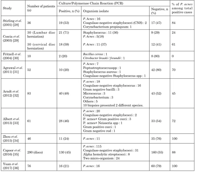
Table 1 presents studies that support the hypothesis that P. acnes has a role in the pathogenesis of Modictype 1 changes and thereby cause low back pain. In the past two decades many authors have identified P. acnes in routine culture of discs. These studies showed that almost 50% of discs cultured were positive for various organism, and in the vast majority of culture-positive discs, P. acnes was the primary organism isolated. Stirling et al. (28) found 16 of 19 culture-positive discsto have isolated P. acnes. Similarly, Agrawal et al. (31) demonstrated 7 P. acnes positive discs out of 10 positive culture results in total. Arndt et al. (32) isolated various organisms out of 40 culture-positive discs but the majority demonstrated P.acnes (18 cases). Albert et al. (33) demonstrated direct correlation between P. acnes and Modic changes where the disc with nucleus with anaerobic bacteria, 80% developed New Modic change in the adjacent to the previous disc herniation. In contrast, none of those with aerobic bacteria and only 44% of patients with negative cultures developed new Modic change. The association between an anaerobic culture and new Modic change was highly statistically significant. In above study isolated anaerobic organism was identified in 22 patients out of which 20 were P. acnes. Similarly, Yuan et al. (36) harvested intervertebral discs from 76 patients with low back pain and / or sciatica without any symptoms of discitis or spon-dylodiscitis, 16 intervertebral discs were found to be positive for P. acnes via 16s rDNA PCR and prevalence was 21.05 % ; among them, 7 samples had visible microbes. They concluded that P. acnes is capable of colonizing some degenerated intervertebral discs without causing discitis.
Capoor et al. (35) conducted a study involving 290 adult pa-tients (mean age 47 ± 13 years) who underwent lumbar microdis-cectomy for symptomatic lumbar disc herniation. The posterior midline approach was used in all cases. The obtained interver-tebral disc samples were then homogenized and used for quan-titative anerobic culture and a sample was frozen and used for quantification of the P. acnes genome by real-time PCR. Bacteria were identified in 130 of 290 disc samples. Threshold was de-fined as ≥ 1 × 10³ CFU / mL for discs with abundant P. acnes. The prevalence of disc samples with abundant P. acnes was 11% (39 cases) and P. acnes was not abundant in the remaining 29% (76 cases) of positive disc samples. The number of P. acnes genomes by PCR in 259 P. acnes-positive discs ranged from 2 to 5831 with median of 260 genomes per 500 ng of total DNA. The authors believed that P. acnes is involved in the pathological process inat least a subset of patients with degenerative disc disease.
In a pilot study conducted by Yuan et al. (37), 76 degenerated intervertebral discs were harvested from patients with low back pain and / or sciatica. After anerobic culture and PCR analysis, 15 intervertebral discs were categorized into the P. acnes-positive group and another 15 discs were selected from the remaining bac-teria-free samples and formed a matched P. acnes-negative group.
IL-8, MIP-1α, MCP-1, IP-10, TNF-α, and neutrophils were much higher in the P. acnes-positive groupthan that in the matched P. acnes-negative group. Furthermore, 7 of 15 P. acnes-positive samples were histologically positive and a subgroup analysis suggested that both histological and PCR-positive samples had the highest concentration of cytokines of IL-8, MIP-1α, TNF-α, and MCP-1 and the greatest number of neutrophils. They con-cluded that latent P. acnes infection was associated with chronic inflammation in degenerated intervertebral discs, especially in samples with visible bacteria on histology, which manifested as increased number of cytokines and neutrophils. Discs with P. acnes infection had extensive severe degeneration and this may be attributed to P. acnes-associated chronic inflammation (37).
STUDIES REFUTING THE ROLE OF
P. ACNES
IN
THE PATHOGENESIS OF DISC DEGENERATION
AND MODIC TYPE 1 CHANGES
Table 2 presents the studies that refute the hypothesis that P. acnes has a role in the pathogenesis of Modictype 1 changes. Ben Galim et al. (38) and Wedderkopp et al. (40) identified only 2 culture-positive cases in their series, andneither of the cases were positive for P. acnes.
Rigal et al. (41) conducted a prospective study in which all the
patients underwentminimallyinvasive video-assisted anterior lumbar fusion or disc prosthesis placement at L4 / L5 and / or L5 / S1 via an anterior retroperitoneal approach to eliminate the risk of contamination on posterior approach. In 385 samples of 313 patients (mean age 47.6 ± 13 months) preoperative MRI demonstrated Modictype 1 changes in 303 cases (78.7%), Modic type 2 in 58 cases (15.1%), and absence of Modic changes in 24 cases (6.2%). ByPfirrmann classification there were 50 cases with grade 2, 211 with grade 3, and 124 with grade 4 changes. Out of 6 patients with a positive culture, 5 patients had Modic 1 changes and 1 patient had type 2 Modic changes. A total of 262 (68%) biopsies were done from the L5 / S1 level and 123 (32%) from the L4 / L5 level. The biopsies were then cultured for 4 weeks with specialized enrichment culture and subjected to his-topathological analysis. All 6 positive samples originated from L5 / S1 disc biopsies. Histological examination of the disc showed non-specific fibrous remodeling of the intervertebral disc with no features of acute inflammatory response, which were similar to that of fibrocartilage tissue. They concluded that the 6 positive samples were related to contamination and emphasized that biological modulators as well as biochemical and genetic mecha-nismswere factors responsible for thedegeneration. The absence of infection atthe 1-year follow-up was an additional argument in favor of no relation between disc degeneration and infection. Table 1 : Studies supporting the role of P. acnes in the pathogenesis of disc degeneration and Modic type 1
Study Number of patients (n)
Culture/Polymerase Chain Reaction (PCR) % of P. acnes among total positive cases Positive, n (%) Organism isolate Negative, n (%)
Stirling et al. (2001) [28] 36 19 (53) P. Acnes : 16 Coagulase-negative staphylococci (CNS) : 2 Corynebacterium propinquum: 1 17 (47) 84 Coscia et al. (2003) [29] 30 (Lumbar disc herniation) 30 (cervical disc herniation) 21 (71) 18 (59) Staphylococcus : 11 (36) P. Acnes : 5(18) P. Acnes : 11 (37) 9 (29) 12 (41) 24 61 Fritzell et al.
(2004) [30] 10 2 (20) Bacillus cereus : 1Citrobacter braaki / freundii : 1 8 (80) 0
Agrawal et al. (2011) [31] 52 10 (20) P. Acnes : 7 Peptostreptococcusspp : 1 Staphylococcus aureus : 1 Coagulase-negative Staphylococcus spp : 1 42 (80) 70 Arndt et al. (2012) [32] 83 40 (48) P. acnes : 18 Coagulase-negative staphylococcus : 16 Gram negative bacilli : 3
Micrococcus : 3 Corynebacterium : 3 Others : 5
10 biopsies presented 2 different species.
43 (52) 45
Albert et al.
(2013) [33] 61 28 (46)
P. acnes : 20
Coagulase-negative staphylococci : 2 P. acnes+ Gram positive cocci : 3 P. acnes+ Neisseria spp : 1 Gram positive cocci : 1 Gram negative rod : 1
33 (54) 72 Zhou et al. (2015) [34] 46 11 (24) P. acnes : 11 35 (76) 100 Capoor et al. (2016) [35] 290 (discs) 130 (45) P. acnes : 115 Coagulase-negative staphylococci : 31 Alpha hemolytic streptococci : 8 Two micro-organism : 24
160 (55) 88
Yuan et al.
EXPERIMENTAL STUDIES DEMONSTRATING THE
ROLE OF
P. ACNES
IN MODIC TYPE 1 CHANGES
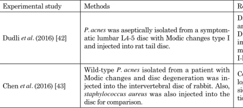
Experimental studies by Dudli et al. (42) and Chen et al. (43) demonstrated that aseptic isolation of P. acnes from the interver-tebral disc of symptomatic patients with Modic type 1 changes and subsequent injection into rat and rabbit intervertebral disc induces Modic type 1 changes in the adjacent bone marrow (Table 3). The intervertebral disc changes were more like discitis if the inoculation was with Staphylococcus aureus, however, chang-es caused by P. acnes would be consideredModic type 1 changes and disc degeneration rather than discitis (43).
STUDIES DEMONSTRATING THE ROLE OF
ANTIBI-OTICS IN LOW BACK PAIN
The role ofantibiotics in low back pain indirectly provides evidence of microbial infestation at the intervertebral disc, followinga course of antibiotic treatment. In 1984 Modic et al. (44)performed MRI of the intervertebral disc in 20 healthy in-dividuals, 8 patients with degenerative lumbar disc disease, 27 with both degenerativedisc disease and herniation, and 5 with disc space infection to demonstrate the diagnostic role of MRI in low back pain. Five patients with suspected disc space infection were found to have markedly increased signal intensity of the entire disc as wellas the adjacent endplates. Follow-up scan after 4 weeks of antibiotic therapy again revealed increased signal intensity in both the bodies and disc, but there was a central area of decreased signal intensity within the disc suggestive of both
healing and degeneration.
Similarly, Albert et al. (45) conducted a pilot study in 2008, where they assessed the clinical effect of antibiotic treatment in a cohort of patients with low back pain and Modic type 1 changes. All the 32 patients with low back pain and Modic changes were treated with amoxicillin-clavulanate (500 mg / 125 mg) 3 × day for 90 days. The authors found clinically important and statis-tically significant improvement in all outcome measures in 29 patients who completed the treatment at the long-term follow-up (mean 10.8 months). They highlighted the need for a randomized controlled trial (RCT) to test the hypothesis that bacterial infec-tion plays a role in low back pain with Modic changes.
In 2013 Albert et al. (22) published the results of theirRCT which aimed to test the efficacy of antibiotic treatment in pa-tients with chronic low back pain ( > 6 months)and Modic type 1 changes. In this double-blind RCT, 162 patients were ran-domized to either 100 days of antibiotic treatment (Bioclavid) or placebo and were blindly evaluated at baseline, end of treatment, and at 1-year follow-up. The 2 groups were similar at baseline and 144 of 162 patients were evaluated at 1-year follow-up. The antibiotic group improvedsignificantly on all outcome measures. At baseline, 100-day follow-up, and 1-year follow-up disease-spe-cific disability using theRoland Morris Disability Questionnaire-score changed as follows : antibiotic 15, 11, 5.7 vs placebo 15, 14, 14. For leg pain : antibiotic 5.3, 3.0, 1.4 vs placebo 4.0, 4.3, 4.3. For lumbar pain : antibiotic 6.7, 5.0, 3.7 vs placebo 6.3, 6.3, 6.3. They concluded that the antibiotic protocol was significantly more effective than placebo in all the primary and secondary outcomes for treatment of patients with chronic low back pain and Modic type 1 changes.
Table 2 : Studies refuting the role of P. acnes in the pathogenesis of disc degeneration and Modic type 1 changes
Study Number of patients (n)
Culture/Polymerase chain reaction (PCR) results % of P. acnes among total positive cases Po s it ive , n
(%) Organism isolate Negative, n (%) Ben-Galim et
al. (2006) [38] 30 2 (7) Coagulase-negative staphylococci : 2 28 (93) 0
Carricajo et al. (2007) [39] 54 2 (4) P. acnes : 2 52 (96) 100 Wedderkopp et al. (2009) [40] 24 2 (8) Staphylococcus epidermidis : 1 Coagulase-negative staphylococci : 1 22 (92) 0 Rigal et al. (2016) [41] 385 samples (313 patients) 6 (1.5) P. acnes : 2 Staphylococcus epidermidis : 2 Citrobacter freundii : 1 Saccharopolyspora hirsute : 1 379 (98.5) 33
Table 3 : Experimental studies demonstrating the role of P. acnes in Modic type 1 changes
Experimental study Methods Results
Dudli et al. (2016) [42] P. acnes was aseptically isolated from a symptom-atic lumbar L4-5 disc with Modic changes type I and injected into rat tail disc.
Day 3 : apparent upregulation of interleukin 1 and interleukin 6.
Day 14 : T-cells and tumor necrosis factor α immunoreactivity were identified at the disc; or marrow junction and MRI showedModic type I-like changes in the adjacent bone marrow.
Chen et al. (2016) [43]
Wild-type P. acnes isolated from a patient with
Modic changes and disc degeneration was in-jected into the intervertebral disc of rabbit. Also,
staphylococcus aureus was also injected into the
disc for comparison.
Compared with staphylococcus aureus, the
patho-logical changes caused by P. acnes would be
con-sidered Modic changes type I and disc degenera-tion rather than discitis.
SUMMARY
Given all the controversy surrounding the role of P. acnes in Modic type 1 changes, many clinical and experimental studies have been conducted to investigate this issue. These studies have clearlydemonstrated the presenceof P. acnes in a subset of patients with low back pain. Identification of P. acnes in culture indicates the infective patho-mechanism in the pathogenesis of Modic type 1 changes, which may be amenable to antibiotic treatment. However, it is still difficult to identify which subset of these patients (patients with low back pain with type 1 Modic change) are infective in nature. Although the use of antibiotics has improved the outcomes significantly, this does not mean that all patients that have low back pain with Modic changes are eligible for long-term antibiotic therapy. Moreover, no studies demonstrates improvement if Modic change in MRI study after antibiotic treatment. Also, further investigations for clear identi-fication of the infective subgroup among low back pain patients in general may be helpful. Thus, more clinical trials are required to arrive at any conclusion.
CONFLICTS OF INTEREST
The authors declare that there are no conflicts of interest.
REFERENCES
1. Chou R, Fu R, Carrino JA, Deyo RA : Imaging strategies for low back pain ; Systemic review and meta-analysis. Lancet 373 : 463-72, 2009
2. Von Korff M : Studying the natural history of back pain. Spine 19 : 2041S-2046S, 1994
3. Krismer M, Van Tulder M : Low back pain group of the bone and joint health strategies for Europe project. Strategies for prevention and management of musculoskeletal conditions. Low Back Pain (non-specific). Best Pract. Res. Clin. Rheu-matol 21 : 77-91, 2007
4. Werber A, Zimmermann-stenzel M, Moradi B, Neubauer E, Schiltenwolf M : Awareness of the German population of common available guidelines of how to cope with lower back pain. Pain Physician 17 : 217-26, 2014
5. Bishop A, Foster NE, Thomas E, Hay EM : How does the self reported clinical management of patients with low back pain relate to the attitudes and beliefs of health care
practi-tioners and physiotherapists. Pain 135 : 187-195, 2008 6. de Roos A, Kressel H, Spritzer C, Dalinka M : MR imaing
of marrow changes adjacent to end plates in degenerative lumbar disc disease. AJR Am J Roentgenol 149(3) : 531-4, 1987. DOI : 10.2214 / ajr.149.3.531
7. Modic MT, Steinberg PM, Ross JS, Masaryk TJ, Carter JR : Degenerative disc disease : assessment of changes in ver-tebral body marrow with MR imaging. Radiology 166(1 Pt 1) : 193-9, 1988. DOI : 10.1148 / radiology.166.1.3336678 8. Modic MT, Masaryk TJ, Ross JS, Carter JR : Imaging of
degenerative disk disease. Radiology 168(1) : 177-86, 1988. DOI : 10.1148 / radiology.168.1.3289089
9. Toyone T, Takahashi K, Kitahara H, Murakami M, Moriya H : Vertebral bone-marrow changes in degenerative lumbar disc disease : an MRI study of 74 patients with low back pain. J Bone Joint surg Br 76(5) : 757-64, 1994. PMID : 8083266 10. Weishaupt D, Zanetti M, Hodler J, Min K, Fuchs B,
PfirrmannCW, Boos N : Painful lumbar disk derange-ment : relevance of endplate abnormalities at MR imaging. Radiology 218(2) : 420-7, 2001. DOI : 10.1148 / radiology.218.2. r01fe15420
11. Kuisma M, Karppinen J, Niinimaki J,Ojala R, Haapea M, Heliovaara M, Korpelainen R, Taimela S, Natri A, Tervonen O : Modic changes in endplates of lumbar vertebral bod-ies : prevalence and association with low back and sciatic pain among middle-ages male workers. Spine 32(10) : 1116-22, 2007. DOI : 10.1097 / 01.brs.0000261561.12944.ff 12. Albert HB, Manniche C : Modic changes following lumbar
disc herniation. Eur Spine J 16(7) : 977-82, 2007. DOI : 10. 1007 / s00586-007-0336-8
13. Modic MT : Modic type 1 and type 2 changes. J Neurosurg Spine 6(2) : 150-1, 2007. DOI : 10.3171 / spi.2007.6.2.150 14. Rahme R, Moussa R : The Modic vertebral endplate and
mar-row changes : pathologic significance and relation to low back pain and segmental instability of the lumbar spine. AJNR Am J Neuroradiol 29(5) : 838-42, 2008. DOI : 10.3174 / ajnr. A0925
15. Karchevsky M, Schweitzer ME, Carrino JA, Zoga A, Montgomery D, Parker L : Reactive endplate marrow changes : a systemic morphologic and epidemiologic evalu-ation : skeletal Radiol 34 : 125-9, 2005
16. Kjaer P, Leboeuf-Yde C, Korsholm L, Sorensen JS, Bendix T : Magnetic resonance imaging and low back pain in adults : a diagnostic imaging study of 40-year-old men and women. Spine 30(10) : 1173-80, 2005. Doi : 10.1097 / 01. brs.0000162396.97739.76
Table 4 : Studies demonstrating the role of antibiotics in low back pain
Study No. of patients Type of antibiotics Duration of treatment Changes observed
Modic et al. (1984) [44] 5 4 weeks Changes in signal intensity ob-served in repeat MRI suggesting healing and degeneration Albert et al.
(Non-ran-domized prospective trial) (2008) [45] 32
Amoxicillin-Clavulanate
(500 mg / 125 mg) Three times a day for 90 days
29 patients reported clinically relevant and statistically signifi-cant improvement on all outcome measures.
Albert et al. (Double-blind
randomized clinical con-trol trial) (2013) [22] 162
Amoxicillin-Clavulanate
(500 mg / 125 mg) Three times a day for 100 days
144 of 162 original patients were evaluated at the 1-year follow-up. The antibiotic group had signifi-cant improvement compared with the placebo group on all outcome measures; improvement contin-ued from 100 days follow-up until the 1-year follow-up.
17. Perry A, Lambert P : Propionibacterium acnes ; infection beyond the skin ; Expert review of Anti-infective therapy 9(12), 1149-56, 2011. DOI : 10.1586 / eri.11.137
18. Zhang Yh, Zhao CQ, Jiang LS, Chen XD, Dai LY : Modic changes : a systemic review of the literature. Eur Spine J 17 : 1289-99, 2008. DOI : 10.1007 / s00586-008-0758-y 19. Brown MF, Hukkanen MV, Mc Carthy ID, Redfem DR,
Batten JJ, Crock HV, Hughes SP, Polak JM : Sensory and symptomatic innervation of the vertebral endplate in pa-tients with degenerative disc disease. J Bone Joint Surg Br 79(1) : 147-53, 1997. PMID : 9020464
20. Burke JG, Watson RW, Mc Cormack D, Dowling FE, Walsh MG, Fitzpatrick JM : Intervertebral disc which cause low back pain secrete high levels of proinflammatory me-diators. J Bone Joint Surg Br 84(2) : 196-201, 2002. doi 10.1302 / 0301-620X.84B2.12511. PMID : 11924650
21. Crock HV : Internal disc disruption : a challenge to disc prolapse fifty years on. Spine 11(6) : 650-3, 1986. PMID : 3787337
22. Albert HB, Sorensen JS, Christensen BS, Manniche C : An-tibiotic treatment in patient with chronic low back pain and vertebral bone edema(Modic type 1 changes) : a double-blind randomized clinical controlled trail of efficacy. Eur Spine J 22(4) : 697-707, 2013. DOI : 10.1007 / s00586-013-2675-y 23. Mitra D, Cassar-Pullicino VN, McCall IW :
Longitudi-nal study of vertebral type-1 end-plate changes on MR of the lumbar Spine. Eur Radiol 14(9) : 1574-81, 2004. DOI : 10.1007 / s00330-004-2314-4
24. Liu PF, Hseih YD, Lin YC,Two A, Shu CW, Huang CM : Pro-pionibacterium acnes in the pathogenesis and immunother-apy of acnes vulgaris. Curr Drug Metab 16(4) : 245-54, 2015. PMID : 26264195
25. Portillo ME, Corvec S, Borens O, Trampuz A : Propionibac-terium acnes : an underestimated pathogen in implant-as-sociated infections. Biomed Res Int 2013 : 804391, 2013. DOI : 10.1155 / 2013 / 804391. PMID : 24308006
26. Chen Z, Cao P, Zhou Z, Yuan Y, Jiao Y, Zheng Y : Over-view : the role of Propionibacterium acnes in non-pyo-genic intervertebral discs. Int Orthop 40 : 1291-8, 2016. DOI : 10.1007 / s00264-016-3115-5
27. Albert Hb, Kjaer P, Jensen TS, Sorensen JS, Bendix T, Manniche C : Modic changes, possible causes and rela-tion to low back pain. Med hypotheses 70(2) : 361-8, 2008. DOI : 10.1016 / j.mehy.2007.05.014
28. Stirling A, Worthington T, Rafiq M, Lambert PA, Elliott TS : Association between sciatica and Propionibacterium acnes. Lancet 357(9273) : 2024-5, 2001. PMID : 11438138 29. Coscia MF, Wack M, Denys G : Low virulence bacterial
infections of intervertebral disc and the resultant spinal disease processes. Abstract from Scoliosis Research Society (SRS) Annual Meeting 2003
30. Fritzell P, Bergstrom T, Welinder-olsson C : Detection of bacterial DNA in painful degenerated spinal discs in patients without signs of clinical infection. Eur Spine J 13(8) : 702-6, 2004. DOI : 10.1007 / s00586-004-0719-z 31. Agarwal V, Golish SR, Alamin TF : Bacteriological
cul-ture of excised intervertebral disc from immunocompe-tent patients undergoing single level primary lumbar microdiscectomy. J Spinal Disord Tech 24(6) : 397-400, 2011. DOI : 10.1097 / BSD.0b013e3182019f3a
32. Arndt J, Charles YP, Koebel C,Bogorin I, Steib JP : Bac-teriology of degenerated lumbar intervertebral disc. J Spi-nal Disord Tech 25(7) : E211-6, 2012. PMID : 22332554 33. Albert HB, Lambert P, Rollason J, Sorensen JS, Worthington
T, Pedersen MB, Norgaard HS, Vernallis A, Busch F,
Manniche C, Elliott T : Does nuclear tissue infected with bacteria following disc herniations lead to Modic changes in the adjacent vertebrae? Eur Spine J 22(4) : 690-6, 2013. DOI : 10.1007 / s00586-013-2674-z
34. Zhou Z, Chen Z, Zheng Y, Cao P, Liang Y, Zhang X, Wu W, Xiao J, Qiu S : Relationship between annular tear and pres-ence of Propionibacterium acnes in lumbar intervertebral disc. Eur Spine J ; 24(11) : 2496-502, 2015. DOI : 10.1007 / s00586-015-4180-y
35. Capoor MN, Ruzicka F, Machackova T, Jancalek R, Smrcka M, Schmitz JE, Hermanova M, Sana J, Michu E, Baird JC, Ahmed FS, Maca K, Lipina R, Alamin TF, Coscia MF, Stonemets JL, Witham T, Ehrlich GD, Gokaslan GD, GokaslanZl, Mavrommatis K, Birkenmajer C, Fischetti VA, Slaby O : Prevalence of Propionibacterium acnes in inter-vertebral discs of patients undergoing lumbar microdiscec-tomy : a prospective cross-sectional study. PLuS One 11(8), 2016. DOI : 10.1371 / journal.pone.0161676
36. Yuan Y, Zhou Z, Jiao Y, Li C, Zheng Y, Lin Y, Xiao J, Chen Z, Cao P : Histological identification of Propionibacterium acnes in non pyogenicdegeneratd intervertebral discs. Biomed Res Int. 2017 : 6192935, 2017. DOI : 10.1155 / 2017 / 6192935 37. Yuan Y, Chen Y, Zhou Z, Jiao Y, Li C, Zheng Y, Lin
Y, Xiao J, Chen Z, Cao P : Association between chron-ic inflammation and latent infection of Propionibacte-rium acnes in non-pyogenic degenerated intervertebral discs ; a pilot study. Eur Spine J. 27 (10) : 2506-17, 2018. DOI : 10.1007 / s00586-017-5363-5
38. Ben-Galim P, Rand N, Giladi M, Schwartz D, Ashkenazi E, Millgram M, Dekel S, Floman Y : Association between sciatica and microbial infection : true infection or culture contamination? Spine (Phila Pa 1976) 31(21) : 2507-9, 2006 39. Carricajo A, Nuti C, Aubert E, Hatem O, Fonsale N, Mallaval
FO, Vautrin AC, Brunon J, Aubert G : Propionibacterium acnes contamination in lumbar disc surgery. J Hosp Infect 66(3) : 275-7, 2007. PMID : 17573158
40. Wedderkopp N, Thomsen K, manniche C,Kolmos HJ, Secher Jensen T, LeboeufYde C : No evidence for presence of bacte-ria in Modic type 1 changes. Acta Radiol 50(1) : 65-70, 2009. DOI : 10.1080 / 02841850802524485
41. Rigal J, Thelen T, Byrne F, Cogniet A, Boissiere L, Aunoble S, Le Huec JC : Prospective study using anterior approach did not show association between Modic 1 changes and low grade infection in lumbar spine. Eur Spine J 25(4) : 1000-5, 2016. DOI : 10.1007 / s00586-016-4396-5
42. Dudli S, Liebenberg E, Magnitsky S, Miller S, Demir-Deviren S, Lotz JC : Propionibacterium acnes infected interverte-bral discs causes verteinterverte-bral bone marrow lesions consistent with Modic changes. J Orthop Res 34(8) : 1447-55, 2016. DOI : 10.1002 / jor.23265
43. Chen Z, Zheng Y, Yuan Y, Jiao Y, Xiao J, Zhou Z, Cao P : Modic changes and disc degeneration caused by inocula-tion of Propionibacterium acnes inside intervertebral disc of rabbit : a pilot study. Biomed Res Int 2016 : 9612437, 2016. DOI : 10.1155 / 2016 / 9612437
44. Modic MT, Pavlicek W, Weinstein MA, Boumphrey F, Ngo F, Hardy R, Duchesneau PM : Magnetic resonance imaging of intervertebral disk disease. Clinical and pulse sequence consideration. Radiology 152(1) : 103-11, 1984. DOI : 10.1148 / radiology.152.1.6729099
45. Albert HB, Manniche C, Sorensen JS, Deleuran BW : An-tibiotic treatment in patients with low back pain associated with Modic changes type 1 (bone edema) : a pilot study. Br J Sports Med 42(12) : 969-73, 2008. DOI : 10.1136 / bjsm.2008 .050369