Angiotensin II regulates islet
microcirculation and insulin secretion in mice
著者
庵谷 千恵子
著者(英)
Ihoriya Chieko
学位名
博士(医学)
学位授与機関
川崎医科大学
学位授与年度
平成25年度
学位授与年月日
2014-03-13
学位授与番号
35303甲第605号
URL
http://doi.org/10.15111/00000012
Title Page
Angiotensin II Regulates Islet Microcirculation and Insulin Secretion in Mice
CHIEKO IHORIYA*, MINORU SATOH*, ATSUNORI KUWABARA*, TAMAKI SASAKI* AND NAOKI KASHIHARA*
*Department of Nephrology and Hypertension, Kawasaki Medical School, Kurashiki, Okayama, Japan
Short running title: Effect of Ang II on islet microcirculation
Sources of support: This work was supported in part by a grant from Novartis Pharmaceuticals
Corporation (Basel, Switzerland) to N. Kashihara. This work was also supported in part by the Ministry of Health, Labour and Welfare of Japan. M. Satoh and N. Kashihara are recipients of a Grant-in-Aid for Scientific Research from the Japan Society for the Promotion of Science.
Address for correspondence:
Minoru Satoh, MD, PhD
Department of Nephrology and Hypertension, Kawasaki Medical School, Kurashiki, Okayama 701-0192, Japan
Tel: +81-86-462-1111 Fax: +81-86-462-1199
ABSTRACT
Objective: Angiotensin II causes potent increases in systemic and local pressure through its
vasoconstrictive effect. Despite the importance of angiotensin II for local blood flow regulation, whether angiotensin II regulates the pancreatic islet microcirculation remains incompletely understood. We hypothesized that angiotensin II directly regulates the pancreatic islet microcirculation and thereby regulates insulin secretion. The aims of this study were to develop a new technique to visualize pancreatic islet hemodynamic changes in vivo and to analyze changes in islet circulation induced by angiotensin II or an angiotensin type 1 receptor blocker.
Methods: Using an in vivo imaging method, we observed the pancreatic islet microcirculation.
Various doses of angiotensin II or an angiotensin type 1 receptor blocker were injected intravenously, and changes in islet microcirculation were observed. Glucose-stimulated insulin secretion from the pancreas was measured from the hepatic portal vein.
Results: We identified islet microcirculation by using a fluorescent dye. Angiotensin II significantly
induced blood vessel contraction in the islets in a dose-dependent manner. In contrast, the angiotensin type 1 receptor blocker induced vasodilation. Glucose-stimulated insulin secretion was decreased by angiotensin II infusion.
Conclusions: These results show that angiotensin II is involved in the regulation of pancreatic islet
microcirculation and insulin secretion.
Key words: Angiotensin II, Angiotensin II type 1 receptor blocker, Islet, Insulin secretion, In vivo
imaging, Microcirculation
Abbreviations used: ARB, angiontensin type 1 receptor blocker; DsRed, Discosoma sp. red
INTRODUCTION
The progression of hypertension is a strong predictor of new-onset type 2 diabetes [14]. Hypertension and diabetes share a common basis and therefore frequently develop concomitantly. Relative to non-hypertensive patients, hypertensive patients show an approximately 3.3-fold increased risk of new-onset diabetes [8]. Hypertension has been shown to cause microvascular damage in the islets as a vascular complication [16]. Some clinical studies have shown that renin-angiotensin inhibitors delay new-onset diabetes independently of the degree of their antihypertensive effect [11].
Angiotensin II is the central product of the renin-angiotensin system and is well known to cause potent increases in systemic and local blood pressure through its vasoconstrictive effect. Thus, angiotensin II decreases blood flow by vasoconstriction and produces ischemic changes in organs [5, 13], including the pancreas [4]. Islet blood flow is important for supplying nutrients and oxygen to islet cells. The regulatory mechanisms that control islet blood flow are complex and include neural,
hormonal, and local mechanisms that are independent of the surrounding exocrine pancreas [3, 22]. Despite the importance of angiotensin II for local blood flow regulation, whether angiotensin II regulates pancreatic islet blood flow remains incompletely understood.
We hypothesized that angiotensin II directly regulates pancreatic islet microcirculation and thus regulates insulin secretion. We have established an in vivo method to visualize the
microcirculation by 2-photon laser microscopy [25, 31]. Using this technique, we developed a novel technique to visualize pancreatic islet microcirculation in vivo. We also evaluated the effect of angiotensin II and an angiotensin type 1 receptor blocker (ARB) on islet microcirculation and insulin secretion.
MATERIALS AND METHODS
Materials
Angiotensin II (A9525) was obtained from Sigma-Aldrich (St Louis, MO, USA). The angiotensin II receptor blocker, valsartan, was purchased from Novartis Pharmaceuticals Corporation (Basel, Switzerland). We obtained 500 kilodalton (kDa) fluorescein-dextran (anionic; excitation, 494 nm; emission, 518 nm) from Invitrogen, Japan (Tokyo, Japan), and stored it at 2 mg mL-1 in
phosphate-buffered saline (pH 7.4). The enzyme-linked immunosorbent assay kit for insulin was from the Morinaga Institute of Biological Science (Yokohama, Japan). The antibody against mouse insulin (#3014) was purchased from Cell Signaling (Danvers, MA, USA). Tetramethyl rhodamine
isothiocyanate-conjugated wheat germ agglutinin (WGA) was purchased from Vector Laboratories (Burlingame, CA, USA). The FITC-labeled anti-rabbit antibody was purchased from Dako Japan (Tokyo, Japan). Antibodies against mouse Akt (#9272) and phospho-Akt (Ser473; #9271) were purchased from Cell Signaling.
Animals
The animal protocol for this experiment was approved by the Animal Care and Use Committee of Kawasaki Medical School (no. 12-048, 2012). Male STOCK Tg(Ins1-DsRed*T4)32Hara/J (Ins1-DsRed) mice [15], which harbor a transgenic construct containing the Discosoma sp. red fluorescent protein (DsRed) gene under the control of the mouse insulin 1 promoter, were purchased from The Jackson Laboratory (Bar Harbor, ME, USA). They were housed in a temperature- and humidity-controlled room with a 12-h light-dark cycle, fed standard laboratory animal chow, and provided free access to tap water.
Male Ins1-DsRed mice were used. A polyethylene catheter (PE50; Nihon Becton Dickinson, Tokyo, Japan) was inserted into the carotid artery under sevoflurane-induced anesthesia to monitor systolic blood pressure (SBP) by using a pressure transducer (RMP-6000; Nihon Kohden, Tokyo, Japan). Next, a catheter was inserted into the left external jugular vein for infusion of angiotensin II (25, 50, and 100 g kg-1
; bolus injection; n = 5) and/or valsartan (20 or 40 mg kg-1; bolus injection; n = 5). A midline incision of the abdomen was then made, and the pancreas was exteriorized. During all procedures and imaging, the core body temperature of the mice was maintained at 37 °C using a homeothermic table. A 0.5-mL volume of 500 kDa fluorescein-dextran solution was infused through the jugular venous catheter immediately before microscopic imaging. Next, for analysis of the change in diameter of islet arterioles, 0.5 mL of angiotensin II and/or valsartan was infused. The diameter was measured at 3 min after infusion. After the pancreatic islet microcirculation was recorded for 30 min, another dose was infused. The diameter of islet arterioles was measured using NIH Image/Image J software [30].
Multiphoton-excitation Laser-scanning Fluorescence Microscopy
The multiphoton microscope used in these studies was a Leica TCS SP2 AOBS MP confocal microscope system (Leica Microsystems Japan, Tokyo, Japan) with the following components: a Leica DM IRE2 inverted microscope powered by a wide-band, fully automated, infrared (710–920 nm) combined photo-diode pump laser and mode-locked titanium-sapphire laser (Mai-Tai,
Spectra-Physics, Mountain View, CA, USA) [25, 31]. Images were collected in time (xyt) series (0.5– 5 Hz) using Leica Confocal Software (LCS 2.61.1537) and analyzed using LCS 3D, Process and Quantify packages. The excitation laser power applied to the sample was attenuated to 2–28 mW by using neutral-density filters.
In Situ Pancreatic Perfusion
experimental procedures were essentially performed as described by Vikman et al [33]. The mice were kept on a heating pad during the entire experiment. After the abdominal cavity was opened and the renal, hepatic, and splenic arteries were ligated, the aorta was tied off above the level of the pancreatic artery. The pancreas was perfused with Hanks' balanced salt solution, pre-heated at 37°C and
containing the indicated glucose concentration, via a silicone catheter placed in the aorta (1 mL min-1). The perfusate was collected through a silicone catheter in the portal vein at 120-s intervals, as indicated, in 1.5-mL microtubes containing a proteinase inhibitor. Insulin concentration in the effluent medium was determined by insulin enzyme-linked immunosorbent assay kit (Morinaga, Yokohama, Japan). There were 3 groups: the vehicle control group, angiotensin II group, and valsartan group (n = 4 in each). Angiotensin II (100 µg kg-1) was infused 10 min before the perfusate was collected. Valsartan (40 mg kg-1) was administered by gavage 30 min before the procedure.
Chronic Experimental Protocol
Male C57BL/6 mice (6 weeks old) were assigned to 1 of the 3 groups: the control group (vehicle infusion, n = 8), the Ang II group (angiotensin II, 1.0 g kg-1
min-1, n = 8), or the Val group (valsartan, 40 mg kg-1 per day, by gavage, n = 8). Angiotensin II or vehicle was infused using an osmotic mini-pump (Model 1004; Muromachi Kikai, Japan) [32]. At 2 weeks after treatment, SBP, oral glucose tolerance, and insulin tolerance were measured. The mice were then killed under anesthesia induced by sevoflurane inhalation and blood samples were immediately obtained. The pancreas was cross-sectioned and the sections were fixed in 4% paraformaldehyde.
Physiological and Biochemical Measurements
The SBP was measured by the tail-cuff method (BP-98A; Softron, Tokyo, Japan). At the time of sacrifice, blood samples were obtained from the right atrium, and serum creatinine, blood urea nitrogen, and fasting serum glucose levels were measured.
Oral Glucose Tolerance Test and Insulin Tolerance Test
The oral glucose tolerance test was performed after 16 h of overnight fasting. Glucose (1.5 g kg-1) was administered orally, and a small amount of blood was collected from the tail vein at 0, 30, 60, and 120 min. The AUC of the blood glucose was calculated for each animal. For the insulin tolerance test, mice were given an intraperitoneal injection of 0.75 U kg-1 insulin solution after 2 h of fasting. The blood glucose level was determined by the glucose dehydrogenase pyrroloquinoline quinone method (Medisafe-Mini; Terumo, Tokyo, Japan). The glucose decay constant (KITT) was calculated using the
formula 0.693/(T1/2), where the T1/2 of plasma glucose was determined from the glucose curve during
its phase of linear decay (0–30 min) [9].
For measurement of phosphorylated Akt in skeletal muscle, 6-h fasted mice were
administered 0.2 U of insulin and killed by anesthesia induced by sevoflurane inhalation after 10 min, and the quadriceps muscle was excised and snap-frozen. Protein samples (50 μg per lane) were subjected to immunoblotting analysis with antibodies against phospho-Akt (Ser473) (Cell Signaling) and Akt (Cell Signaling). Signals were detected using an enhanced chemiluminescence system (GE Healthcare Japan, Tokyo, Japan).
Histological Examination and Immunohistochemistry
The pancreas was cut into 4-m sections and subjected to Azan staining. Histological changes of islets were evaluated by light microscopy. Paraffin-embedded tissue was cut into 4-m sections that were then deparaffinized and hydrated. The sections were incubated in goat serum for 1 h at room temperature. The primary antibody (dilution, 1:200 in phosphate-buffered saline) was added and
incubated overnight at 4 °C. After the slides were washed in phosphate buffered saline, they were incubated in a goat anti-rabbit FITC-conjugated secondary antibody and tetramethyl rhodamine isothiocyanate-conjugated WGA (dilution: 1:1000) for 1 h at room temperature.
Statistical Analysis
Values are expressed as the mean ± SEM. All parameters were evaluated using two-tailed unpaired Student’s t-test or one-way analysis of variance for comparison of multiple means. A p value of <0.05 was considered to represent a significant difference.
RESULTS
Effect of Angiotensin II and ARB on Pancreatic Islet Blood Flow
Islet vasculature was visualized using 500 kDa FITC-labeled dextran under a confocal microscope. The islet was identified by local existence of beta cells that expressed DsRed under the control of the mouse-insulin 1 promoter (Figure 1A, 1B). We confirmed that some arterioles flowed into and perfused the islets.
We then evaluated the reaction of the islet vasculature to angiotensin II infusion (25, 50, 100 µg kg-1). SBP was increased by angiotensin II in a dose-dependent manner (Figure 1C). After angiotensin II infusion (100 µg kg-1), the islet vasculature gradually constricted, and blood flow decreased with vasoconstriction (Supporting Movie 1). Angiotensin II significantly induced the contraction of blood vessels in the islets in a dose-dependent manner (% change of vascular diameter: 8.2% ± 1.9% by 25 µg kg-1, 19.5% ± 1.9% by 50 µg kg-1, and 20.3% ± 2.4% by 100 µg kg-1; Figure. 1d). Finally, SBP and the diameter of the islet arterioles returned to the basal levels by valsartan administration (Figure 1C, 1D).
We also examined the reaction of the islet vasculature to valsartan (20 and 40 mg kg-1). The islet microvasculature was dilated at 30 min after infusion of 40 mg kg-1 valsartan (Figure 2B),
compared to vehicle infusion (Figure 2A). SBP was decreased by valsartan in a dose-dependent manner (Figure 2C). Valsartan induced vasodilation in mice under normal conditions (% change of vascular diameter: 16.5% ± 5.2% by 20 µg kg-1 and 25.0% ± 7.1% by 40 µg kg-1; Figure 2D).
Effect of Angiotensin II and ARB on Pancreatic Insulin Secretion
To evaluate the effect of angiotensin II and ARB on insulin release dynamics, we performed in situ pancreatic perfusion experiments (Figure 3). When the glucose concentration was increased from 5.5 mM to 16.7 mM in vehicle-treated pancreata, insulin release increased in a biphasic manner consisting
of a first increase followed by a nadir and a second increase (Figure 3A). In angiotensin II-infused pancreata, high-glucose-stimulated insulin secretion was decreased in both the first and second phase (Figure 3B). Pretreatment with valsartan increased insulin release in the first phase of the response to high glucose (Figure 3C). The first phase lasted 7.5 ± 0.9 min in vehicle-treated mice and 6.5 ± 0.5 min in angiotensin II-pre-infused mice. In the valsartan pre-treated mice, the first phase was significantly extended to 10.5 ± 0.5 min (p < 0.05 vs. vehicle group). A comparable reduction of total insulin release as measured by the AUC was observed in the Angiotensin II-treated group. In contrast, valsartan treatment did not affect total insulin secretion (Figure 3D. Ang II, 26.1% ± 3.5%; Val, 109.2% ± 28.4%).
Effect of Chronic Angiotensin II and ARB on Islet Morphology and Glucose Tolerance
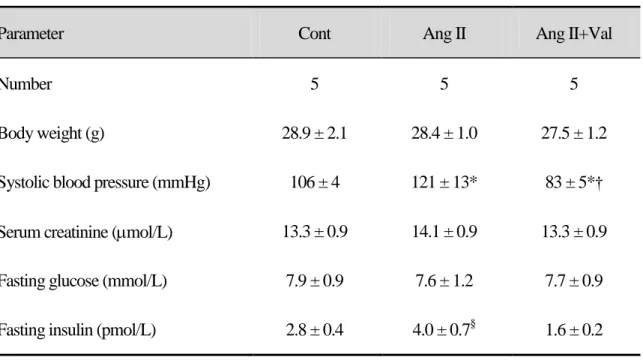
Angiotensin II increased the SBP at 14 days. Valsartan treatment reduced SBP to the level in the control group (Table 1). There was no difference in body weight or serum creatinine levels among the 3 groups. Fasting glucose levels also did not differ among the 3 groups. The Ang II group showed a tendency toward increased insulin levels; however, this increase was not significant (Table 1).
Azan staining showed the absence of fibrotic changes and inflammatory cell infiltration in the 3 groups (Figure 4A upper). We evaluated the insulin content and endothelial volume in islets in detail (Figure 4A lower). The area of positive insulin staining did not differ among the 3 groups (Figure 4A, B. Control, 63.5% ± 1.6%; Ang II, 57.5% ± 1.8%; and Ang II+Val, 58.1% ± 1.2%), but the endothelium volume as detected by WGA-lectin staining was significantly lower in the Ang II group than in the control group (Figure 4A, C. Control, 16.8% ± 1.6%, Ang II, 8.0% ± 0.6%; p < 0.05). Valsartan treatment prevented the decrease in endothelium volume induced by angiotensin II (Figure 4A, C. Ang II+Val, 12.5% ± 0.9%, p < 0.05 vs. Ang II).
Finally, we investigated oral glucose tolerance and insulin tolerance. The oral glucose tolerance test revealed that the Ang II group showed an exacerbation of hyperglycemia after glucose
administration that was normalized in the ARB Val group (Figure 5A, B). However, the insulin tolerance test (Figure 5C, D) and the evaluation of insulin-stimulated phosphorylation of Akt in skeletal muscle (Figure 5E) did not show a significant difference among the groups. After insulin injection, Akt-Ser473 phosphorylation increased in all groups, but there was no significant difference.
DISCUSSION
The aims of this study were to visualize mouse islet microcirculation by using an in vivo imaging method and to evaluate a potential association of the renin-angiotensin system with the regulation of islet blood vasoconstriction and insulin secretion. In the present study, we showed that angiotensin II significantly induced blood vessel contraction in the islets and that an angiotensin type 1 receptor blocker induced vasodilation in mice under normal conditions. Angiotensin II directly decreased insulin secretion from the pancreas and the angiotensin II receptor blocker increased the initial insulin secretion in response to high glucose stimulation. However, in the normal condition, chronic angiotensin II infusion did not induce glucose intolerance or insulin resistance.
We previously developed an in vivo imaging method to visualize and analyze the microcirculation of islets in a living mouse. The pancreatic islets are among the most vascularized organs of the body [10, 28]. Using 2-photon laser microscopy, we established an in vivo imaging method to visualize the renal microcirculation [31]. We attempted to visualize mouse islet
microcirculation using this in vivo imaging method. We confirmed that pancreatic islets have a dense network of sinusoidal capillaries with a morphology resembling that of the glomerulus.
We observed some inflow from blood vessels into the islets. There are 3 models of islet perfusion [2]. The central concept of the first model is that secretory products of non-beta cells affect insulin secretion by beta cells. The second model describes a pattern of microcirculation in which beta cells are perfused first and insulin released into the circulation by these cells affects the secretion of non-beta cells. The third model describes a feeding arteriole that enters 1 side of the islet and
immediately branches into capillaries to perfuse endocrine cells of all types. In this study, however, we could not identify the mechanism underlying islet perfusion in detail because time resolution of the 2-photon laser microscope was too low to permit observation of the movement of red blood cells. Further examinations are required concerning the measurement of islet blood flow by using another type of microscope that can measure islet blood flow.
We measured microvessel diameter in the islets to assess changes of islet microcirculation. Previous studies that examined islet blood flow used microsphere methods [17, 19, 20]. The
microsphere method is an important technique in cardiovascular research for the estimation of blood flow distribution within organs. Regarding pancreatic flow changes in response to angiotensin II and ARB, our findings agree with those of previous studies that used the microsphere method [4]. Thus, the angiotensin system plays a pivotal role in islet blood perfusion to cause adequate insulin release by controlling vasoconstriction.
Two possibilities exist for the mechanism underlying impaired glucose-induced insulin release with angiotensin II. The first mechanism is the disturbance of (pro)insulin biosynthesis regulation by angiotensin II [26], as angiotensin II type 1 receptor receptors are expressed in beta cells and participate in insulin biosynthesis. The second mechanism involves a defect in islet blood flow [4]. We showed that angiotensin II induces the constriction of islet arterioles. The existence of the
pancreatic tissue renin-angiotensin system has now been confirmed in various species, including mice, rats, dogs, and humans [29]. Expression of angiotensin II type 1 receptors has been identified in islet arterioles, microvessels, and beta cells [6, 7, 27]. In this study, we considered that angiotensin II directly affected the islet arterioles through the contraction of vascular smooth muscle cells in the islet. This
contraction would have decreased blood flow and thus decreased the secretion of insulin.
We showed that ARB induced vasodilation in mice under normal conditions, which may have resulted in increased islet blood flow and increased insulin secretion. However, ARB only increased the first phase of insulin secretion in response to glucose stimulation in our study. A previous study indicated that pancreatic islet blood flow was significantly and preferentially enhanced after the administration of an angiotensin-converting enzyme inhibitor or ARB [4, 18]. However, regarding insulin release caused by an renin-angiotensin system inhibitor, both increase [18] and no change [4] have been reported. A previous study showed that in transplanted islets, ARB markedly increases blood perfusion and the first phase of glucose-stimulated insulin secretion [24]. Reasons for these discrepant results may include differences between angiotensin-converting enzyme inhibitors and angiotensin II type 1 receptor blockers, the timing of drug administration, or a difference in animal species. We used normal mice, but use of a disease model such as mouse models of type 1 or type 2 diabetes would have been helpful.
We have observed that chronic angiotensin II infusion induced glucose intolerance in normal mice in vivo. Juan et al. reported that angiotensin II pretreatment leads to increased insulin-stimulated glucose transport in isolated rat adipocytes, and administration of angiotensin II increases insulin sensitivity in rats [23]. In contrast, other studies have suggested that angiotensin II may negatively modulate the effect of insulin by regulating multiple levels of the insulin-signaling cascade [12]. Our data indicate that angiotensin II reduced endothelial cell defects in islets but had no effect on peripheral insulin sensitivity in normal mice. Some studies have shown that defective in vivo insulin secretion is caused by islet vascular abnormalities that result in decreased islet blood flow without
impairment of beta cell function [1, 21]. Much more long-term infusion of angiotensin II may result in insulin resistance in peripheral tissues.
In conclusion, we visualized mouse islet microcirculation in vivo for the first time. We also identified an association of the renin-angiotensin system with the regulation of islet blood constriction and insulin secretion. Angiotensin II has a marked vasoconstrictive effect on islet vasculature.
Decreased islet blood flow could be important for impairments of insulin release. In contrast, ARB vasodilates islet arterioles and increases insulin release rapidly in response to high glucose levels. This process may be involved in new-onset diabetes and progressive islet damage in hypertensive patients.
PERSPECTIVE
The development of a murine model to examine the islet microcirculation provide investigators with a new technique to study islet pathology and analyze potential treatments. We confirmed that the renin-angiotensin system is involved in not only islet microcirculation but also insulin secretion. This study shed light on the pathophysiology of diabetes in hypertensive patients.
ACKNOWLEDGMENTS
All authors sincerely thank E. Yorimasa, M. Ishihara, and M. Yokohata at Kawasaki Medical School for their excellent secretarial assistance. This work was supported in part by a grant from Novartis Pharmaceuticals Corporation (Basel, Switzerland) to N. Kashihara. This work was also supported in part by the Ministry of Health, Labour and Welfare of Japan. M. Satoh and N. Kashihara are recipients of a Grant-in-Aid for Scientific Research from the Japan Society for the Promotion of Science. The sponsor had no role in the design or conduct of the study.
REFERENCES
1. Anneren C, Welsh M, Jansson L. Glucose intolerance and reduced islet blood flow in transgenic mice expressing the FRK tyrosine kinase under the control of the rat insulin promoter. Am J Physiol Endocrinol Metab 292: E1183-1190, 2007.
2. Ballian N, Brunicardi FC. Islet vasculature as a regulator of endocrine pancreas function.
World J Surg 31: 705-714, 2007.
3. Bonner-Weir S, Orci L. New perspectives on the microvasculature of the islets of Langerhans in the rat. Diabetes 31: 883-889, 1982.
4. Carlsson PO, Berne C, Jansson L. Angiotensin II and the endocrine pancreas: effects on islet blood flow and insulin secretion in rats. Diabetologia 41: 127-133, 1998.
5. Chai W, Wang W, Liu J, Barrett EJ, Carey RM, Cao W, Liu Z. Angiotensin II type 1 and type 2 receptors regulate basal skeletal muscle microvascular volume and glucose use. Hypertension 55: 523-530, 2010.
6. Chappell MC, Millsted A, Diz DI, Brosnihan KB, Ferrario CM. Evidence for an intrinsic angiotensin system in the canine pancreas. J Hypertens 9: 751-759, 1991.
7. Chu KY, Lau T, Carlsson PO, Leung PS. Angiotensin II type 1 receptor blockade improves beta-cell function and glucose tolerance in a mouse model of type 2 diabetes. Diabetes 55: 367-374, 2006.
8. Conen D, Ridker PM, Mora S, Buring JE, Glynn RJ. Blood pressure and risk of developing type 2 diabetes mellitus: the Women's Health Study. Eur Heart J 28: 2937-2943, 2007.
9. Durham HA, Truett GE. Development of insulin resistance and hyperphagia in Zucker fatty rats. Am J Physiol Regul Integr Comp Physiol 290: R652-658, 2006.
10. Eberhard D, Kragl M, Lammert E. 'Giving and taking': endothelial and beta-cells in the islets of Langerhans. Trends Endocrinol Metab 21: 457-463, 2010.
11. Elliott WJ, Meyer PM. Incident diabetes in clinical trials of antihypertensive drugs: a network meta-analysis. Lancet 369: 201-207, 2007.
12. Folli F, Kahn CR, Hansen H, Bouchie JL, Feener EP. Angiotensin II inhibits insulin signaling in aortic smooth muscle cells at multiple levels. A potential role for serine phosphorylation in insulin/angiotensin II crosstalk. J Clin Invest 100: 2158-2169, 1997.
13. Fujimoto S, Satoh M, Nagasu H, Horike H, Sasaki T, Kashihara N. Azelnidipine exerts renoprotective effects by improvement of renal microcirculation in angiotensin II infusion rats.
Nephrol Dial Transplant 24: 3651-3658, 2009.
14. Gress TW, Nieto FJ, Shahar E, Wofford MR, Brancati FL. Hypertension and antihypertensive therapy as risk factors for type 2 diabetes mellitus. Atherosclerosis Risk in Communities Study.
N Engl J Med 342: 905-912, 2000.
15. Hara M, Dizon RF, Glick BS, Lee CS, Kaestner KH, Piston DW, Bindokas VP. Imaging pancreatic beta-cells in the intact pancreas. Am J Physiol Endocrinol Metab 290: E1041-1047, 2006.
16. Hayden MR, Sowers JR. Treating hypertension while protecting the vulnerable islet in the cardiometabolic syndrome. J Am Soc Hypertens 2: 239-266, 2008.
captopril, irbesartan and pravastatin, and suppressed by palmitate. Biochem Biophys Res
Commun 346: 26-32, 2006.
18. Huang Z, Jansson L, Sjoholm A. Vasoactive drugs enhance pancreatic islet blood flow, augment insulin secretion and improve glucose tolerance in female rats. Clin Sci (Lond) 112: 69-76, 2007.
19. Iwase M, Tashiro K, Uchizono Y, Goto D, Yoshinari M. Pancreatic islet blood flow in conscious rats during hyperglycemia and hypoglycemia. Am J Physiol Regul Integr Comp
Physiol 280: R1601-1605, 2001.
20. Iwase M, Uchizono Y, Tashiro K, Goto D, Iida M. Islet hyperperfusion during prediabetic phase in OLETF rats, a model of type 2 diabetes. Diabetes 51: 2530-2535, 2002.
21. Iwashita N, Uchida T, Choi JB, Azuma K, Ogihara T, Ferrara N, Gerber H, Kawamori R, Inoue M, Watada H. Impaired insulin secretion in vivo but enhanced insulin secretion from isolated islets in pancreatic beta cell-specific vascular endothelial growth factor-A knock-out mice. Diabetologia 50: 380-389, 2007.
22. Jansson L. The regulation of pancreatic islet blood flow. Diabetes Metab Rev 10: 407-416, 1994.
23. Juan CC, Chien Y, Wu LY, Yang WM, Chang CL, Lai YH, Ho PH, Kwok CF, Ho LT. Angiotensin II enhances insulin sensitivity in vitro and in vivo. Endocrinology 146: 2246-2254, 2005.
24. Kampf C, Lau T, Olsson R, Leung PS, Carlsson PO. Angiotensin II type 1 receptor inhibition markedly improves the blood perfusion, oxygen tension and first phase of glucose-stimulated
insulin secretion in revascularised syngeneic mouse islet grafts. Diabetologia 48: 1159-1167, 2005.
25. Kuwabara A, Satoh M, Tomita N, Sasaki T, Kashihara N. Deterioration of glomerular endothelial surface layer induced by oxidative stress is implicated in altered permeability of macromolecules in Zucker fatty rats. Diabetologia 53: 2056-2065, 2010.
26. Lau T, Carlsson PO, Leung PS. Evidence for a local angiotensin-generating system and dose-dependent inhibition of glucose-stimulated insulin release by angiotensin II in isolated pancreatic islets. Diabetologia 47: 240-248, 2004.
27. Leung PS, Chan HC, Fu LX, Wong PY. Localization of angiotensin II receptor subtypes AT1 and AT2 in the pancreas of rodents. J Endocrinol 153: 269-274, 1997.
28. Olsson R, Carlsson PO. The pancreatic islet endothelial cell: emerging roles in islet function and disease. Int J Biochem Cell Biol 38: 710-714, 2006.
29. Paul M, Poyan Mehr A, Kreutz R. Physiology of local renin-angiotensin systems. Physiol Rev 86: 747-803, 2006.
30. Rasband WS. ImageJ, U. S. National Institutes of Health, Bethesda, Maryland, USA, http://rsb.info.nih.gov/ij/, 1997-2012.
31. Satoh M, Kobayashi S, Kuwabara A, Tomita N, Sasaki T, Kashihara N. In vivo visualization of glomerular microcirculation and hyperfiltration in streptozotocin-induced diabetic rats.
Microcirculation 17: 103-112, 2010.
32. Satoh M, Ogita H, Takeshita K, Mukai Y, Kwiatkowski DJ, Liao JK. Requirement of Rac1 in the development of cardiac hypertrophy. Proc Natl Acad Sci U S A 103: 7432-7437, 2006.
33. Vikman J, Jimenez-Feltstrom J, Nyman P, Thelin J, Eliasson L. Insulin secretion is highly sensitive to desorption of plasma membrane cholesterol. FASEB J 23: 58-67, 2009.
Figure legends
Figure 1. Effect of angiotensin II on pancreatic islet microcirculation. (A, B) Representative islet
microvascular morphology before (A) and at 3 min after (B) angiotensin II (100 µg kg-1) infusion. Green, 500 kDa FITC-labeled dextran. Red, Ds-Red. Scale bar, 80 µm. (C, D) Changes of SBP (c) and percent (%) change of vascular diameter (D) after angiotensin II (25, 50, 100 µg kg-1) and valsartan (Val; 40 mg kg-1) administration. Values represent mean ± SEM. *p < 0.05 vs. vehicle.
Figure 2. Effect of valsartan on pancreatic islet vascular diameter. (A, B) Representative islet
microvascular morphology before (A) and at 30 min after (B) valsartan (40 mg kg-1) administration. Green, 500 kDa FITC-labeled dextran. Red, Ds-Red. Scale bar, 80 µm. (C, D) Changes of SBP (C) and percent (%) change of vascular diameter (D) after valsartan (20 and 40 mg kg-1) administration. Values represent mean ± SEM. *p < 0.05 vs. vehicle.
Figure 3. Relative insulin secretion by in situ pancreatic islet perfusion. (A–C) High-glucose-induced
insulin secretion after pretreatment with vehicle (A), angiotensin II (Ang II) (B), or valsartan (Val) (C). Values represent mean ± SEM of 4–6 experiments. (D) Comparison of AUC among groups. Values represent mean ± SEM. *p < 0.05 vs. vehicle.
Figure 4. Histological changes of islets upon chronic angiotensin II infusion. (A) Upper, Azan staining.
Lower, double immunohistochemical staining for insulin (Green) and endothelium (Red). Cont, vehicle-treated mice. Ang II, angiotensin II-treated mice. Ang II +Val, Ang II+valsartan-treated mice. Scale bar, 80 µm. (B) Percentage of insulin-positive area in islet. (C) Percentage of endothelium volume in islet. Values represent mean ± SEM. *p < 0.05 vs. Cont; †p < 0.05 vs. Ang II.
Figure 5. Changes in glucose tolerance upon chronic angiotensin II infusion. (A) Oral glucose
tolerance test in mice treated with vehicle (Cont), angiotensin II (Ang II), and Ang II+valsartan (Val). n = 6–8 in each group. Values represent mean ± SEM. (B) Comparison of the AUC for glucose among groups. Values represent the mean ± SEM. (C) Insulin tolerance in mice from Cont, Ang II, and Ang II+Val groups; n = 6–8 in each group. Values represent mean ± SEM. *p < 0.05 vs. Cont; †p < 0.05 vs. Ang II. (D) Comparison of glucose decay rate (KITT). Values represent mean ± SEM. (E) Western blot
analysis for phospho-Akt (Ser473) in skeletal muscle after insulin injection.
SUPPORTING INFORMATION
Table 1. Physiological data
Data are the mean ± SEM. *p < 0.05 vs. Cont. †p < 0.05 vs. Ang II. §p = 0.14 vs. Cont.
Parameter Cont Ang II Ang II+Val
Number 5 5 5
Body weight (g) 28.9 ± 2.1 28.4 ± 1.0 27.5 ± 1.2
Systolic blood pressure (mmHg) 106 ± 4 121 ± 13* 83 ± 5*† Serum creatinine (mol/L) 13.3 ± 0.9 14.1 ± 0.9 13.3 ± 0.9
Fasting glucose (mmol/L) 7.9 ± 0.9 7.6 ± 1.2 7.7 ± 0.9