Identification of Novel Genes Related to Tooth Morphogenesis
14
0
0
全文
(2) Nakagawa et al., Novel Genes Related to Tooth Morphogenesis. facilitate these roles. Tooth morphologies are defined by the width of the crown and length of the tooth (macro-morphologies) and by the number and locations of the cusp and roots (micro-morphologies)3, 4. Teeth are established by complex structures, including multiple tissues, such as enamel, dentin, cementum, pulp and periodontal tissue. In addition, teeth have crown and root morphologies that depend on their roles, such as incisors for biting and molars for grinding. Thus, teeth perform their functions in the occlusal system due to their morphologically complex structures and play an indispensable role in adequate quality of life. Similar to most other ectodermal organs, the tooth arises from organ germ through reciprocal interactions between the epithelium and mesenchyme during organogenesis5-8. The area of tooth development is determined according to bodily development, and tooth germ development is initiated by dental lamina and placode formation due to epithelium thickening and neural crest-derived mesenchymal cell aggregation. The dental epithelium invaginates the dental mesenchyme (bud stage) and progresses to a tooth germ-specific cap-shaped morphology (cap stage). The first enamel knots formed at the cap stage form a signaling center that expresses various signaling molecules and coordinates the tooth macro-morphology by spatiotemporally controlling the proliferation and apoptosis of tooth epithelial cells. Subsequently, secondary enamel knots are formed and act as a signaling center to regulate tooth micro-morphology, such as the number and form of cusps and roots (bell stage). Root formation and tooth eruption begin following crown formation, and tooth development is completed by occlusion. The complex morphology of teeth is formed by the spatiotemporal control of cell proliferation, differentiation, and migration caused by epithelial-mesenchymal interactions.. The spatiotemporal regulation of complex morphogenesis is explained by changes in gene expression, and various genes are involved in the process of tooth development. In the lamina stage, as explained by the Turing model, the position of tooth development is determined by the balance of activator and inhibitor gene expression levels. Downregulation of the inhibitor gene Ectodin or overexpression of the activator gene Eda causes supernumerary tooth formation9. The first enamel knots that regulate macro-morphology express many signaling molecules, such as Shh, Wnts, Fgfs, and Bmps, promoting the proliferation of surrounding epithelial and mesenchymal cells and suppressing the proliferation of enamel knot cells, whereas enamel knots promote tooth bell-shaped morphogenesis10, 11. The secondary enamel knot acts as a signaling center that coordinates the detailed and final morphogenesis and controls cell proliferation in the cusp according to position, and in the process, Shh, Bmps (Bmp 2, 4, and 7), Fgfs (Fgf 3, 4, 9, and 20), and Wnt show similar gene expression patterns as in primary enamel knots6. Although many of other genes involved in tooth development are known, the molecular mechanism underlying tooth morphogenesis is not completely understood. In this study, to identify the unknown genes involved in tooth germ development, a comprehensive analysis of gene expression at each stage in the developmental process of mouse molar tooth germ, namely, lamina, bud, cap, early bell, and late bell, was performed. We analyzed 327 genes with high levels of expression and large changes in expression by using in situ hybridization, and we found 17 genes with characteristic expression, such as in the whole tooth germ, enamel knot, tooth germ epithelium and mesenchyme. Furthermore, by analyzing the spatiotemporal gene expression of these 17 genes by qPCR, the change in the expression. 42.
(3) J Oral Tissue Engin 2020;18(2):41-54. level of each gene during the tooth germ developmental process revealed the same result as in situ hybridization. In this study, we revealed the spatiotemporal expression of 17 genes that have not been previously described in tooth development.. Array scanning was performed using a DNA MicroArray Scanner (Agilent Technologies, Santa Clara, CA, USA), digitation was performed using Feature Extraction (Agilent Technologies, Santa Clara, CA, USA), and clustering was performed using Gene Spring GX (Agilent Technologies, Santa Clara, CA, USA) and Multi experiment Viewer (MeV). Experimental design and resulting microarray files have been deposited in the NCBI GEO database with reference GSE161851.. MATERIALS AND METHODS 1. Animals C57BL/6 mice were purchased from SLC Inc. (Shizuoka, Japan). The animals were housed in environmentally controlled rooms. All the mice care and handling procedures complied with the NIH guidelines for animal research, and all the experimental procedures using animals were approved by the Institutional Animal Care and Use Committee of RIKEN Kobe Branch (Permit Number: A2014-02-14). Female mice pregnant at various stages (day 11, 12, 14, 16, 18) were sacrificed by cervical dislocation and male and female embryos quickly removed from the amnion sac and decapitated.. 4. Histological analysis Frozen tissue sections (10 μm) were stained with hematoxylin and eosin and observed using light microscope Axio Imager A1 (Carl Zeiss, Oberkochen, Germany) with an AxioCAM MRc5 (Carl Zeiss, Oberkochen, Germany) microscope camera. 5. In situ hybridization The head of mouse embryo was embedded in OCT compounds (Sakura Finetek, Tokyo, Japan) immediately after extirpation, and sliced into 10-μm frozen sections using a Cryostat (Leica, Wetzlar, Germany). Digoxygenin-l abeled RNA probes for specific transcripts were transcribed with an in vitro transcription reaction designed using published sequences (Table 1). The specimens were postfixed for 10 min in 4% paraformaldehyde in 400 mM phosphate buffer (PB, sodium dihydrogen phosphate 2-water, disodium hydrogen phosphate 12-water = 39:61, pH 7.0) and washed thrice in PBS. After acetylation by 1.5% triethanol amine and 0.175% hydrochloric acid in Milli-Q water, prehybridization was carried out at room temperature for 1 hr in prehybridization buffer (50% deionized formamide, 5× SSC, in Milli-Q water). The prehybridization buffer was replaced with fresh hybridization buffer (50% deionized formamide, 5× SSC, 5×Denhardot’s solution, 10mM EDTA, 0.1% Tween 20 in Milli-Q water). 2. Stereo microscope analysis Tooth germs were observed using stereo microscope Stemi 2000-CS (Carl Zeiss, Oberkochen, Germany) with a microscope camera AxioCAM MRc5 (Carl Zeiss, Oberkochen, Germany). 3. Microarray analysis Total RNA was isolated from the first molar of the mandible at embryonic days (E) 11, 12, 14, 16 and 18 with TRIzol reagent (Life Technologies, Carlsbad, CA, USA) and then purified using a RNeasy Mini kit (Qiagen, Venlo, Netherlands) according to the manufacturer’s protocol. RNA quality was verified using a Nano Drop (Nano Drop Technologies, Wilmington, DE, USA) and 2100 Bioanalyzer (Agilent Technologies, Santa Clara, CA, USA). Labeling and array hybridization were performed according to standard protocols at the DNA Chip Research Inc.. 43.
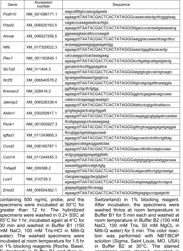
(4) Nakagawa et al., Novel Genes Related to Tooth Morphogenesis. Table 1 Primers used for in situ hybridization. Accession Gene Sequence number aagcattttgtccaacgagaata Pcdh10 NM_001098171.1 agaagcttTAATACGACTCACTATAGGGcaaatcatactgcttcgggtaag cagacccaaagaaatcacttgtc Pdzd2 XM_006520163.5 agaagcttTAATACGACTCACTATAGGGttgaccccactaatgataaaacg ggaaaagtaacattccccaagat Ahnak XM_006527256.5 agaagcttTAATACGACTCACTATAGGGaaggtaccaaacttcagctttcc acaaaggaaaagaggaagactgg Nfib XM_017320022.3 agaagcttTAATACGACTCACTATAGGGaaactgggttacacactgPlec1. NM_001163540.1. Slc7a5. NM_011404.3. Nr2f2. XM_006540578.2. Kremen2. NM_028416.2. Jakmip2. XM_006526338.4. Ablim1. XM_030250917.1. Penk1. NM_001002927.3. Igfbp3. XM_011243665.3. Ccnd2. XM_036165787.1. Cxcl14. XM_011244545.3. Tnfaip6. NM_009398.2. Loxl1. NM_010729.3. Emid2. XM_006504362.1. caccaaggcctcactaaagaag agaagcttTAATACGACTCACTATAGGGtccttgattgcattgatgtactg gacaactctcctttggaagatca agaagcttTAATACGACTCACTATAGGGtatgtgtcgtccatctgtcagtc tgcaagaattaaaactgagtcca agaagcttTAATACGACTCACTATAGGGttgtcatgctgatttcaatggta ggttatgcctgcttctgtgg agaagcttTAATACGACTCACTATAGGGggtctcgagaatcagccaac cataccctcagaaggcaaatgct agaagcttTAATACGACTCACTATAGGGttattcctcgtgcttcattaccc gctgtagagactcatgctggatt agaagcttTAATACGACTCACTATAGGGcaagaggcgtgagatccatatac ttcatgagaagcctcaaaagaag agaagcttTAATACGACTCACTATAGGGgtttcgtcaggagagatgaggta ttccgtggctgatatagacaaat agaagcttTAATACGACTCACTATAGGGagccactcctctttcctgtttag tggagaccatagacggaatctaa agaagcttTAATACGACTCACTATAGGGaacctttctttccatgtccaaa actgcgaggagaagatggttatc agaagcttTAATACGACTCACTATAGGGatgtgtggtgacatatggacaaa aaaaattggattccatgtctgtg agaagcttTAATACGACTCACTATAGGGcatgacatttcctgtgctaatga ctacgaacagggctacgtgtact agaagcttTAATACGACTCACTATAGGGcaagtaaggtggctccagca gagagatggtgcttccaagg agaagcttTAATACGACTCACTATAGGGttttgtagtgccctgatgtcttt. containing 500 ng/mL probe, and the specimens were incubated at 50°C for greater than 12 hr. The hybridized specimens were washed in 0.2× SSC at 65°C for 1 hr, incubated again at 4°C for 30 min and washed in Buffer B1 (150 mM NaCl, 100 mM Tris-HCl in Milli-Q water). The washed specimens were incubated at room temperature for 1.5 hr in 1% blocking reagents (Roche, Basel, Switzerland) in Buffer B1 and then incubated at 4°C for 12 hr with 1/5,000 anti-DIG antibody (Roche, Basel,. Switzerland) in 1% blocking reagent. After incubation, the specimens were washed thrice at room temperature in Buffer B1 for 5 min each and washed at room temperature in Buffer B2 (100 mM NaCl, 100 mM Tris, 50 mM MgCl2 in Milli-Q water) for 5 min. The color reaction was performed with NBT/BCIP solution (Sigma, Saint Louis, MO, USA) in Buffer B2 at 30°C. The stained specimens were cleared, mounted in 90% glycerol with propyl gallate, and examined by a microscope.. 44.
(5) J Oral Tissue Engin 2020;18(2):41-54. Biosystems QuantStudio 12K Flex (Life Technologies, Carlsbad, CA, USA) using SYBR Premix Ex Taq II (TaKaRa Bio, Shiga, Japan). Reactions were run in triplicate in three independent experiments. The data were normalized to Gapdh expression. The primer pairs used for real-time qPCR are listed in Table 2.. 6. Quantitative real-time polymerase chain reaction (qPCR) Total RNA was extracted from tooth germs using the RNeasy Plus Micro kit (Qiagen, Venlo, Netherlands) according to the manufacturer’s protocol and reverse transcribed using SuperScript VILO (Life Technologies, Carlsbad, CA, USA) for cDNA synthesis. Real-time qPCR was performed on the Applied Table 2. Primers used for real-time qPCR.. Gene. NCBI Gene ID. Pcdh10. 18526. Pdzd2. 68070. Ahnak. 66395. Nfib. 18028. Plec1. 18810. Slc7a5. 20539. Nr2f2. 11819. Kremen2. 73016. Jakmip2. 76217. Ablim1. 226251. Penk1. 18619. Igfbp3. 16009. Ccnd2. 12444. Cxcl14. 57266. Tnfaip6. 21930. Loxl1. 16949. Emid2. 140709. Gapdh. 14433. Sequence ACCGAGGAATGTAAAGCACTGG TCAAAGACCTCGGTGTCTGGAAC GGCTGTGCTCTTTAGAACCCAAC ACTCTGTTGCCACCATTCACAGG AAGGCAAGTGGGAAGAGTCTG AATGGATGCTTCAGGTGAGC CAGCATTGCAGCACTTACAGTC TCCCAGCGGACTTCATGTAAC GCACAGCAGCCAGTATTCAAC TCCAGCAACTGAGTGACACGTTC AGGCCTGGACTTTCTGACTTTC TTGACCCAAATGCACGCTAC CCCATACCATGACAAACCTAGC CCTTGGCTGCCAATAAATTCCAC TTCTGTGGCTCTGAAAGTGACC ATAGATGCCTAGTCGTCCATCGC TTCAGCCGTGGAACCAGTTTCAG AGTGTTTCCATGGGGTGTGGTG AGCACTTGGATTCACCCCCAATG TTGCTCTGTGTTACTGCAGGTG TGAGCAACTGCCTTGTCAATG TCACAGCTTTCAGGCAGTGTAG AATAAGTGCAGGCCCTATGGAG AGGCATATGCTTCCAGATGTCC ATCCTCATCCCAGCATTCTTCG ATCCTTCTAAGCCATCACAATGC AAAACTCCAGGCCAGTTGAG AACTGACCCTGGTAAGAAGAGC ATGACAACCAGGTCTGCTACTG AAGCAGCCTGGATCATGTTC TACGAACAGGGCTACGTGTAC ACCTCCGTAGTCCTCGTAAC CCTTGAGCATATGATTGGAGTCC CTCGCTTCATCTTGAGATTGGC TCCTCGTCCCGTAGACAAAATG AAATGGCAGCCCTGGTGACC. 45.
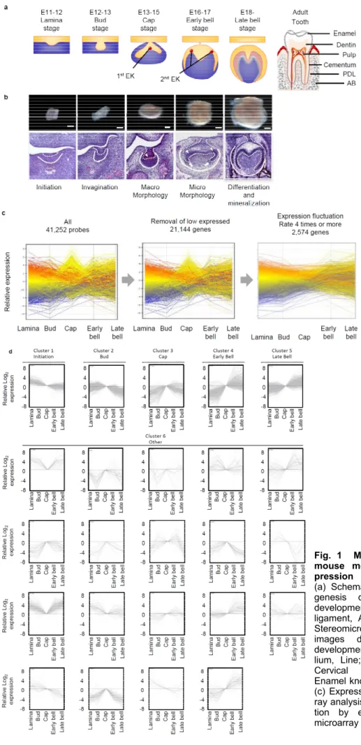
(6) Nakagawa et al., Novel Genes Related to Tooth Morphogenesis. Fig. 1 Microarray analysis of mouse molar tooth gene expression (a) Schematic image of morphogenesis during mouse molar development. PDL; Periodontal ligament, AB; Alveolar bone. (b) Stereomicroscopic images and HE images during mouse molar development. Dotted line; Epithelium, Line; Mesenchyme, Arrow; Cervical Loop, Arrow head; Enamel knot. Scale bars, 200 μm. (c) Expression pattern of microarray analysis. (d) Cluster classification by expression pattern of microarray analysis.. 46.
(7) J Oral Tissue Engin 2020;18(2):41-54. characters as previously reported6, is confirmed to classify as Cluster2. Cluster 3 is characterized by peak expression in the cap stage (Fig. 1d), and we confirmed the expression pattern of Myc, which character is similar to this cluster genes as reported in past study13, is classified as Cluster3. Cluster 4 is highly expressed in the early bell stage (Fig. 1d) and contains and we compared with the expression pattern of Shh, which is known to highly express in the similar stage in past study14, is classified as Cluster4. Cluster 5 is highly expressed in the late bell stage (Fig. 1d), and we confirmed that Cluster 5 includes Ambn, which have the similar characters as previously reported15. Cluster 6 is not classified as Clusters 1-5. For example, it maintains high expression from the initiation stage to the late bell stage and we confirmed the Tlx1 expression, which is known to express as similar pattern in past study15, is classified as Cluster6. These results suggest that the gene expression patterns obtained by this analysis reflect the expression patterns of genes in the tooth development process.. 7. Statistical analyses Student’s t-test was used to calculate P values on Microsoft Excel, with Twotailed tests. RESULT 1. Microarray analysis of mouse molar tooth gene expression To understand the comprehensive gene expression profile during molar development, we investigated the gene expression pattern during the early stage of molar tooth germ development. First, to comprehensively analyze the expression of genes at each stage of the tooth development process, we performed DNA microarray analysis. We isolated tooth germ at the placode (E11), bud (E12), cap (E14), early bell (E16), and late bell (E18) stages and performed genome-wide analysis using the Agilent Whole Mouse Genome 44k Array. We found 41,252 probes that were differentially expressed in the tooth germ of the E11 to E18 stages (Fig. 1a-c). The results of the analysis of gene expression were subject to data mining using Gene Spring software. To identify genes with greater than baseline expression, we selected 21,144 genes based on the expression intensity at each stage of tooth development (Fig. 1c). To classify genes by expression pattern, using 2,574 genes changes in gene expression level during tooth development that were four-fold or greater (Fig. 1c), and the genes was classified into 6 clusters by the K-means method (Fig. 1d). To determine whether these cluster classifications are appropriate for the classification of gene expression patterns in tooth development, they were compared with gene expression patterns that have been reported in the course of tooth development in the past. Cluster 1 is highly expressed in the initiation stage and weakly expressed as development progresses (Fig. 1d), and we confirmed that Fgf8, which previously reported to have the similar characters12, is classified as Cluster1. Cluster 2 exhibits peak expression in the placode stage (Fig. 1d). Msx1, which have the similar. 2. Spatiotemporal gene expression analysis during tooth development by in situ hybridization Many genes known to play an important role in tooth development are highly expressed from the early stages of development. Thus, we selected 327 genes with high levels of expression and large changes in expression from 1,077 genes with high expression in the early tooth developmental stage (clusters 1, 2, 3). To confirm the microarray results, we analyzed the gene expression of teeth at each developmental stage of bud (E12), cap (E14), and early bell (E16) by in situ hybridization. As a result, we found 17 novel genes with characteristic expression, such as expression in the whole tooth germ, enamel knot, tooth germ epithelium and mesenchyme. Pcdh10 was expressed on the buccal side of the tooth germ from the bud stage to the bell stage (Fig. 2a). Pdzd2, Ahnak, Nfib,. 47.
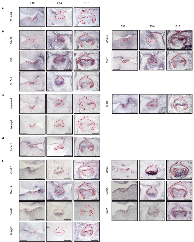
(8) Nakagawa et al., Novel Genes Related to Tooth Morphogenesis. Fig. 2 Localization of the expression of 17 novel genes during development as detected by in situ hybridization Detection of 17 novel genes was carried out in the dissected sections of tooth germs at the bud (E12), cap (E14), and bell (E16) stages of development. Expression patterns of specific genes around the tooth germ (a), the entire tooth germ (b), enamel knot (c), tooth germ epithelium (d), and tooth germ mesenchyme (e). Scale bars, 200μm.. 48.
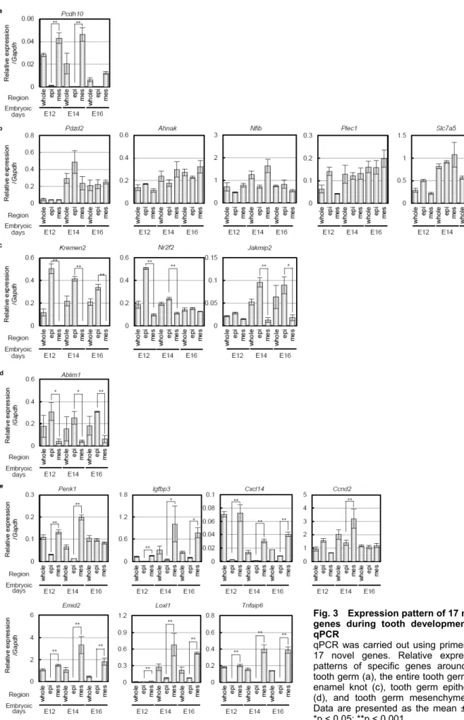
(9) J Oral Tissue Engin 2020;18(2):41-54. and is expressed in the mesenchyme to face secondary enamel knot in the bell stage. Loxl1 and Tnfaip6 was expressed in the dental papilla mesenchyme in the cap to bell stage. These results suggest that expression of these genes at the appropriate time at each site, such as near or in the entire tooth germ, enamel knots, epithelium, and mesenchyme, may be important in tooth development.. Plec1, and Slc7a5 were expressed in the entire tooth germ throughout early to late development (Fig. 2b). Pdzd2 was expressed in both the epithelium and mesenchyme in the cap to bell stage. Ahnak was observed inside the epithelium, which is similar to the growth arrest region10, and mesenchyme from the bud to bell stage. Nfib was widely expressed in the tooth germ epithelium and mesenchyme of the bud stage. The strongest expression was observed in the mesenchymal cap stage, and its expression was maintained up to the bell stage. Plec1 and Slc7a5 were highly expressed in the epithelium in the bud stage but were also expressed in the epithelium and mesenchyme in the cap to bell stage. Kremen2, Nr2f2, and Jakmip2 were expressed in enamel knots (Fig. 2c). Kremen2, Nr2f2, and Jakmip2 were strongly expressed in the epithelium of the bud stage and localized to primary and secondary enamel knots in the cap to bell stage. Ablim1 was expressed exclusively in the epithelium of the tooth germ (Fig. 2d). Ablim1 was observed in the entire tooth germ epithelium through the bell stage from the bud, but almost no expression was observed in the mesenchyme. Penk1, Igfbp3, Cxcl14, Ccnd2, Emid2, Loxl1, and Tnfaip6 were expressed in mesenchyme (Fig. 2e). Penk1 was expressed in the mesenchyme in the bud to cap stage and expressed in the epithelium and mesenchyme in the bell stage. Igfbp3 exhibited strong expression in the mesenchyme of the cap stage and strong expression in the dental papilla up to the bell stage. Cxcl14 was strongly expressed in the mesenchyme of the buccal side in the bud stage, and its expression was maintained in the dental papilla mesenchyme to cap to bell stage. Ccnd2 was highly expressed in the epithelium in the bud stage, highly expressed mesenchyme in the bud stage, and expressed in the inner enamel epithelium and dental papilla mesenchyme. Emid2 is expressed in the dental mesenchyme from the bud stage, is strongly expressed in the dental papilla mesenchyme in the cap stage,. 3. Gene expression analysis of 17 novel genes by qPCR To confirm the microarray and in situ hybridization results, we also analyzed the gene expression of teeth at each developmental stage, including bud (E12), cap (E14), and early bell (E16), by qPCR. The peak expression of the Pcdh10 gene was in the bud stage and was expressed only in the mesenchyme (Fig. 3a). Pdzd2, Ahnak, Nfib, Plec1 and Slc7a5 were more highly expressed in the cap and early bell stages than the bud stage and were expressed in both the epithelium and mesenchyme during tooth development (Fig. 3b). Kremen2, Nr2f2, Jakmip2 and Ablim1 exhibit higher gene expression in epithelial tissue than mesenchymal tissue (Fig. 3c and d). In contrast, Penk1, Igfbp3, Cxcl14, Ccnd2, Emid2, Loxl1, and Tnfaip6 exhibit high gene expression in mesenchymal tissues. These results indicate that 17 novel genes involved in tooth development could be identified and classified into several expression patterns. DISCUSSION In this study, we performed a comprehensive analysis of the early stage of tooth organogenesis using microarrays and in situ hybridization. The identification of new genes that are expressed in a particular region and time during tooth germ development indicates that our method is useful for obtaining spatiotemporal gene expression profiles. Our findings will contribute to a comprehensive understanding of tooth embryogenesis development, especially the morphogenesis process regulated by signaling centers.. 49.
(10) Nakagawa et al., Novel Genes Related to Tooth Morphogenesis. Fig. 3 Expression pattern of 17 novel genes during tooth development by qPCR qPCR was carried out using primers for 17 novel genes. Relative expression patterns of specific genes around the tooth germ (a), the entire tooth germ (b), enamel knot (c), tooth germ epithelium (d), and tooth germ mesenchyme (e). Data are presented as the mean ± SD. *p < 0.05; **p < 0.001.. 50.
(11) J Oral Tissue Engin 2020;18(2):41-54. In the development process, the ectodermal organ forming region is determined by the Turing model of activator and inhibitor16, 17. Osr2, Spry2 and 4 act as inhibitors of the regional determination of teeth and specify the lingual and front regions, respectively18-20. Deficiency in these genes results in the formation of supernumerary teeth in each region. On the other hand, the Bmp, Shh, and Fgf signals act as activators. Fgf signaling promotes tooth formation by activating the PI3K/Akt signaling pathway21. Deficiency in Bmp4 causes defects in tooth germs, whereas inactivation of Osr2 rescues molar morphogenesis in Bmp4-/- mutant mice18. In this study, we found that Pcdh10 is expressed in the mesenchyme of the buccal region. In addition, Pdzd2, Nfib, and Slc7a5 expression is observed in the entire tooth germ, including the epithelium and mesenchyme. Although there is no report that Pcdh10 is involved in organ-forming field determination in any organ, it has been reported that Pcdh10 has the ability to inhibit PI3K/Akt signaling in cancer cells22. Moreover, previous studies suggested that Nfib activates Fgf signaling in lung development23, Slc7a5 activates Shh signaling in nerves and eyes24, and Pdzd2 suppresses Shh signaling in limb development25. These facts suggest that Pcdh10 acts as an inhibitor by suppressing the Fgf signals of the buccal region, and Nfib, Pdzd2, and Slc7a5 act as activators to determine the tooth forming field and tooth development by activating the Fgf and Shh signals (Fig. 4). The reciprocal interaction between the epithelium and mesenchyme is vital for the induction and morphogenesis of tooth germ, and enamel knots in the epithelium function as a signaling center to control cooperative proliferation, migration, and differentiation of surrounding cells by releasing cytokines, such as Shh, Bmp, Fgf, and Wnt ligands6. Moreover, Bmp4 is expressed in the mesenchyme underlying enamel knots and contributes to the formation and maintenance of enamel knots via the p21 pathway26. Previous studies. demonstrated that Pax6 inhibits the differentiation of signaling centers to nerve cells and maintains its function as a signaling center in brain development27. In this study, we found that the following genes are expressed in primary and secondary enamel knots: Kremen2, an inhibitor of Wnt/β-catenin signaling28; Jakmip2, which acts with Klf4 to maintain the undifferentiated status of PSCs29; and Nr2f2, a component of the Pluri Network30. These findings suggest that these genes may play a role in preventing the differentiation of enamel knots and allow them to function as a signaling center (Fig. 4). Control of cell proliferation is important for tooth development, and the formation of the cusp and cervical loop is inhibited by deficiency of integrin β131, which is involved in cell proliferation via Ccnd132. In a recent study, using 4D cell trucking analysis, we demonstrated that the growth arrest of primary enamel knots and proliferation of surrounding epithelial cells yield bell-shaped morphogenesis via invagination and elongation of the cervical loop10. In this study, we observed that Emid2 expression is confined to the mesenchyme underlying enamel knots. Emid2 is expressed in mesenchyme and has been suggested to play a role in epithelial-mesenchymal interactions in the development of salivary glands, inner ear, and kidneys33. We also observed that Cxcl14, Ccnd2, and Igfbp3 are expressed in the tooth germ mesenchyme. Cxcl14 and Ccnd2 act on cell proliferation by maintaining the cell cycle in heart development and brain development34, 35 , respectively. Igfbp3, a binding protein of IGF, plays a role in the determination of the size and cusp number of teeth36. Furthermore, expression of Loxl1, which promotes crosslinking of collagen and elastin in lung development37, was observed in the position of prop-upping the primary enamel knot. These results suggest that these genes expressed in the mesenchyme may control mesenchymal cells through the specialization of enamel knots facing the mesenchyme and the control or maintenance of the cell cycle via ECM in tooth development (Fig. 4).. 51.
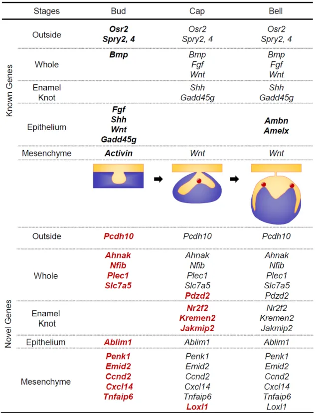
(12) Nakagawa et al., Novel Genes Related to Tooth Morphogenesis. Fig. 4 Spatiotemporal gene expression profiles in mouse molar development Schematic image of spatiotemporal gene expression of previously known and novel genes related to mouse molars. Gene expression was first observed from indicated stage. Known Genes; Black Bold, Novel Genes; Red Bold.. 52.
(13) J Oral Tissue Engin 2020;18(2):41-54. Genetics 2004: 499-508. 8) Sharpe PT, Young CS. Test-tube teeth. Sci Am 2005: 34-41. 9) Wang XP, Fan J. Molecular genetics of supernumerary tooth formation. Genesis. 2011: 261-277. 10) Morita R, Kihira M, Nakatsu Y, Nomoto Y, Ogawa M, Ohashi K, Mizuno K, Tachikawa T, Ishimoto Y, Morishita Y, Tsuji T. Coordination of cellular dynamics contributes to tooth epithelium deformations. PLoS One 2016; e0161336-e. 11) Marin-Riera M, Moustakas-Verho J, Savriama Y, Jernvall J, Salazar-Ciudad I. Differential tissue growth and cell adhesion alone drive early tooth morphogenesis: An ex vivo and in silico study. PLoS Comput Biol 2018; e1005981. 12) Neubüser A, Peters H, Balling R, Martin GR. Antagonistic interactions between FGF and BMP signaling pathways: a mechanism for positioning the sites of tooth formation. Cell 1997; 247- 255. 13) Yamada S. Expression of c-myc and N-myc in mouse embryos during craniofacial development. Kokubyo Gakkai Zasshi 1990: 83-105. (in Japanese) 14) Iseki S, Araga A, Ohuchi H, Nohno T, Yoshioka H, Hayashi F, Noji S. Sonic hedgehog is expressed in epithelial cells during development of whisker, hair, and tooth. Biochem Biophys Res Commun 1996; 688-693. 15) Landin MA, Shabestari M, Babaie E, Reseland JE, Osmundsen H. Gene expression profiling during murine tooth Development. Front Genet 2012; 139. 16) Economou AD, Ohazama A, Porntaveetus T, Sharpe PT, Kondo S, Basson MA, Gritli-Linde A, Cobourne M T, Green JBA. Periodic stripe formation by a Turing mechanism operating at growth zones in the mammalian palate. Nat Genet 2012; 348-351. 17) Sick S, Reinker S, Timmer J, Schlake T. WNT and DKK determine hair follicle spacing through a reaction-diffusion mechanism. Science 2006; 314 (5804): 1447-1450. 18) Kwon HJ, Park EK, Jia S, Liu H, Lan Y, Jiang R. Deletion of Osr2 Partially Rescues Tooth Development in Runx2 Mutant Mice. J Dent Res 2015; 1113-1119. 19) Jia S, Kwon H-JE, Lan Y, Zhou J, Liu H, Jiang R. Bmp4-Msx1 signaling and Osr2 control tooth organogenesis through antagonistic regulation of secreted Wnt antagonists. Developmental Biology 2016; 110-119. 20) Klein OD, Minowada G, Peterkova R,. In conclusion, in this study, we identified 15 stage- and region-specific novel genes by combining comprehensive genetic analysis using microarray and spatiotemporal high-throughput screening using in situ hybridization. Although the function of genes, such as Plec1, Ablim1, Penk1, and Tnfaip6, in tooth development cannot be surmised based on known information, these genes are expressed in important and specific stages and regions, such as the buccal region of tooth germ and enamel knots. These genes may play important roles in the field determination and morphogenesis of the tooth development process. In the future, functional analysis of the discovered genes is expected to lead to a more comprehensive understanding of tooth development. ACKNOWLEDGEMENTS We thank the members of the lab in RIKEN BDR, especially Dr. A. Noma, Ms. Y. Morioka and Ms. M. Takase. This work was supported by JSPS KAKENHI (Grant number: 19H01180). REFERENCES. 1) James. K. Avery NA. Oral Development and Histology. Third Edition ed: Theime; 2002 Pauline F.Steele. New York, 226-291. 2) Dawson PE. Functional Occlusion From TMJ to Smile Design: Elsevier Health Sciences; 2006. Amsterdam, 27-32. 3) Ishida K, Murofushi M, Nakao K, Morita R, Ogawa M, Tsuji T. The regulation of tooth morphogenesis is associated with epithelial cell proliferation and the expression of Sonic hedgehog through epithelial-mesenchymal interactions. Biochem Biophys Res Commun 2011 : 455-461. 4) Cai J, Cho SW, Kim JY, Lee MJ, Cha YG, Jung HS. Patterning the size and number of tooth and its cusps. Dev Biol 2007: 499-507. 5) Pispa J, Thesleff I. Mechanisms of ectodermal organogenesis. Dev Biol 2003; 195-205. 6) Thesleff I. Epithelial-mesenchymal signalling regulating tooth morphogenesis. J Cell Sci 2003;116(Pt 9): 1647-1648. 7) Tucker A, Sharpe P. The cutting-edge of mammalian development; how the embryo makes teeth. Nature Reviews. 53.
(14) Nakagawa et al., Novel Genes Related to Tooth Morphogenesis. 21). 22). 23). 24). 25). 26). 27) 28). 29). 30). Kangas A, Yu BD, Lesot H, Peterka M, Jernvall J, Martin GR. Sprouty genes control diastema tooth development via bidirectional antagonism of epithelial-mesenchymal FGF signaling. Developmental Cell 2006; 181-190. Liu C, Gu S, Sun C, Ye W, Song Z, Zhang Y, et al. FGF signaling sustains the odontogenic fate of dental mesenchyme by suppressing β-catenin signaling. Development 2013; 4375- 4385. Ye M, Li J, Gong J. PCDH10 gene inhibits cell proliferation and induces cell apoptosis by inhibiting the PI3K/Akt signaling pathway in hepatocellular carcinoma cells. Oncol Rep 2017; 3167-3174. Hsu YC, Osinski J, Campbell CE, Litwack ED, Wang D, Liu S, et al. Mesenchymal nuclear factor I B regulates cell proliferation and epithelial differentiation during lung maturation. Dev Biol 2011; 242-252. Katada T, Sakurai H. Xenopus slc7a5 is essential for notochord function and eye development. Mechanisms of Development 2019; 48-59. Yeung ML, Tam TS, Tsang AC, Yao KM. Proteolytic cleavage of PDZD2 generates a secreted peptide containing two PDZ domains. EMBO Rep 2003; 412-418. Jernvall J, Aberg T, Kettunen P, Keränen S, Thesleff I. The life history of an embryonic signaling center: BMP-4 induces p21 and is associated with apoptosis in the mouse tooth enamel knot. Development 1998; 161-169. Takahashi M, Osumi N. Pax6 regulates boundary-cell specification in the rat hindbrain. Mech Dev 2011; 289- 302. Mao B, Wu W, Davidson G, Marhold J, Li M, Mechler BM, Delius H, Hoppe D, Stannek P, Walter C, Glinka A, Niehrs C. Kremen proteins are Dickkopf receptors that regulate Wnt/beta-catenin signalling. Nature 2002; 417(6889): 664-667. Bernardo AS, Jouneau A, Marks H, Kensche P, Kobolak J, Freude K, Hall V, Feher A, Polgar Z, Sartori C, Bock I, Louet C, Faial T, Kerstens HHD, Bouissou C, Parsonage G, Mashayekhi K, Smith JC, Lazzari G, Hyttel P, Stunnenberg HG, Huynen M, Pedersen RA, Dinnyes A. Mammalian embryo comparison identifies novel pluripotency genes associated with the naïve or primed state. Biol Open 2018; 1-17. Som A, Harder C, Greber B, Siatkowski M, Paudel Y, Warsow G, Cap C, Schöler H, Fuellen G. The PluriNetWork: an electronic representation of. 31). 32). 33). 34). 35). 36). 37). the network underlying pluripotency in mouse, and its applications. PLoS One 2010; e15165. Chen B, Goodman E, Lu Z, Bandyopadhyay A, Magraw C, He T, Raghavan S. Function of beta1 integrin in oral epithelia and tooth bud morphogenesis. J Dent Res 2009; 539-544. Moreno-Layseca P, Streuli CH. Signalling pathways linking integrins with cell cycle progression. Matrix Biol. 2014; 144-153. Leimeister C, Steidl C, Schumacher N, Erhard S, Gessler M. Developmental expression and biochemical characterization of Emu family members. Dev Biol 2002; 204- 218. Waldemer-Streyer RJ, Reyes-Ordoñez A, Kim D, Zhang R, Singh N, Chen J. Cxcl14 depletion accelerates skeletal myogenesis by promoting cell cycle withdrawal. NPJ Regen Med. 2017; 16017. Chen Z, Duan RS, Zhu Y, Folkesson R, Albanese C, Winblad B, Jie Z. Increased cyclin E expression may obviate the role of cyclin D1 during brain development in cyclin D1 knockout mice. J Neurochem 2005; 1281-1284. Oyanagi T, Takeshita N, Hara M, Ikeda E, Chida T, Seki D,Yoshida M, Seiryu M, Takano I, Kimura S, Oshima M, Takashi T, Takano-Yamamoto T. Insulin-like growth factor 1 modulates bioengineered tooth morphogenesis. Sci Rep 2019; 368-382. Mižíková I, Morty RE. The Extracellular Matrix in Bronchopulmonary Dysplasia: Target and Source. Front Med 2015; 2 1-20. (Received, October 29, 2020/ Accepted, November 30, 2020). Corresponding author: Dr. Takashi Tsuji, Ph.D. 2-2-3, Minatojima-Minamimachi, Chuo-Ku, Kobe, Hyogo, 650-0047, Japan Laboratory for Organ Regeneration RIKEN Center for Biosystems Dynamics Research (BDR) TEL: +81 78 306 3447 FAX: +81 78 306 3449 E-mail: [email protected]. 54.
(15)
図
+2
関連したドキュメント
In this, the first ever in-depth study of the econometric practice of nonaca- demic economists, I analyse the way economists in business and government currently approach
For staggered entry, the Cox frailty model, and in Markov renewal process/semi-Markov models (see e.g. Andersen et al., 1993, Chapters IX and X, for references on this work),
Keywords: continuous time random walk, Brownian motion, collision time, skew Young tableaux, tandem queue.. AMS 2000 Subject Classification: Primary:
Then it follows immediately from a suitable version of “Hensel’s Lemma” [cf., e.g., the argument of [4], Lemma 2.1] that S may be obtained, as the notation suggests, as the m A
Definition An embeddable tiled surface is a tiled surface which is actually achieved as the graph of singular leaves of some embedded orientable surface with closed braid
Section 3 is first devoted to the study of a-priori bounds for positive solutions to problem (D) and then to prove our main theorem by using Leray Schauder degree arguments.. To show
This paper presents an investigation into the mechanics of this specific problem and develops an analytical approach that accounts for the effects of geometrical and material data on
discrete ill-posed problems, Krylov projection methods, Tikhonov regularization, Lanczos bidiago- nalization, nonsymmetric Lanczos process, Arnoldi algorithm, discrepancy
