Adjusting serum urate level by affecting membrane transporters involved in the disposition of urate
著者 盧 楊
著者別表示 Lu Yang journal or
publication title
博士論文要旨Abstractおよび要約Outline 学位授与番号 13301甲第3967号
学位名 博士(薬学)
学位授与年月日 2013‑09‑26
URL http://hdl.handle.net/2297/37261
doi: 10.2133/dmpk.DMPK-12-RG-070
Creative Commons : 表示 ‑ 非営利 ‑ 改変禁止 http://creativecommons.org/licenses/by‑nc‑nd/3.0/deed.ja
Adjusting serum urate level
by affecting membrane transporters involved in the disposition of urate
輸送体を介した血清尿酸値調節
Graduate School of
Natural Science and Technology Kanazawa University
Major subject: Life Sciences Course: Molecular Effects
School registration number: 1023032534 Name: Lu Yang
Chief advisor: Ikumi Tamai
Abstract:
This thesis investigates roles of urate-related transporters (e.g. URAT1/SLC22A12, URATv1/SLC2A9, and BCRP/ABCG2) in adjusting serum urate (SUA) level under physiological and disease conditions. In Chapter 2, functional cooperation of sodium-dependent monocarboxylate transporters (SMCTs) and urate transporter 1 (URAT1) was demonstrated indicating that SMCTs indirectly regulates urate reabsorption mediated by URAT1 function; therefore SMCT may serve as a potential target for the alteration of renal urate handling. In Chapter 3, interaction of whisky congeners with urate transporters was studied as a new rationale for their SUA-lowering effect. Since
URAT1-mediated urate uptake was significantly inhibited by whisky congeners, it is conceivable that SUA-lowering effect of drinking whisky may be caused by blocking URAT1 by congeners abundantly contained in whisky. In Chapter 4, association of increased uremic toxins with extra-renal elimination of urate mediated by urate efflux transporter, breast cancer resistance protein (BCRP). In patients with chronic kidney disease, although renal function is greatly reduced, SUA level is relatively maintained.
Indoxyl sulfate, as a model uremic toxin, induced significantly transport activity of BCRP in Caco-2 cells, such compensatory increase of extra-renal urate excretion may contribute to balance SUA. In conclusion, SUA can be adjusted directly or indirectly by urate
transporters, providing their physiological significance in urate homeostasis.
Chapter 1: Introduction
Urate is a weak organic acid with a pKa value of 5.8. It is the end product of purine degradation in humans and some higher primates. Due to the mutation of urate oxidase gene occurred in Miocene epoch, urate in humans cannot undergo further oxidation catalyzed by urate oxidase (UOX1 or uricase) to form allantoin, a more water-soluble substance with being easily excreted. Loss of urate oxidase results in higher serum urate level (SUA) in humans, compared with those in other mammals. Epidemiologic studies show that abnormal SUA is associated with several diseases. For example, hyperuricemia (SUA over 420 μM for men, over 360 μM for women) may increase risk of gout attack as reported by many studies. It might also play a role in the development of coronary heart disease, stroke, hypertension, diabetes mellitus, renal diseases and other cardiovascular diseases. Because urate has anti-oxidant property, reduced SUA may cause harmful effect in humans. Indeed, hypouricemia (SUA lower than 120 μM) has already been linked to Hodgkin’s disease and Alzheimer’s disease. Thus, maintenance of normal SUA is crucial to human health. Due to the close relationship of urate with a number of diseases, it has been used as a biomarker for many diseases and is now involved in regular clinical blood test. Clinical and animal studies have also suggested the participation of urate in the generation and development of diseases, and the importance of controlling urate level as a preventing and/or treating method of such diseases.
Generation of urate primarily occurs in liver, muscles and intestine, while excretion of urate mainly occurs from the kidney and intestine. Of the urate daily produced, about two thirds is excreted from kidney and the rest is mainly eliminated directly across intestinal epithelial cells from blood. In kidney, there is a urate transport system located on renal proximal tubule which plays an important role in the regulation of SUA. Most of urate is filtrated at the glomerulus and reabsorbed by this transport system. Urate transporter 1 (URAT1, encoded by SLC22A12) was identified as a transporter responsible for
reabsorption of urate at the apical membranes of proximal tubule cells, where it plays a predominant role in urate uptake from urine. Loss-of-function mutations of human URAT1 have been reported to result in hypouricemia. Previous studies found that many drugs (such as benzbromarone and losartan) could decrease SUA by exhibiting inhibitory effects on URAT1. These studies suggest an essential role of URAT1 in the transport of urate.
Similarly, organic onion transporter 4 (OAT4, encoded by SLC22A11) and OAT10, encoded by SLC22A13, are highly expressed at the apical side of proximal tubular cells and also involved in the reabsorptive transport of urate from luminal side into tubular cells.
On the basolateral membranes of proximal tubular cell, a voltage-driven urate transporter, URATv1 (GLUT9) encoded by SLC2A9 gene is recently reported as a solute carrier responsible for the urate transport from the tubular cells into blood. Hypouricemia was also found in patients with the loss-of-function mutations in URATv1, independent of genetics of URAT1. URAT1, OAT4, and OAT10 at the apical side of renal proximal tubule and URATv1 at the basolateral side of proximal tubule cells together consist of the vectorial transport form the urine to blood (as is shown in Fig. 1).
Since urate simultaneously undergoes secretion from blood to urine, other transporter system may also important to consider renal handling of urate in kidney. The system is possibly consisted of organic onion transporter 1 (OAT1, encoded by SLC22A6), and organic onion transporter 3 (OAT1, encoded by SLC22A8) on the basolateral side of proximal tubule cells, and sodium phosphate transporter 1 (NPT1, encoded by SLC17A1), NPT4 (encoded by SLC17A3), multidrug resistance protein 4 (MRP4, encoded by ABCC4), and breast cancer resistance protein (BCRP, encoded by ABCG2). SUA is also influenced by non-renal urate transport pathway. In humans, expression of BCRP in kidney is relatively low compared with other organs. On the contrary, expression of BCRP in intestine is high where one third of urate daily generated is excreted, compared with the expression in kidney. BCRP is expressed at the apical side of intestinal epithelial cells and mediates the urate efflux transport across the intestinal epithelial wall. Reduction of BCRP function is closely related to gout and hyperuricemia as is demonstrated by recent genome-wide association studies. Thus, BCRP can be regarded as an important efflux transporter mediating the non-renal excretion of urate.
With the development of economy, and westernization of lifestyle, the past several decades have witnessed an obvious increase in the prevalence of diseases such as cardiovascular disease, obesity, and diabetes mellitus, and the prevalence of these disease will continue to increase in the following several decades. SUA level seems to be closely associated with these diseases. Because of the important role of urate transporters in regulation of SUA level, adjusting of these urate transporters to control SUA is of critical significance. The present thesis will focus on this topic and investigate the modulation of urate transporters to affect SUA level.
Fig. 1 Transporter mediated urate transport in renal proximal tubule. On the apical side of renal proximal tubule, URAT1, OAT4, and OAT10 are responsible for the reabsorptive transport of urate from luminal side into renal proximal tubular cell. On the basolateral side, URATv1 is responsible for the transport of urate from proximal tubular cell into blood.
OAT1 and OAT3 on the basolateral side mediate the urate excretory transport from blood into proximal tubular cell. MRP4, BCRP, NPT1 and NPT4 located on the apical side functions as excretory transporter and transport urate from proximal tubular cell into urine.
Urine Proximal tubular cell Blood GFR
Urate
URATv1
OAT3 OAT1 BCRP
URAT1
OAT10
NPT1/4 OAT4
MRP4
Chapter 2: Functional cooperation of SMCTs and URAT1 for renal reabsorption transport of urate
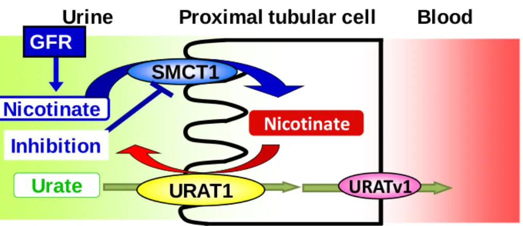
SUA can be regulated by a urate transport system located on renal proximal tubule. Urate transporter 1 (URAT1) is located on the apical side of proximal tubules and is responsible for the reabsorption of urate from luminal side into tubular cells. At the same site, it has been hypothesized that sodium-coupled monocarboxylate transporters (SMCTs) are responsible for the transportation of monocarboxylates such as lactate and nicotinate, which are driving force for urate transport via URAT1 by exchange transport. Accordingly, SMCTs can enhance URAT1-mediated urate reabsorption by providing monocarboxylates for the exchange.
Chapter 2 aimed to clarify the hypothesized functional cooperative relationship between URAT1 and SMCTs in the reabsorptive transport of urate. By preloading nicotinate in SMCT1-URAT1 co-expressing Xenopus oocytes, URAT1-mediated urate transport was stimulated. Nicotinate was taken up by SMCT1 but not by URAT1. When removing sodium ion from the uptake medium, the stimulation effect of nicotinate was decreased.
When adding SMCT1 inhibitors, the stimulation effect was also reduced. The results from this study indicate the cooperative relationship of URAT1 and SMCT1, and that SMCT1 is a potential target for the alteration of renal handling of urate indirectly (Fig. 2).
Fig. 2 Indirect regulation of SUA by affecting SMCT1.
Urine Proximal tubular cell Blood
Nicotinate GFR
Nicotinate
Urate URAT1
Inhibition
URATv1 SMCT1
Chapter 3: A putative mechanism of lowered SUA by whisky
Lifestyle and dietary factors are closely related to human health. Daily exposure of food is inevitable for all human beings. Influence of different kinds of food and beverage on human health has been realized since ancient times. In Chapter 3, a putative mechanism for the lowered SUA after whisky consumption was proposed and examined.
Consumption of alcoholic beverages has been a feature of many cultures throughout the world since ancient times, and especially in some Asian countries, is often associated with social gatherings. Many studies have shown that regular alcohol consumption is a risk factor for hyperuricemia and gout attack. However, recent clinical studies show that effect of moderate whisky consumption is quite different from the effect of consumption of other alcoholic beverages. Instead of increasing SUA as many other beverages do, it results in increased renal excretion of urate into urine and decreased SUA. Considering the
prevalence of alcohol and whisky consumption worldwide, it is of critical significance to understand why this different effect happens. Since renal reabsorption influences SUA, we examined the effects of whisky congeners on urate transporters, URAT1 and URATv1, involved in reabsorptive transport of urate. In transporter-expressing Xenopus oocytes, 12- and 18-year old whisky congeners inhibited urate uptake by URAT1 with IC50 values of 0.084 ± 0.011 and 0.042 ± 0.006 mg/mL, respectively, while urate uptake by URATv1 was inhibited at 1 mg/mL. Results of this study suggested that decreased SUA after whisky consumption may be mainly due to inhibition of URAT1 by congeners (Fig. 3). Daily intake of food and beverage might influence SUA by affecting urate transporters.
Fig. 3 Proposed mechanism for lowered SUA after whisky consumption. Whisky congeners may reduce SUA by inhibition of URAT1-mediated urate reabsorptive transport.
Urine Proximal tubular cell Blood GFR
Urate URAT1
congeners
URATv1
Chapter 4: Indoxyl sulfate upregulates BCRP expression in intestinal cell line
In Chapter 4, role of indoxyl sulfate on an important efflux urate transporter, BCRP, was investigated. At chronic kidney disease (CKD), renal function is greatly reduced. This leads to the tremendous increase of the concentrations of a great number of solutes.
However, the rise of SUA is not much, compared with other solutes. This phenomenon can be explained by the compensatory increase of extra-renal urate excretion. Previous study shows that intestinal Bcrp is important in controlling SUA and its expression is increased in CKD rats. But the molecular mechanism remains unknown. In this report, we examined whether indoxyl sulfate (IS) is involved in the upregulation of BCRP in intestine at CKD state. After exposure to IS, mRNA level, and protein level of BCRP in Caco-2 cells, which are used as human intestinal cultured cell model, were assayed by real-time PCR and flow cytometry, respectively. BCRP mRNA level was increased by exposure to IS for 24 h in a concentration dependent manner and reached steady state at 0.2 mM, a clinically relevant concentration. BCRP expression in the plasma membrane was also increased by 1.8 folds after treatment with 0.2 mM for three days. Basolateral to apical transcellular transport of urate in Caco-2 cells had 22% increase after IS treatment.
Intracellular accumulation of pheophorbide a, a functional marker of BCRP, was also decreased by 22% after IS treatment. IS was also observed to increase BCRP mRNA expression in LS180 cells and HepG2 cells, which are human enterocytes and hepatocytes cultured cell mode, respectively. However, no protein was expressed in plasma membrane of LS180 cells. BCRP protein expression in plasma membrane of HepG2 cells was increased after IS treatment. However, no function of BCRP was observed in HepG2 cells. Results indicate that indoxyl sulfate might be involved in the up-regulation of BCRP in intestine at CKD state, thereby minimizing an increase of serum uric acid level.
Fig. 4 Possible involvement of indoxyl sulfate in the induction of BCRP to increase intestinal urate excretion.
Blood
BCRP Urate
indoxyl sulfate
Intestine
Chapter 5: Conclusion
This thesis shows that SUA can be regulated by directly or indirectly influencing urate transporters and humans have a self-compensatory mechanism to regulate SUA at disease state by modulation of urate transporters. The results of this thesis indicate that transporters involved in urate transport can be used as targets for developing novel compounds to control SUA.