福島県立医科大学 学術機関リポジトリ
This document is downloaded at: 2021-11-08T00:16:00Z
Title Predicting Factors of Transmural Thermal Injury after Cryoballoon Pulmonary Vein Isolation( 本文 )
Author(s) 松本, 善幸
Citation
Issue Date 2019-03-22
URL http://ir.fmu.ac.jp/dspace/handle/123456789/992
Rights
© 2018 Springer Nature. This is a post-peer-review, pre- copyedit version of an article published in [Journal of Interventional Cardiac Electrophysiology]. The final authenticated version is available online at:
https://doi.org/10.1007/s10840-018-0454-8 DOI
Text Version ETD
Predicting Factors of Transmural Thermal Injury after Cryoballoon Pulmonary Vein Isolation
9
Predicting Factors of Transmural Thermal Injury after Cryoballoon Pulmonary Vein Isolation
Yoshiyuki Matsumoto, M.D.1; Takashi Kaneshiro, M.D.1,2; Naoko Hijioka. M.D.1; Minoru Nodera, M.D.1; Shinya Yamada, M.D.1; Masashi Kamioka, M.D.1; Akiomi Yoshihisa, M.D.3; Hiroshi Ohkawara, M.D.4; Takuto Hikichi, M.D.5; Hitoshi Suzuki, M.D.1; Yasuchika Takeishi, M.D.1,2
1Department of Cardiovascular Medicine, Fukushima Medical University
2Department of Arrhythmia and Cardiac Pacing, Fukushima Medical University
3Department of Advanced Cardiac Therapeutics, Fukushima Medical University
4Department of Hematology, Fukushima Medical University
5Department of Endoscopy, Fukushima Medical University
9
3 9 4
9 3
3
9 94 5 3
3
4 3 9 3
9 94
3 8 4
2015 2 2017 11
110 4 9
2 93 4
3 3 3 3
4 CT 3
3 3 41. Left atrium(LA)-Aorta(Ao) angle 32. Left inferior pulmonary vein (LIPV) angle 33. LA-Ao distance -
19 4 2 3 17
8 4 4 3
9 LIPV angle LA-Ao distance 4
LA-Ao distance 8 4
3 3
9 4
Journal of Interventional Cardiac Electrophysiology, Epub ahead of print
Abstract
Purpose: Transmural thermal injury (TTI), such as esophageal erosion/ulcer and periesophageal nerve injury leading to gastric hypomotility, is not rare complications associated with pulmonary vein isolation (PVI). However, the mechanism and predicting factors of TTI have not yet been fully elucidated with second-generation cryoballoon (CB) PVI.
Methods: One hundred ten consecutive patients, who underwent CB PVI for atrial fibrillation and received esophagogastroduodenoscopy 2 days later, were investigated. The relationships between TTI and both clinical and anatomical parameters were examined. We measured the following parameters based on the computed tomography data: the angle of the left atrial (LA) posterior wall to the descending aorta (Ao) (LA-Ao angle); the branching angle of the left inferior pulmonary vein (LIPV) to the coronal plane (LIPV angle); and the minimum distance between the LA posterior wall and descending Ao enclosing the esophagus (LA-Ao distance).
Results: TTIs occurred in 19 patients (esophageal erosion in 2 and gastric hypomotility in 17). The patients with TTI were significantly older than those without TTI. In the anatomical parameters, the LIPV angle was larger and the LA-Ao distance was shorter in the TTI (+) group compared to the TTI ( ) group. With the multivariate logistic regression analysis, the age (odds ratio [OR] 2.148, P=0.022) and LA-Ao distance (OR 0.430, P=0.013)
were independent predictors of TTI.
Conclusions: The occurrence of TTI in CB PVI was associated with aging, suggesting compromised periesophageal circulation, and the anatomical proximities between the LA and the descending Ao, which enclose the esophagus.
Keywords: Atrial fibrillation . Cryoballoon pulmonary vein isolation . Transmural thermal injury . Esophageal erosion . Periesophageal nerve injury
Introduction
After the pulmonary vein (PV) was identified as the trigger for atrial fibrillation (AF), PV isolation (PVI) has been a common method for catheter ablation of AF, especially in patients with paroxysmal AF [1, 2]. Transmural thermal injury (TTI), such as esophageal erosion/ulcer and periesophageal nerve injury leading to gastric hypomotility, is important in catheter ablation of AF. TTI has potential to advance to the left atrial-esophagus fistula, which is a fatal complication associated with PVI [3, 4]. Various factors, such as left atrium (LA) enlargement, distance between the LA and esophagus, additional ablation to PVI, and body mass index (BMI), have been reported to be associated with TTI in radiofrequency catheter ablation (RFCA) [5, 6]. We previously reported that the factors concerning the anatomical proximities of the structures surrounding the esophagus were strongly related to the occurrence of TTI in RFCA [7]. PVI with cryoballoon (CB) has recently been widely accepted as a treatment for AF. PVI with second-generation CB (Arctic Front Advance, Medtronic, Inc., Minneapolis, MN) especially has a high cooling effect that can achieve durable lesions of PV antrum and better clinical outcomes [8, 9]. However, the mechanism and predictors of TTI have yet to be fully evaluated in CB-PVI.
The aims of the current study were to reveal the mechanism, and investigate the predictors, of TTI in CB-PVI.
Methods
Study population
We retrospectively analyzed 110 consecutive patients, who had undergone their first CB- PVI for drug refractory AF at our institution between February 2015 and November 2017, and evaluated for the prevalence and predictors of CB-PVI-associated TTI. The study protocol was approved by the ethical committee of Fukushima Medical University. Written informed consent was obtained from all study subjects. As the risk factor of atherosclerosis, the prevalence of hypertension, diabetic mellitus, dyslipidemia, hyperuricemia, smoking, and chronic kidney disease (CKD) were evaluated. CKD was defined as an estimated glomerular filtration ratio < 60 ml/min/1.73 m2. CHA2DS2-VASc score was also calculated in each study subject: 1 point for age 65–74 years, female gender, heart failure, hypertension, diabetic mellitus, and vascular disease; and 2 points for age 75 years or older, previous stroke/transient ischemic attack.
Cryoballoon pulmonary vein isolation for atrial fibrillation
Catheter ablation was performed for AF following the cessation of all antiarrhythmic drugs for over five half-lives before the procedure. Preprocedural electrocardiogram-gated computed tomography (CT) was performed in all study subjects 1 day before the procedure and the anatomy of the LA and PV in each patient was confirmed to be suitable for CB-PVI.
Unsuitable anatomical forms, such as common PV, were excluded from the CB-PVI procedure. CB-PVI was performed using second-generation CB while the patients were
sedated with dexmedetomidine hydrochloride. Immediately after vascular access, 3000 IU heparin was administered. The periprocedural activated clotting time was maintained at 300–400 s with a bolus infusion of 5000 IU heparin and a continuous infusion of heparin at a rate of 2000–3000 IU/h after a transseptal approach. A single transseptal puncture was performed using an 8.5 Fr long sheath (SL8.5, AF division, St. Jude Medical, Inc., Minneapolis, MN) and an RF needle (Japan Lifeline Co., Ltd., Tokyo, Japan). Two SL8.5 sheaths were inserted into the LA via a single puncture site, and left arteriography was performed during right ventricle burst pacing at a rate of 200 bpm.
The transseptal sheath was exchanged with a 15-Fr steerable sheath (Flexcath Advance, Medtronic), and a 10-polar spiral mapping catheter (Achieve, Medtronic) was used for PV mapping. A cryoballoon catheter (Arctic Front Advance, Medtronic) was advanced to each PV ostium over the Achieve catheter, and complete occlusion of the PV ostium was confirmed with contrast medium injection. One CB application consisted of a 3-min freezing, and an additional CB was not applied after isolation. As for the PV that could not be completely occluded, two CB applications were performed at the upper and lower portions of the PV ostium. If the PV was not isolated with several CB ablations, additional touch-up radiofrequency catheter ablation (RFCA) was applied for isolation by a 7.5-Fr irrigation catheter with a 3.5-mm distal electrode and real-time contact force monitoring (ThermoCool ContactForce, Biosense Webster, Inc., Diamond Bar, CA). The endpoint of PVI was defined as the creation of a bidirectional conduction block between the LA and PVs. For safety, CB ablation was terminated when the balloon temperature fell below 60 °C [9]. We then monitored the diaphragmatic compound motor action potential during phrenic nerve pacing
to avoid phrenic nerve injury, especially during CB ablations for the right PVs.
After completion of PVI, non-inducibility of AF was confirmed with coronary sinus burst pacing at a minimum cycle length of 200 ms during continuous intravenous administration of isoproterenol (1.0–3.0 µg/min).
Luminal esophageal temperature monitoring
We used a luminal esophageal temperature (LET) monitoring probe (Esophaster, Japan Lifeline Co., Ltd.) during the PVI procedures in all patients. When the LET fell below 15 °C, CB ablation was terminated [10].
Esophagogastroduodenoscopy after cryoballoon ablation
All study subjects underwent esophagogastroduodenoscopy (EGDS) to check for the presence of TTI 2 days after PVI. As a result, esophageal erythema, erosion, and ulceration were detected with direct observation. Gastric hypomotility was defined as when gastric residue was observed even with overnight fasting for 12 h.
Evaluation of anatomical structures surrounding the esophagus based on computed tomography data
Electrocardiogram-gated CT data were acquired using a 64-row multidetector CT scanner (Aquilion 64, Toshiba Medical Systems Co., Ltd., Tochigi, Japan) with a collimation of 64 × 0.5 mm, a pixel size of 0.39 × 0.39 mm, a gantry rotation time of 350 ms, and a tube voltage of 120 kV. Based on the CT data, we evaluated the anatomical proximities between the
esophagus and the surrounding structures using a computer workstation (Ziostation, Ziosoft Inc., Tokyo, Japan). We defined and calculated the following parameters to evaluate the proximities of the esophagus to its surrounding structures, as we previously reported [7].
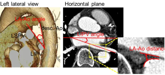
(1) The angle of the left atrial (LA) posterior wall to the descending aorta (Ao): LA-Ao angle (Fig. 1). The surface of the LA posterior wall bends forward and touches the anterior aspect of the esophagus. If the LA-Ao angle is small, the contact area of the LA posterior wall and esophagus might be larger. (2) The branching angle of the left inferior PV (LIPV) to the coronal plane: LIPV angle (Fig. 1). As mentioned above, the anterior aspect of the esophagus touches the LA posterior wall in the lower portion. The LIPV branches backwards; therefore, if the LIPV angle is larger, the ostium of the LIPV becomes closer to the esophagus. (3) The distance between the descending Ao and LA posterior wall enclosing the esophagus: LA-Ao distance (Fig. 1). The esophagus is a mobile organ because it is not fixed in the posterior mediastinum, and therefore the distance from the LA posterior wall to the esophagus, which can vary, is an unreliable parameter to use in the evaluation of the proximity between the esophagus and these structures. The esophagus is located between the LA posterior wall and the descending Ao; thus, if the LA-Ao distance is smaller, the proximity between the LA posterior wall and esophagus could be closer with a sandwiching mechanism between the LA posterior wall and the descending aorta during CB ablation. These parameters were calculated by averaging the values evaluated by two investigators, who were blinded to the existence of TTI in each study subject.
Statistical analysis
All data are shown as means ± standard deviation, numbers, or percentages. Differences between the two groups were investigated using a Mann-Whitney U test for continuous data.
All categorical data were compared using Fisher’s exact probability test. All parameters with a significance of < 0.10 in the univariate logistic regression analysis were entered into a multivariate logistic regression analysis. Receiver-operating characteristics (ROC) analysis was performed for each parameter to determine the optimal cut-offs for the prediction of TTI. A two-sided P value of < 0.05 was considered statistically significant.
These analyses were performed using a statistical software package (SPSS ver. 21.0, IBM, Armonk, NY, USA).
Results
Baseline characteristics and catheter ablation of atrial fibrillation
The clinical characteristics of the study subjects are summarized in Table 1. The mean age was 64 ± 9 years, and 80 of the 110 subjects (73%) were male. The mean LA diameter (LAD) and LA volume index (LAVI) in echocardiography were 36.8 ± 5.9 mm and 31.3 ± 14.9 ml/m2, respectively. In all subjects, complete PVI was achieved in both sides of the PV. Additional touch-up ablation was performed in 19 study subjects (17%). Said ablation was performed for the left superior PV in 3 patients (roof), left inferior PV in 2 (anterior-inferior and posterior inferior), right superior PV in 2, and right inferior PV in 15. In all study subjects, the esophagus was located in the left or central portion of the LA, meaning that the esophagus overlay the left-sided CB ablation lines.
No patients had a history of open chest surgery, which can influence the prevalence of TTI through the esophageal fixation in the mediastinum.
Prevalence of transmural thermal injury
All study subjects underwent EGDS to check for the presence of TTI 2 days after the PVI procedure. Nineteen of the 110 patients (17%) had TTIs, including esophageal erosion in 2 patients and gastric hypomotility in 17 patients. No patients had TTI-associated symptoms.
In the two patients with esophageal erosion, re-evaluation with EGDS after 1 week revealed that the esophageal erosion had healed or disappeared in all patients. Representative cases are shown in Figs. 2 and 3.
Comparisons between the groups with and without transmural thermal injury
The details of comparisons between the two groups, with and without TTI, are shown in Table 1. LAD and LAVI in echocardiography and BMI, which have been previously reported to be predictors of TTI, were not different between the two
groups in the present study. However, the age was significantly higher in patients with TTI than those without TTI (68 ± 8 vs. 63 ± 9 mm, P = 0.026). In the anatomical parameters suggesting the proximity between the esophagus and its surrounding structures, the LIPV angle in the TTI (+) group was significantly larger (24.5 ± 11.2 vs. 20.0 ± 8.1 mm, P = 0.047) and the LA-Ao distance was significantly smaller (4.9 ± 1.3 vs. 5.8 ± 1.6 mm, P = 0.030) compared to those in the TTI ( ) group. There was no difference in LA-Ao angle between the two groups with and without TTI (Table 1). As risk factors of atherosclerosis, each prevalence of hypertension, diabetic mellitus, dyslipidemia, hyperuricemia, smoking, and CKD did not differ between the groups. There was no difference in CHA2DS2-VASc score between the two groups. (Table 1).
No difference was observed in nadir balloon temperature during CB ablation, total freezing time, or prevalence of termination of CB ablation due to the LET falling below 15 °C, between the two groups. The prevalence of touch-up ablation for left PV did not differ between the groups. There was no patient in whom the CB ablation was stopped due to the balloon temperature falling below 60 °C. (Table 2).
Logistic regression analysis and ROC analysis to predict transmural thermal injury
The results of the logistic regression analysis for predicting TTI are shown in Table 3. The multivariate logistic regression analysis revealed that age (odds ratio [OR] 2.148, 95%
confidential interval [95% CI] 1.113–4.140, P = 0.022) and LA-Ao distance (OR 0.430, 95%
CI 0.219–0.841, P = 0.013) were independent predictors of TTI.
We analyzed the ROC curve to identify the best cut-off value of these predictors to distinguish the risk of TTI. According to the ROC curve analysis, we identified the cut-off age as 65.5 years with 63% sensitivity, 54% specificity, and an area under the curve of 0.643 (95% CI 0.508–0.778). We also identified the cut-off LA-Ao distance to be 5.9 mm, with 41%
sensitivity, 90% specificity, and an area under the curve of 0.651 (95% CI 0.525–0.777) to predict TTI.
Discussion
The major finding of the current study was that the age and anatomical proximity of the structures surrounding the esophagus were associated with TTI incidence. In addition, our results revealed that the age and LA-Ao distance were independent predictors of TTI. From the results of the present study, aging, which may suggest a compromised periesophageal circulation, and the anatomical proximity of the esophagus and surrounding structures, were important regarding the prevalence of TTI.
Mechanism of transmural thermal injury in cryoballoon pulmonary vein isolation
The mechanism of TTI in CB PVI remains unclear; however, its mechanism in RFCA is speculated to be direct thermal injury by a radiofrequency ablation catheter, since the anatomical distortion of the LA wall occurs during catheter mapping with a high contact force (> 10 g). [11] During catheter contact with the LA wall with a high contact force, tenting of the LA wall within about 2 mm beyond the LA surface occurs. Both LA wall tenting to the esophagus during RF application and the anatomical proximities of surrounding structures could make the LA wall and the esophagus closer, resulting in an increased risk of TTI.
However, it is noteworthy that the esophageal erosions showed a linear, longitudinal form in the present study, although the esophageal erosion showed a spot lesion in the RFCA [7].
This suggests that the mechanism of TTI is different between CB-PVI and RFCA-PVI. Not only the energy source but also the lesion size might affect the mechanism of TTI in CB-PVI and RFCA-PVI. Second-generation CB can create a large PV antral lesion compared to that
by first-generation CB and RFCA PVI [12, 13]. This larger lesion created by the second- generation CB might have contributed to the development of the esophageal lesions observed in the current study. Moreover, it is interesting that the esophageal lesions showed a circumferential form in the present study, although the spot lesion in RFCA-PVI appeared in the anterior aspect of the esophagus, which touches the LA posterior wall. This suggests that the mechanism of esophageal erosion is not only direct thermal injury by CB. We speculate that the mechanism of esophageal erosion in CB-PVI could be the additional impairment of periesophageal circulation by cryo-energy. Therefore, aging, which suggests compromised periesophageal circulation, could be a predictor of esophageal erosion in CB- PVI.
The fibers of periesophageal vagal nerve plexus descend along the anterior and posterior aspects of the esophagus. Therefore, strong cryo-energy using second-generation CB could injure the periesophageal nerve, resulting in gastric hypomotility. Moreover, the periesophageal nerve is fed by the perforators of periesophageal circulation; therefore, the cryo-energy-induced impairment of the periesophageal circulation, in addition to the compromised periesophageal circulation, could also increase the risk of gastric hypomotility.
We should consider the influence of additional touch-up ablations on the prevalence of TTI.
As mentioned above, each esophagus was located at the left or central portion of the LA, meaning that the esophagus overlay the left-sided CB ablation lines in all study subjects.
Only one out of five touch-up ablations in the left PVs was located at the left posterior- inferior area, adjacent to the esophagus. Therefore, we believe that the contribution of the touch-up ablations to the TTI should be extremely low.
Anatomical proximity of esophagus to surrounding structures
As the esophagus is not fixed in the posterior mediastinum, it is considered a mobile organ [14, 15]. Therefore, evaluation of the proximities between the surrounding structures, such as the LA posterior wall and the esophagus, has little meaning. In the present study, we focused on the proximity between the esophagus and the fixed structures surrounding the esophagus, such as the LA posterior wall and descending Ao. In the anatomical parameters, only LA-Ao distance was a predictor of TTI in the present study. The left-sided esophagus is located within the narrow “triangle zone”, surrounded by the LA posterior wall, descending Ao, and spinal column. The diameter of the triangle zone is approximated to the LA-Ao distance; mean LA-Ao distance was 5.7 ± 1.6 mm in the current study. We speculate that the high level of cryo-energy by CB might cool this “triangle zone” diffusely, resulting in the additional impairment of circumferential periesophageal circulation and periesophageal nerve injury leading to TTI.
Clinical implications
In the clinical setting, EGDS after PVI procedure is not routinely performed. However, in the present study, the prevalence of TTI was relatively high, and all patients with TTI were asymptomatic. Esophageal erosions in our patients healed or disappeared 1 week after their first EGDS evaluation. However, it is worth noting that esophageal erosions and ulcers have a strong potential to develop into LA-esophageal fistulae, which have a mortality rate of over 75% [16–19]. There are several limitations in LET monitoring in AF ablations [20–23].
Moreover, Miyazaki et al. reported that the use of LET monitoring is a predictor of esophageal erosion in CB-PVI [24]. Therefore, it is important to forecast the risk of TTI before performing CB-PVI based on the clinical profiles and anatomical relationship between the esophagus and its surrounding structures. It is also necessary to prevent TTI, for example, administration of proton pump inhibitor during the periprocedural period.
Study limitations
The present study has several limitations. First, it is a single-center observational study, and its findings should be evaluated in a multicenter, randomized control study. Second, we did not evaluate EGDS before the CB-PVI procedure in all study subjects. Because all the patients with TTI in this study were asymptomatic, we should have performed the EGDS before the CB-PVI procedure to confirm that the TTI resulted from the CB-PVI procedure.
We defined gastric hypomotility as the presence of gastric residue observed after overnight fasting. Therefore, motility tests before and after the ablation would be useful [25].
Conclusions
Aging, which suggests compromised periesophageal circulation, and the anatomical proximities of the LA posterior wall and descending Ao surrounding the esophagus, are strongly associated with TTI incidence. It is important to consider the risk of TTI based on the clinical profile and the anatomical relationship between the esophagus and its surrounding structures.
Compliance with ethical standards
Conflict of interest
We have no conflict of interest.
Informed consent
Informed consent was obstained from all individual participants included in this study.
Acknowledgments
We thank Drs. Takashi Kaneshiro, Naoko Hijioka, Minoru Nodera, Shinya Yamada, Masashi Kamioka, Akiomi Yoshihisa, Hiroshi Ohkawara, Takuto Hikichi, Hitoshi Suzuki, Yasuchika Takeishi for their important contribution on the present study.
References
1. Haïssaguerre M, Jaïs P, Shah DC, Takahashi A, Hocini M, Quiniou G, et al. Spontaneous initiation of atrial fibrillation by ectopic beats originating in the pulmonary veins. N Engl J Med. 1998;339:659–66.
2. Calkins H, Reynolds MR, Spector P, Sondhi M, Xu Y, Martin A, et al. Treatment of atrial fibrillation with antiarrhythmic drugs or radiofrequency ablation: two systematic literature reviews and meta-analyses. Circ Arrhythm Electrophysiol. 2009;2:349–61.
3. Pappone C, Oral H, Santinelli V, Vicedomini G, Lang CC, Manguso F, et al. Atrio- esophageal fistula as a complication of percutaneous transcatheter ablation of atrial fibrillation. Circulation. 2004;109:2724–6.
4. Dagres N, Kottkamp H, Piorkowski C, Doll N, Mohr F, Horlitz M, et al. Rapid detection and successful treatment of esophageal perforation after radiofrequency ablation of atrial fibrillation: lessons from five cases. J Cardiovasc Electrophysiol. 2006;17:1213–5.
5. Martinek M, Meyer C, Hassanein S, Aichinger J, Bencsik G, Schoefl R, et al. Identification of a high-risk population for esophageal injury during radiofrequency catheter ablation of atrial fibrillation: procedural and anatomical considerations. Heart Rhythm. 2010;7:1224–
30.
6. Yamasaki H, Tada H, Sekiguchi Y, Igarashi M, Arimoto T, Machino T, et al. Prevalence and characteristics of asymptomatic excessive transmural injury after radiofrequency catheter ablation of atrial fibrillation. Heart Rhythm. 2011;8:826–32.
7. Kaneshiro T, Matsumoto Y, Nodera M, Kamioka M, Kamiyama Y, Yoshihisa A, et al.
Anatomical predisposing factors of transmural thermal injury after pulmonary vein isolation. Europace. 2018;20: 1122–8.
8. Ciconte G, de Asmundis C, Sieira J, Conte G, Di Giovanni G, Mugnai G, et al. Single 3- minute freeze for second-generation cryoballoon ablation: one-year follow-up after pulmonary vein isolation. Heart Rhythm. 2015;12:673–80.
9. Su W, Kowal R, Kowalski M, Metzner A, Svinarich JT, Wheelan K, et al. Best practice guide for cryoballoon ablation in atrial fibrillation: the complication experience of more than 3000 procedures. Heart Rhythm. 2015;12:1658–166.
10. Fürnkranz A, Bordignon S, Böhmig M, Konstantinou A, Dugo D, Perrotta L, et al.
Reduced incidence of esophageal lesions by luminal esophageal temperature-guided second- generation cryoballoon ablation. Heart Rhythm. 2015;12:268–74.
11. Anjo N, Nakahara S, Okumura Y, Hori Y, Nagashima K, Komatsu T, et al. Impact of catheter tip-tissue contact on three-dimensional left atrial geometries: relationship between the external structures and anatomic distortion of 3D fast anatomical mapping and high contact force guided images. Int J Cardiol. 2016;222:202–8.
12. Kenigsberg DN, Martin N, Lim HW, Kowalski M, Ellenbogen K. Quantification of the cryoablation zone demarcated by pre and postprocedural electroanatomic mapping in atrial fibrillation patients using the 28-mm second-generation cryoballoon. Heart Rhythm.
2015;12:283–90.
13. Bordignon S, Fürnkranz A, Dugo D, Perrotta L, Gunawardene M, Bode F, et al. Improved lesion formation using the novel 28mm cryoballoon in atrial fibrillation ablation: analysis of biomarker release. Europace. 2014;16:987–93.
14. Good E, Oral H, Lemola K, Han J, Tamirisa K, Igic P, et al. Movement of the esophagus during left atrial catheter ablation for atrial fibrillation. J Am Coll Cardiol. 2005;46:2107–
10.
15. Sánchez-Quintana D, Cabrera JA, Climent V, Farré J, Mendonça MC, Ho SY. Anatomic relations between the esophagus and left atrium and relevance for ablation of atrial fibrillation. Circulation. 2005;112:1400–5.
16. Scanavacca MI, D'ávila A, Parga J, Sosa E. Left atrial-esophageal fistula following radiofrequency catheter ablation of atrial fibrillation. J Cardiovasc Electrophysiol.
2004;15:960–2.
17. Ren JF, Lin D, Marchlinski FE, Callans DJ, Patel V. Esophageal imaging and strategies for avoiding injury during left atrial ablation for atrial fibrillation. Heart Rhythm.
2006;3:1156–61.
18. Ghia KK, Chugh A, Good E, Pelosi F, Jongnarangsin K, Bogun F, et al. A nationwide survey on the prevalence of atrioesophageal fistula after left atrial radiofrequency catheter ablation. J Interv Card Electrophysiol. 2009;24:33–6.
19. Dagres N, Hindricks G, Kottkamp H, Sommer P, Gaspar T, Bode K, et al. Complications of atrial fibrillation ablation in a high-volume center in 1,000 procedures: still cause for concern? J Cardiovasc Electrophysiol. 2009;20:1014–9.
20. Singh SM, d'Avila A, Doshi SK, Brugge WR, Bedford RA, Mela T, et al. Esophageal injury and temperature monitoring during atrial fibrillation ablation. Circ Arrhythm Electrophysiol. 2008;1:162–8.
21. Rillig A, Meyerfeldt U, Birkemeyer R, Wiest S, Sauer BM, Staritz M, et al. Oesophageal
temperature monitoring and incidence of oesophageal lesions after pulmonary vein isolation using a remote robotic navigation system. Europace. 2010;12:655–61.
22. Perzanowski C, Teplitsky L, Hranitzky PM, Bahnson TD. Real-time monitoring of luminal esophageal temperature during left atrial radiofrequency catheter ablation for atrial fibrillation: observations about esophageal heating during ablation at the pulmonary vein ostia and posterior left atrium. J Cardiovasc Electrophysiol. 2006;17:166–70.
23. Martinek M, Bencsik G, Aichinger J, Hassanein S, Schoefl R, Kuchinka P, et al.
Esophageal damage during radiofrequency ablation of atrial fibrillation: impact of energy settings, lesion sets, and esophageal visualization. J Cardiovasc Electrophysiol. 2009;20:
726–33.
24. Miyazaki S, Nakamura H, Taniguchi H, Hachiya H, Takagi T, Igarashi M, et al. Gastric hypomotility after second-generation cryoballoon ablation—unrecognized silent nerve injury after cryoballoon ablation. Heart Rhythm. 2017;14:670–7.
25. Obioha C, Erickson J, Suseela S, Hajri T, Chung E, Richards W, et al. Effect of body mass index on the sensitivity of magnetogastrogram and electrogastrogram. J Gastroenterol Hepatol Res. 2013;2:513–9.
23
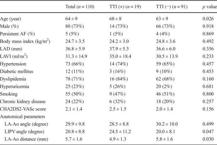
Table.1 Baseline characteristics and comparisons between the groups with and without transmural thermal injury
Data are shown as the means ± standard deviation or number of patients (%)
Ao aorta, AF atrial fibrillation, CF contact force, LAD left atrial diameter, LAVI left atrial volume index, LIPV left inferior pulmonary vein, RF radiofrequency, TTI transmural thermal injury
complete PVI was achieved in both sides of the PV.
Additional touch-up ablation was performed in 19 study sub- jects (17%). Said ablation was performed for the left superior PV in 3 patients (roof), left inferior PV in 2 (anterior-inferior and posterior inferior), right superior PV in 2, and right infe- rior PV in 15. In all study subjects, the esophagus was located in the left or central portion of the LA, meaning that the esophagus overlay the left-sided CB ablation lines.
No patients had a history of open chest surgery, which can influence the prevalence of TTI through the esophageal fixa- tion in the mediastinum.
3.2 Prevalence of transmural thermal injury
All study subjects underwent EGDS to check for the presence of TTI 2 days after the PVI procedure.
Nineteen of the 110 patients (17%) had TTIs, including esophageal erosion in 2 patients and gastric hypomotility in 17 patients. No patients had TTI-associated symptoms. In the two patients with esophageal erosion, re-evaluation with EGDS after 1 week revealed that the esophageal erosion had healed or disappeared in all patients. Representative cases are shown in Figs.2and3.
Table 1 Baseline characteristics and comparisons between the groups with and without transmural thermal injury
Total (n= 110) TTI (+) (n= 19) TTI (−) (n= 91) pvalue
Age (year) 64 ± 9 68 ± 8 63 ± 9 0.026
Male (%) 80 (73%) 14 (73%) 66 (73%) 0.918
Persistent AF (%) 5 (5%) 1 (5%) 4 (4%) 0.869
Body mass index (kg/m2) 24.7 ± 3.5 24.2 ± 3.0 24.8 ± 3.6 0.492
LAD (mm) 36.8 ± 5.9 37.9 ± 5.3 36.6 ± 6.0 0.356
LAVI (ml/m2) 31.3 ± 14.9 35.0 ± 18.4 30.5 ± 13.9 0.233
Hypertension 73 (66%) 14 (74%) 59 (65%) 0.457
Diabetic mellitus 12 (11%) 3 (16%) 9 (10%) 0.453
Dyslipidemia 78 (71%) 16 (84%) 62 (68%) 0.160
Hyperuricemia 25 (23%) 5 (26%) 20 (2%) 0.681
Smoking 55 (50%) 9 (47%) 46 (51%) 0.800
Chronic kidney disease 24 (22%) 6 (32%) 18 (20%) 0.257
CHA2DS2-VASc score 2.1 ± 1.4 2.5 ± 1.5 2.0 ± 1.4 0.156
Anatomical parameters
LA-Ao angle (degree) 29.9 ± 9.8 28.5 ± 8.8 30.2 ± 10.0 0.499
LIPV angle (degree) 20.8 ± 8.8 24.5 ± 11.2 20.0 ± 8.1 0.047
LA-Ao distance (mm) 5.7 ± 1.6 4.9 ± 1.3 5.8 ± 1.6 0.030
Data are shown as the means ± standard deviation or number of patients (%)
Aoaorta,AFatrial fibrillation,CFcontact force,LADleft atrial diameter,LAVIleft atrial volume index,LIPVleft inferior pulmonary vein,RFradiofrequency,TTItransmural thermal injury
Fig. 2 Representative case of transmural thermal injury. The left panel shows esophageal erosion and the right panel shows gastric hypomotility,
J Interv Card Electrophysiol
Table.2 Procedural characteristics and comparisons between the groups with and without transmural thermal injury
Data are shown as the means ± standard deviation or number of patiens (%)
CB cryoballoon, LET luminal esophageal temperature, LIPV left inferior pulmonary vein, LSPV left superior pulmonary vein, LPV left pulmonary vein, RIPV right inferior pulmonary vein, RSPV right superior pulmonary vein
3.3 Comparisons between the groups
with and without transmural thermal injury
The details of comparisons between the two groups, with and without TTI, are shown in Table1. LAD and LAVI in echo- cardiography and BMI, which have been previously reported to be predictors of TTI, were not different between the two
groups in the present study. However, the age was significant- ly higher in patients with TTI than those without TTI (68 ± 8 vs. 63 ± 9 mm,P= 0.026). In the anatomical parameters sug- gesting the proximity between the esophagus and its surround- ing structures, the LIPV angle in the TTI (+) group was sig- nificantly larger (24.5 ± 11.2 vs. 20.0 ± 8.1 mm, P= 0.047) and the LA-Ao distance was significantly smaller (4.9 ± 1.3
Fig. 3 Representative case with transmural thermal injury. The patient had a short LA-Ao distance.Aoaorta,LAleft atrium, LIPVleft inferior pulmonary vein
Table 2 Procedural
characteristics and comparisons between the groups with and without transmural thermal injury
Total (n= 110) TTI (+) (n= 19) TTI (−) (n= 91) pvalue Nadir balloon temperature (°C)
LSPV −48.3 ± 11.2 −46.8 ± 5.9 −48.6 ± 12.0 0.515
LIPV −43.7 ± 13.8 −44.1 ± 3.7 −43.7 ± 15.1 0.898
RSPV −48.7 ± 6.5 −47.8 ± 6.7 −48.9 ± 6.5 0.539
RIPV −40.4 ± 8.0 −41.2 ± 8.9 −40.2 ± 7.8 0.617
Total freezing time (s)
LSPV 244 ± 129 271 ± 138 238 ± 126 0.308
LIPV 230 ± 116 220 ± 79 231 ± 122 0.711
RSPV 224 ± 120 236 ± 147 221 ± 113 0.614
RIPV 311 ± 171 261 ± 127 321 ± 177 0.166
CB termination due to LET below 15 °C 21 (19%) 3 (16%) 16 (18%) 0.613
Touch up ablation in LPV 5 (5%) 0 (0%) 5 (5%) 0.295
Data are shown as the means ± standard deviation or number of patients (%)
CB cryoballoon, LET luminal esophageal temperature, LIPV left inferior pulmonary vein, LSPV left superior pulmonary vein, LPV left pulmonary vein, RIPV right inferior pulmonary vein, RSPV right superior pulmonary vein
J Interv Card Electrophysiol
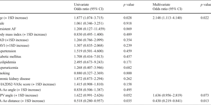
Table.3 Results of logistic regression analysis for predicting transmural thermal injury
Ao aorta, AF atrial fibrillation, CF contact force, CI confidential interval, LAD left atrial diameter, LAVI left atrial volume index, LIPV left inferior pulmonary vein, RF radiofrequency, SD standard deviation, TTI transmural thermal injury
vs. 5.8 ± 1.6 mm,P= 0.030) compared to those in the TTI (−) group. There was no difference in LA-Ao angle between the two groups with and without TTI (Table1). As risk factors of atherosclerosis, each prevalence of hypertension, diabetic mellitus, dyslipidemia, hyperuricemia, smoking, and CKD did not differ between the groups. There was no difference in CHA2DS2-VASc score between the two groups. (Table1).
No difference was observed in nadir balloon temperature during CB ablation, total freezing time, or prevalence of ter- mination of CB ablation due to the LET falling below 15 °C, between the two groups. The prevalence of touch-up ablation for left PV did not differ between the groups. There was no patient in whom the CB ablation was stopped due to the bal- loon temperature falling below−60 °C. (Table2).
3.4 Logistic regression analysis and ROC analysis to predict transmural thermal injury
The results of the logistic regression analysis for predicting TTI are shown in Table3. The multivariate logistic regression analysis revealed that age (odds ratio [OR] 2.148, 95% confi- dential interval [95% CI] 1.113–4.140,P= 0.022) and LA-Ao distance (OR 0.430, 95% CI 0.219–0.841,P= 0.013) were independent predictors of TTI.
We analyzed the ROC curve to identify the best cut-off value of these predictors to distinguish the risk of TTI.
According to the ROC curve analysis, we identified the cut- off age as 65.5 years with 63% sensitivity, 54% specificity, and an area under the curve of 0.643 (95% CI 0.508–0.778).
We also identified the cut-off LA-Ao distance to be 5.9 mm, with 41% sensitivity, 90% specificity, and an area under the curve of 0.651 (95% CI 0.525–0.777) to predict TTI.
4 Discussion
The major finding of the current study was that the age and anatomical proximity of the structures surrounding the esoph- agus were associated with TTI incidence. In addition, our results revealed that the age and LA-Ao distance were independent predictors of TTI. From the results of the present study, aging, which may suggest a compromised periesophageal circulation, and the anatomical proximity of the esophagus and surrounding structures, were important re- garding the prevalence of TTI.
4.1 Mechanism of transmural thermal injury in cryoballoon pulmonary vein isolation
The mechanism of TTI in CB PVI remains unclear; however, its mechanism in RFCA is speculated to be direct thermal injury by a radiofrequency ablation catheter, since the anatom- ical distortion of the LA wall occurs during catheter mapping with a high contact force (> 10 g). [11] During catheter contact with the LA wall with a high contact force, tenting of the LA wall within about 2 mm beyond the LA surface occurs. Both LA wall tenting to the esophagus during RF application and the anatomical proximities of surrounding structures could
Table 3 Results of logistic regression analysis for predicting transmural thermal injury
Univariate pvalue Multivariate pvalue
Odds ratio (95% CI) Odds ratio (95% CI)
Age (+ 1SD increase) 1.877 (1.074–3.715) 0.028 2.148 (1.113–4.140) 0.022
Male 1.061 (0.346–3.251) 0.918
Persistent AF 1.208 (0.127–11.459) 0.869
Body mass index (+ 1SD increase) 0.830 (0.495–1.400) 0.489
LAD (+1SD increase) 1.266 (0.766–2.099) 0.354
LAVI (+1SD increase) 1.307 (0.835–2.068) 0.239
Hypertension 1.519 (0.501–4.600) 0.459
Diabetic mellitus 1.708 (0.416–7.013) 0.457
Dyslipidemia 2.495 (0.673–9.243) 0.171
Hyperuricemia 1.268 (0.407–3.946) 0.682
Smoking 0.880 (0.327–2.369) 0.800
Chronic kidney disease 1.872 (0.873–2.294) 0.262
CHA2DS2-VASc score (+ 1SD increase) 1.415 (0.908–1.810) 0.158
LA-Ao angle (+ 1SD increase) 0.838 (0.506–1.387) 0.495
LIPV angle (+ 1SD increase) 1.622 (0.991–2.626) 0.052 1.636 (0.956–2.819) 0.073
LA-Ao distance (+ 1SD increase) 0.518 (0.280–0.957) 0.035 0.430 (0.219–0.841) 0.013
Aoaorta,AFatrial fibrillation,CFcontact force,CIconfidential interval,LADleft atrial diameter,LAVIleft atrial volume index,LIPVleft inferior pulmonary vein,RFradiofrequency,SDstandard deviation,TTItransmural thermal injury
J Interv Card Electrophysiol
Fig. 1 The parameters used to evaluate the proximities of the esophagus to its surrounding structures on the computed tomography data. LA-Ao angle: the angle of the LA posterior wall to the descending Ao. LIPV angle: the branching angle of the LIPV to the coronal plane.
LA-Ao
distance: the distance between the descending Ao and LA posterior wall.
Ao aorta, LA left atrium, LIPV left inferior pulmonary vein, LSPV left superior pulmonary vein
Fig. 2 Representative case of transmural thermal injury. The left panel shows esophageal erosion and the right panel shows gastric hypomotility, which were revealed by esophagogastroduodenoscopy performed 1 day after PVI.
PVI pulmonary vein isolation
Fig.3 Representative case with transmural thermal injury. The patient had a short LA-Ao distance.
Ao aorta, LA left atrium, LIPV left inferior pulmonary vein