Relationship of Grade of Malignant Brain Tumor to Cancer Stem Cells and Survivin Expression
Yusuke K
OBAYASHI1), Akiko S
ASAKI2), Mayumi T
SUJI2), Yuko U
DAKA2), Hideto O
YAMADA2), Yuko T
SUNODA3), Shinichi I
WAI4), Masayuki S
OMEI2), Junichiro K
IZAKI2), Yasuyuki K
ONDO2), Katsuyoshi S
HIMIZU1)and Tohru M
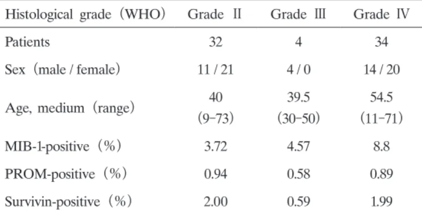
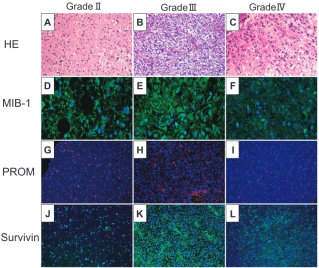
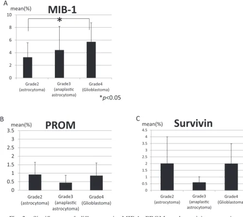
IZUTANI1)Abstract : Glioblastoma (GBM) is difficult to completely cure by surgical treatment alone, and it is generally treated with a combination of surgery, radiotherapy, and chemotherapy. However, GBM is resistant to radiotherapy and chemotherapy, and complete cure cannot be achieved. Cancer stem cells (CSC) and survivin, which inhibit apoptosis, are considered as factors underlying tumor recurrence and the radiation- and drug-resistance of these tumors. We analyzed CSC and survivin expression in surgically excised specimens of malignant brain tumors to establish the relationships between the grades and CSC and survivin expression and between MIB-1(Ki-67) expression and resistance. No relationship was noted between the grades and CSC or survivin expression, or between MIB-1 and CSC expression or between Grade 3 and 4 MIB-1 and survivin expression, although a correlation was noted between MIB-1 and survivin expression in Grade Ⅱ tumors. These findings suggested that CSC are consistently contained in tumor tissue at a specific rate regardless of the histological grade, and the apoptosis of cells with low-level prolif- erative and cell cycling activities does not occur because these cells do not respond to chemotherapy or radiation, being resistant to treatment.
Key words : cancer stem cells, survivin, glioblastoma
Introduction
Among primary brain tumors, highly malignant, high-grade glioblastoma (59%) and medulloblastoma (62%) show a high incidence in males and frequently develop in the 40〜
50s age group. Glioblastoma (GBM) and medulloblastoma of grade Ⅳ, based on the WHO classification, grow rapidly and are fatal if untreated. Histologically, these brain malignancies show abundant cell division and necrosis, rapidly infiltrate the surrounding tissue, and disseminate to the meninx
1). GBM and medulloblastoma are therefore highly malignant tumors associated Original
1)
Department of Neurosurgery, Showa University School of Medicine, 1—5-8 Hatanodai, Shinagawa-ku, Tokyo 142-8666, Japan.
2)
Department of Pharmacology, Showa University School of Medicine, 1-5-8 Hatanodai, Shinagawa-ku, Tokyo 142-8555, Japan.
3)
Breast Center, Kameda Medical Center, 929 Higashi-cho, Kamogawa City, Chiba 296-8602, Japan.
4)