J. Pack. Sci. Tech. Vol. 12 No. 6 (2003)
A p p l i c a t i o n o f a c o n t a c t d e h y d r a t i n g s h e e t t o t h e p r e p a r a t i o n o f a n i m m o b i l i z e d - e n z y m e
m e m b r a n e f o r b i o s e n s o r
Sachie OGAWA, Naoko HAMADA-SATO* , Takeshi KOBAYASHI, C h i a k i I M A D A a n d E t s u o W A T A N A B E
Some enzymes were immobilized in an inner layer of a filter paper using the osmotic action of a contact dehydrating sheet (CDS). CDS was examined to determine if the efficiency and rapidity of an enzyme reaction could be improved, thereby making better of the preparation of an immobilized-enzyme membrane. The activity of the membrane, in which an enzyme exists in its layer, was compared with that of a conventional enzyme adsorption membrane. pH was examinedmodulated for the enzyme to obtain a higher migration rate into the membrane. The membrane in which layer an enzyme exists and the enzyme adsorption membrane were placed on the oxygen electrode. The glucose sensor, the D-amino acid sensor and the sarcosine sensor were constructed. The use of CDS successfully increased enzyme migration into the membrane and enhanced the signal intensity per unit of enzyme. From this point of view, there is a very strong possibility that biosensors utilizing an immobilized-enzyme membrane prepared with CDS can be developed. This is the first report on the application of CDS to bioengineering fields e x c e p t f o r f o o d w r a p p i n g .
Keywords : Immobilized-enzyme membrane, contact dehydrating sheet (CDS), enzyme accumulation me mbrane, enzyme adsorption me mbrane.
1 . I n t r o d u c t i o n
A c o n t a c t d e h yd ra t i n g s h e e t ( C D S ) w a s i n v e n t e d a s a ma t e r i a l fo r re mo v i n g wa t e r f ro m f o o d s s u c h a s f i s h a n d m e a t b y o s m o t i c a c t i o n w i t h o u t h e a t i n g , f r e e z e - d r y i n g a n d r e l a t e d p r o c e s s e s . C D S c o n s i s t s o f t wo p i e c e s o f p o l yv i n yl a l c o h o l a t e f i l m c o mp o s e d o f e d i b l e s u g a r placed between two layers of a semi-perme able me mb rane made of polyvinyl alcohol1 )~3 ). The f i l m h a s m a n y p o r e s t h a t e n a b l e t h e p e r m e a t i o n o f s m a l l m o l e c u l e s s u c h a s w a t e r a n d t r i me t h y l a m i n e b u t r e t a i n s r e l a t i v e l y l a r g e mo l e c u l e s s u c h a s p r o t e i n s a n d e n z y me s4 ). T o u s e CDS, for instance, a piece of meat should be wrapped completely allowing full contact between C D S a n d t h e e n t i r e s u r f a c e o f t h e me a t ( f o r a p r e - d e t e r mi n e d p e r i od of storage at 0~5℃) . Even at such low temperatures, CDS can sel ectively remove water fro m fo od through its semi p e r m e a b l e m e m b r a n e .
___________________________________________________________________________________________
" C o r r e s p o n d i n g a u t h o r . E - m a i l : h s n a o k o @ s . k a i y o d a i . a c . j p
Laboratory of Applied Microbiology, Graduate School of Marine Science and Technology, Tokyo University of Marine Science and Technology , 5-7, Konan 4, Minato-ku, Toky o 108-8477, JAPAN
一般論文
When compared to conventional chemical analytical techniques, biosensor has a number of advantages that include the direct and accurate detection of components in samples, rapidity of signaling, convenience in operation, and high biospecificity. One disadvantage of biosensors is their lack of stability. The progress in enzyme immobilization technology has assisted in further improvement to obtain a stable enzyme activity5 ) 6 ). Conventionally, an enzyme is immobilized on a membrane. In order for an enzyme to function as a receptor, chemical modification methods are used, for example, 1) covalent bonding, 2) crosslinking, 3) covering with polymers, and 4) adsorption7 ). However, an enzyme is damaged by chemical agents during covalent bonding or crosslinking. Furthermore, the disadvantages of these methods include the need for a large amount of enzyme and their high cost.
In this work, we aimed to develop a novel method of immobilizing enzymes inside a membrane using CDS, which has been used so far in only field of food industries as wrapping m a t e r i a l s . C o n v e n t i o n a l l y a n e n z y m e i s i m m o b i l i z e d r a n d o m l y i n a m e m b r a n e . T h e immobilized enzyme system using CDS is developed to improve the efficiency and rapidity of an enzyme reaction. Furthermore, the CDS modification is considered to causes minimal damage to the enzyme. It is also expected that the advantage of the new method lies in its s i mp l e p rep arati on and lo w co nsu mp t ion o f e nz yme s .
2 . M a t e r i a l s a n d M e t h o d s
2 . 1 . M a t e r i a l s
Glucose, D-alanine, sarcosine and glucose oxidase (GO) (E.C.I.1.3.4 from Aspergillus sp.) (pI 4.2) were obtained from Wako Co. (Osaka, Japan). D-amino acid oxidase (AO) (E.C.1.4.2.2 from porcine kidney) (pis 4.0-7.0) and sarcosine oxidase (SO) (E.C.I.5.3.1 from Arthrobacter sp.)(pl 4.9) were purchased from Funakoshi Co. (Tokyo, Japan) and Toyobo Co.
(N.Y., U.S.A.), respectively. All the materials were used as received.
2.2. Preparation of enzy me solution
GO and SO (0.15 U each) were dissolved in 6 μl of 0.1 M phosphate buffer at pHs 5.8 and 7.0, respectively. AO (0.15U) was also dissolved in 6 μl of 0.1 M Tris-HCl buffer at pHs 7 . 0 a n d 8 . 2 .
2.3. Preparation of enzyme adsorption membrane (AM) and membrane in which an enzyme e x i s t s i n i t s l a y e r ( L M )
The enzyme adsorption membrane (AM) was prepared using a filter membrane from Application of a contact dehydrating sheet to the preparation of an immobilized-cnzyme niembrare for biosensor
J.Pack.Sci. Tech- Vol. 12 No.6 (2003)
ADVANTEC Co. (No. 51, depth 0.17 mm, Tokyo, Japan), which was cut into 5×5 mm pieces.
Each piece was dipped in 6 μl of the above enzyme solution. To prepare the membrane in which layer an enzyme exists, the above AM was placed on CDS (Showa Denko Plastic Products Co., Ltd, Tokyo, Japan) overnight in a refrigerator at 4°C. The longer the membrane remains on the surface of the CDS, the further the enzyme migrates into the membrane, which should facilitate efficient enzyme reaction.
2.4. Enzyme electrode preparation
The oxygen electrode employed in this study was a Clark-type electrode consisting of a platinum cathode, a lead anode, an alkaline electrolyte (KOH) and an oxygen-permeable Teflon membrane. The enzyme membranes were placed on the Teflon membrane of the oxygen electrode and covered with a dialysis membrane. The LM was carefully placed on the Teflon membrane of the oxygen electrode, with the enzyme layer touching the cathode.
2.5. Apparatus for sensor system
The measurement system consisted of an enzyme electrode, a flowcell, an electrometer (Hokuto Denko, Co., Tokyo, Japan, HE-104), a recorder (Rikadenki, Co., Tokyo, Japan, U- 1), an airpump (Nihon Suisoukougyou Co., Tokyo, Japan, a4000), a peristaltic pump (Rikadenki, Co., Tokyo, Japan, MP-3N), and a buffer tank. The enzyme electrode was fixed in the flowcell, and measurement using this system was continuously performed (Fig. 1).
2.6. Assay procedures
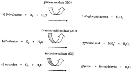
A phosphate-buffered solution was transferred continuously through the sensor by the peristaltic pump. The dissolved oxygen in the buffer was always saturated by aeration. Since the concentration of dissolved oxygen was proportional to the current decrease, 1 kΩ resistance was connected between the enzyme electrode and the recorder in parallel, and current was calculated based on the recorded potential difference. After the output current became steady, a substrate solution was injected into the flow line and current decrease was recorded. The assay was performed with 0.1M phosphate buffer (pH 7.0) for glucose sensor8) and sarcosine sensor9). Tris-HCl buffer (0.1M, pH 8.2) was used as D-amino acid sensorl0). The principle of the above enzyme reactions is shown in Fig. 2. This study was performed at a flow rate of 0.1 ml/min. The temperature was controlled at 30"C during the enzyme reaction. A 50 μl aliquot of the substrate solution was injected into the enzyme sensor system.
Fig. 2. Principle of enzyme reaction, a) Glucose sensor, b) D-amino acid sensor, c) Sarcosine sensor
2.7. Inspection of LM activity
After the enzyme was immobilized in the layer, the membrane was separated into two pieces lengthwise. The side facing CDS was named the lower membrane and the other side of the membrane that did not contact with CDS, was named the upper membrane. These membranes were respectively placed on the oxygen electrode and then the enzyme electrode was placed in the sensor system following the above procedure. A substrate solution was injected to obtain a response. When the lower membrane had a larger response than the upper membrane, it was considered that the LM was established. To gain a response curve, the chart speed was adjusted to either 3 cm/h or 10 cm/h.
Application of a contact dehydrating sheet to the preparation of an immobilized-enzyme membrane for biosensor
J.Pack.Sci. Tech. Vol. 12 No.6 (2003)
3. Results and Discussion
3.1. Preparation of LM
Fig. 3 shows the schematic diagram of the preparation of the LM. Firstly, 5×5 mm of the AM was placed on CDS and then the interface was attached overall. The water migrated inside CDS from the filter membrane due to osmotic pressure. The enzyme can not pass through the semipermeable membrane, which was accumulated at the bottom of the filter m e m b r a n e .
As shown in Fig.4, it was considered that most of the enzyme accumulated in the lower membrane since there was a larger response detected in the lower membrane than in the upper membrane. The LM was prepared in this manner.
The optimal pH for the reaction of glucose with GO to produce gluconolactone is 7.0
Fig. 3. Schematic diagram of enzyme accumulated on one side of intrafilter membrane using CDS.
● H 2 O . ○ E n z y m e ( O x i d a s e ) .
Fig. 4. Relationship between enzyme activities of the inner and outer of membranes determined
with the isoelectric point (pI) observed at pH 5.6 11). The pHs 7.0 and 5.8 were examined using the CDS-immobilized- enzyme system. O2 consumption was higher near the pi than near the pH optimum when examining the lower portion of the membrane, as shown in Fig. 4. pH slightly higher than the pI was used to ensure the solubility of the enzyme but also to decrease the net charge on the protein. At pH 7.0, the enzyme will have a larger net negative charge.
This elevated net charge probably slowed enzyme migration into the membrane due to the electrostatic properties of the membrane. In other words, at a higher net charge (pH 7.0), the interaction between the enzyme and cellulose increases, which leads to slowing migration. On the other hand, enzyme migration near the pi should be faster due to minimized electrostatic and hydrogen bonding interactions of GO with the functional hydroxyl and ether groups of the cellulose membrane. Therefore, it appears that two factors control biosensor efficiency : 1) pH optimum of the enzyme and 2) migration rate.
In the upper membrane, the sensor had a stronger signal at the pH optimum than at the pI (Fig. 4). This result is opposite to that observed in the lower membrane but is still expainable. A greater amount of enzyme should reside in the upper membrane at pH 7.0 (slow migration) that at pH 5.8 (fast migration). This is further evidence that an enzyme migrates into the membrane interior more efficiently at pH 5.8 than at pH 7.0. Thus, GO accumulated in the lower membrane more efficiently at pH 5.8 than at pH 7.0, which also increased the obtainable signal per unit of enzyme. pH 5.8 lies in the range of stable pHs for GO since this enzyme is active between pHs 4.0 and 7.013). The order of efficacy was as follows: pH 5.8 (lower membrane)>pH 7.0 (lower membrane)>pH 7.0 (upper membrane)> pH 5.8 (upper membrane).
It was likewise shown that when examining D-amino acid oxidase, a stronger signal was obtained in the lower membrane compared with the upper membrane at pHs 7.2 and 8.2 (Fig.
4b). Although the optimal pH of this enzyme is 8.2, the LM membrane functioned better when the pH was adjusted to 7.2 (Fig. 4b). pH 7.2 is closer to the pi of the enzyme than pH 8.2.
Sarcosine oxidase was also investigated. Sarcosine oxidase accumulated in the lower membrane at pHs 5.8 and 7.0 (Fig. 4c). A higher signal was obtained at pH 5.8, which is closer to the pi of this protein than pH 7.0. The optimum pH of this enzyme is between 7.5 and 8.5, and the pH at which sarcosine oxidase is stable between 6.5 and 9.0. Thus, the pH 5.8 is far from both the optimum pH and pH range at which this enzyme is stable. However, the LM was prepared in better condition more efficient at pH 5.8 than at pH 7.0, which implies the importance of approaching the pI when an enzyme migrates into the membrane using CDS.
When the immobilized- enzyme-membrane was separated into the upper and lower membranes using a pair of tongs, it was considered to increase errors over the depth of the
Application of a contact dehydrating sheet to the preparation of an immobilized-enzymc membrane for biosensor
J . P a c k . S c i . T e c h . V o l . 1 2 N o . 6 ( 2 0 0 3 )
membranes. However, the larger response was continuously observed at the lower membrane.
Thus, enzymes were considered to accumulate on a close limit at the bottom of the lower m e m b r a n e .
The above results suggest that (1) the enzyme LM is obtained as enzymes accumulated on a close limit at the bottom of the membrane by CDS and (2) the optimal pHs of the enzyme solution for enzyme LM preparation are 5.8 for the glucose sensor, 7.2 for the D-amino acid sensor and 5.8 for the sarcosine sensor.
3.2. Differences between AM and LM of enzyme activity
Fig. 5 shows the schematic diagram of both LM and AM placed on the tip of oxygen electrode. Once substrate get into the enzyme immobilization membrane, the oxidation of substrate by enzyme causes the oxygen consumption. Simultaneously, the consumption of o x y g e n a r o u n d t h e m e m b r a n e r e s u l t s t h e c u r r e n t d e c r e a s e s . T h a t i s , t h e s u b s t r a t e concentration is calculated from the amount oxygen consumed. When substrate diffuses into the enzyme immobilization membrane, the diffusion velocity (DV) is shown as the follow f o r m u l a .
Width of filter membrane (x) : 5 mm Length of filter membrane (y) : 5 mm Thickness of filter membrane (z) : 0.17 mm Diffusion Time (t) The number of substrates (N)
Reaction Volume (V) : X, Y, Z
Namely, the diffusion velocity is proportional to the number of substrates per an unit volume of the enzyme membrane. On the other hand, the frequency in a collision between enzymes and substrate was far higher in LM than AM. Because, in the case of AM, enzymes were considered to be immobilized randomly and the reaction distance z1 was 0.17 mm, which equals to the thickness of filter membrane as shown in Fig. 5.
In the case of LM, enzymes were considered to assembled at the bottom (most interior) of the membrane close to the tip of oxygen electrode and the reaction distance z2 was considered to be reach almost to zero. Therefore, the volume of the field which substrate reacts with enzyme was also considered to reach almost to zero. From the results mentioned above, it was concluded that the enzyme reaction advanced more rapidly in the case of LM compared to AM.
Application of a contact dehydrating sheet to the preparation of an immobilized-enzyme membrane for biosensor
Fig. 5. Diagram of substrate diffusion through filter membrane.
3.3. Calibration curve of enzyme sensor
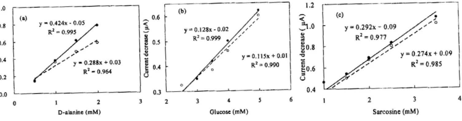
Fig. 6 presents the calibration curve for each of the three sensors. For the D-alanine sensor, as shown in Fig. 6-a, a linear relationship between the concentration of D-alanine and the current decrease was obtained for 0.5~2.0 mM D-alanine. The correlation coefficient (R2) were 0.995 on the enzyme LM and 0.964 on the AM. The slope (x = 0.424) of the calibration curve of the enzyme LM was about 1.5 times larger than the slope (x = 0.288) of the calibration curve of the AM. This indicated that an approximately 50% increase in biosensor efficiency was obtained using the CDS technology. Similarly, using the glucose and sarcosine sensors shown in Fig. 6-b and Fig. 6-c, respectively, linear relationships between the concentrations of glucose and sarcosine and their current decreases were obtained for glucose and sarcosine in the range of 0.5-4.5 mM and 0.05-3.50 mM, respectively. The R2 value for the enzyme LM were 0.999 and 0.977 for the glucose sensor and the sarcosine sensor, respectively. The R2
Fig. 6. Calibration curve for enzyme sensors. ●LM (with CDS). ○AM (without CDS), a) D-alanine sensor, b) Glucose sensor, c) Sarcosine sensor.
J.Pack.Sci. Tech. Vol. 12 No.6 (2003)
value for the AM were 0.990 and 0.985, respectively. Sensors signal was improved by 10% and 20% in the sarcosine and glucose sensor systems, respectively, when using the CDS.
C o n c l u s i o n
A novel method for preparing immobilized enzyme membrane of biosensors using CDS was proposed. By use of CDS, enzyme was migrated into the membrane interior and it was possible to prepare for enzyme accumulation membrane. Enzyme migration rate was optimized by a change of pH in enzyme solution. Enzyme accumulation membrane was examined to determine if it increase accuracy of biosensor signal compared with traditional enzyme adsorption membrane. Improvement of signals by use of CDS was observed among three different type of enzyme membranes, therefore it was expected that CDS successfully increased migration of enzyme into the membrane interior and enhanced signal intensity per unit of enzyme in order to obtain improvement in biosensor efficiency.
A c k n o w l e d g e m e n t s
We would like to thank Mr. K. Saito and Ms. K. Takashima for valuable suggestions.
We thank Showa Denko Plastic Products Co., Ltd for donating CDS.
R e f e r e n c e s
1) Orita, S.. Nihonshokuhinkagakukougakukaishi, 44(12), 86-87 (1997).
2) Takuno, M.: Kagaku, 46(4), 264 (1991).
3) Satou. A.: Shinsozai, 8, 77-78 (1991).
4) Kikuchi, K.: Kagakugijutushi MOL., 5, 1-6 (1987).
5) Skladal, P.: Electroanalysis, 9, 737 (1997).
6) Yabuki, S.: Denki Kagaku, 9, 912-917 (1998).
7) Karube, I.: Biosensor. Kyouritusyuppan, 19 (1986).
8) Jung-Ho , K., Tai-Jin, K., Dong-Hee, R. and Bong-Soo, N.: Food Science and Biotechnology, 7(1), 28-34 (1998).
9) Zeller. H. D., Hille R. and Jornes. H. S. : Biochemistry, 28, 5145-5134 (1989).
10) Chibrongodze,H., Hayashi,K. and Toko, K. : Sensors and Materials, 13(2), 99-106 (2001).
11) Swoboda, B.E.P. and Massey, V. J.BioI.Chem., 240, 2209 (1965).
12) John, S. W. and Dorothy, C. W. : Source book of Enzyme, 112-113 (1997).