Abbreviations: ABP, ambulatory blood pressure; ECG, electrocardiogram; HF, high frequency heart rate fluctuation; LF, low frequency heart rate fluctuation; MESOR, midline estimating statistic of rhythm; MI, mean interval; RMSSD, root mean square successive differences; RR, between two QRS complexes in ECG; %RR50, the proportion of cycles during which the RR difference is > 50 ms; SD, standard deviation Autonomic dysfunction in migraineurs is of
broad interest. In a patient with migraine, symp-toms of autonomic disturbance can be observ-ed, associated with or without migraine attack, such as blood flow in the cranial artery, Horner’s syndrome, etc. However, there is some contro-versy in the results of various studies on the autonomic nervous system. Abnormalities in the sympathetic nervous system have been fairly well documented in migraineurs during the headache-free phase, such as pupillary sym-pathetic hypofunction (Fanciullacci, 1979; Herman, 1983; Takeshima et al., 1987a), ortho-static hypotension (Gotoh et al., 1984;
Havanka-A 48-Hour Holter ECG Study in Migraineurs during Usual Daily Havanka-Activities
Masako Tabata, Naoto Burioka* and Takao TakeshimaDivision of Neurology, Institute of Neurological Sciences and *Third Department of Internal Medicine, Faculty of Medicine, Tottori University, Yonago 683-0826, Japan
In order to clarify the condition of cardiac autonomic nervous activity in migraineurs, 48-h Holter electrocardiograms (ECGs) were carried out under free activity on 20 mi-graineurs during headache free periods and 20 healthy controls. We calculated hourly standard deviation (SD), root mean square successive differences (RMSSD), the propor-tion fo cycles during which the RR difference is > 50 ms (%RR50), low frequency heart rate fluctuation (LF), high frequency heart rate fluctuation (HF) and LF/HF ratio from heart rate variability, and obtained serial 24 data of these parameters. SD, RMSSD, %RR50 and HF are parasympathetic parameters, and LF/HF is sympathetic. These parameters were fit to cosines curve and analyzed circadian rhythms using the cosinor method. By group mean cosinor analysis, the vectors of the amplitude-acrophases of SD, RMSSD, %RR50, LF, HF and LF/HF significantly differed in the migraineurs from those in the controls. The significant differences of vectors of SD, RMSSD, %RR50 and HF indicate cardiac parasympathetic dysfunction. Midline estimating statistic of rhythms (MESORs) of those parameters did not significantly differ except in LF and LF/HF between both groups. MESORs of LF and LF/HF were significantly decreased in the migraineurs, which indicates cardiac sympathetic hypofunction. We concluded that migraineurs might have cardiac parasympathetic dysfunction and sympathetic hypofunction. By evaluating circadian rhythms using cosinor methods, the parameters of heart rate variability in migraineurs were significantly different from controls. Migraineurs may have some disturbance in their rhythm generator.
Key words: autonomic nervous system; circadian rhythm; headache; parasympathetic nervous system; sympathetic nervous system
Kanniainen et al., 1986; Mikamo et al., 1989; Pogacnik et al., 1993), a decrease in overshoot in Valsalva’s maneuver (Gotoh et al., 1984; Havanka-Kanniainen et al., 1986), a low level of plasma norepinephrine (Gotoh et al., 1984; Mikamo et al., 1989) and the cold pressor test (Rubin et al., 1985; Takeshima et al. 1989). However, Thomsen and colleagues (1995) recently reported that there was no significant sympathetic dysfunction in migraineurs. On the other hand, dysfunction of the parasym-pathetic nervous system in migraineurs has not yet been confirmed at all. Some reports showed hyperfunction of parasympathetic activity in
migraineurs (Gotoh et al., 1984) and others reported hypofunction (Havanka-Kanniainen et al., 1986a; Thomsen et al., 1995), in which short period beat-to-beat variations and spectral analysis in a supine position, standing and dur-ing free movement were examined (Appel et al., 1992; Pogacnik et al., 1993). The tone of the sympathetic and parasympathetic auto-nomic nervous systems is influenced not only by emotional, environmental and physical fac-tors, but also by circadian rhythms. In earlier works, many efforts were made to exclude the diurnal variation of autonomic nervous sys-tems, by fixing the examination hour at a cer-tain time of day. In general, sympathetic activi-ties rise from morning to afternoon and para-sympathetic activities rise during night time (Furlan et al., 1990). Depending on the time when the examinations are done, the results of the examinations on the autonomic nervous sys-tem may differ and these may be the reason why reported results are contradictory. Recently, we performed a 2-day measurement of ambulatory blood pressure (ABP) in migraineurs and found a decrease in the circadian amplitude of ABP, which suggests sympathetic hypofunction and dysfunction of the central circadian rhythm generator (Takeshima et al., 1997). Measuring serial data and applying rhythm analysis tech-nology for a 2-day period can reveal a subject’s state of autonomic function including diurnal variations. Heart rate fluctuations are regulated mainly by the activities of the parasympathetic nervous system. In this study, we evaluated heart rate variability in migraineurs under free daily activity for 2 days, by means of Holter electrocardiogram (ECG) recordings, time
domain statistical analysis and spectral analysis of heart rate fluctuations, and cosinor analysis techniques. The aims of this study were: i) to evaluate cardiac parasympathetic tone on the basis of circadian rhythm analysis; and ii) to reveal some specific circadian pattern, if pres-ent, of parameters of heart rate variability in mi-graineurs.
Materials and Methods Subjects
Twenty subjects who were diagnosed as migrai-neurs according to the criteria developed by the Headache Classification Committee of the International Headache Society (1988) had Holter ECG recordings (Table 1) performed on them. They were examined during headache-free periods. The mean period between the last attack and the Holter recording was 18.8 ± 19.1 days (mean ± SD, range 1–65 days). During the examinations, they were free from prophylactic medication. Before starting this study, they took anti-migraine drugs only when they had attacks. Two of the migraineurs used a mix-ture of analgesics 1.0 g (Sedes G, Shionogi, Tokushima, Japan) and metoclopramide 5 mg, 2 days before the examination. The others were free from the abortive use of anti-migraine drugs by at least 7 days prior to testing. The control group consisted of 20 healthy volun-teers (Table 1). All participants were neuro-logically normal, free from respiratory disease and gave their consent after fully understanding the procedure, nature and scope of the study.
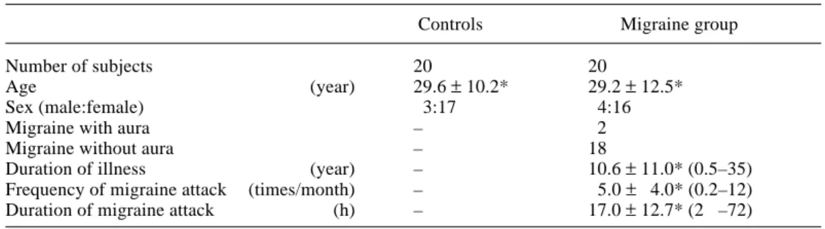
Table 1. Subjects
Controls Migraine group
Number of subjects 20 20
Age (year) 29.6 ± 10.2* 29.2 ± 12.5*
Sex (male:female) 3:17 4:16
Migraine with aura – 2
Migraine without aura – 18
Duration of illness (year) – 10.6 ± 11.0* (0.5–35)
Frequency of migraine attack (times/month) – 5.0 ± 4.0* (0.2–12)
Duration of migraine attack (h) – 17.0 ± 12.7* (2 –72)
( ), range. *Mean ± SD.
No examinees were admitted to the hospital. All subjects were permitted normal daily acti-vity during the 48-h Holter ECG recordings. They were advised to spend their 48-h period as usual, but were prohibited from hard exercise or the intake of alcohol. They were also required to take note of their activities.
The 48-h Holter recordings
During the examination, the ECGs were record-ed continuously using a Space Labs Mrecord-edical- Medical-Holter recorder 90205. Five disposable Ag/Ag-Cl electrodes were located so as to record ECG signals corresponding to the V1 and V5 chest leads. In this way, the 48-h sequential ECGs were recorded. The 2 recorded ECG leads were transferred from the Holter recorder to a Holter ECG analysis system (FT2000A Medical Analysis and Review Station, Space Labs Medical, Inc., Redmond, WA).
Time domain analysis and spectral analy-sis
Following the removal of arrhythmia and noise, we carried out, on an hourly basis, time domain statistical analyses which consisted of the RR mean interval (MI), standard deviation (SD), root mean square successive differences (RMSSD) and the proportion of cycles during
which the RR difference is > 50 ms (%RR50) (Fig. 1). These parameters reflect mainly para-sympathetic nervous system activity. Spectral analysis of heart rate fluctuations was perform-ed as follows. Spectral decomposition was imposed on the heart rate time series by a Fast Fourier transformer. The power spectrum of heart rate fluctuations was calculated with a computer-assisted analysis system, a subtrace every 300 s, which was taken every 60 min from the 48-h ECG signal. The analog signal was sampled by an analog-to-digital converter with a frequency of 128 Hz, and a moving win-dow using a straight line was applied for digital smoothing. The heart rate power spectrum was computed in a frequency band from 0.02–0.40 Hz. Hourly low frequency heart rate fluctuation (LF) (0.02–0.15 Hz), high frequency heart rate fluctuation (HF) (0.15–0.40 Hz) and LF/HF ratio (LF/HF) were calculated. The significance of the circadian rhythm of these parameters was tested using the single and the group-mean cosinor methods (Nelson et al., 1979).
Single and group mean cosinor analyses and comparison of heart rate related vari-ables between groups
Hourly averaged values of MI, SD, RMSSD, %RR50, LF, HF and LF/HF were calculated in each subject. As an example, RMSSD values Fig. 1. Definition of time domain analysis: MI, SD, RMSSD and %RR50. MI, mean interval; n, number of beats in 60 min; SD, standard deviation; RMSSD, root mean square successive differences; %RR50, the proportion of cycles during which the RR difference is > 50 ms; ti, RR interval in ms.
MI = i = 1 n ti n (ms) SD = (MI – ti) 2 n (ms) RMSSD = (ti – ti–1) 2 n – 1 (ms) %RR50 = (Number of (ti – ti – 1) > 50) n – 1 (%) 60 min R1 R2 R3 Rn t1 t2 t3 tn
for Day 1 from 0900 to 0959, and for Day 2 from 0900 to 0959 were averaged and this value was employed as the RMSSD for 0900. Thus we obtained 24 hourly values of each parameter for each examinee. The significance of the averages of these hourly parameters was exa-mined with the unpaired t-test among groups. The significance of the circadian rhythm of these parameters was tested using the single and the group-mean cosinor methods. We applied cosinor analysis to the 24 serial data using BASIC programming language. Following all the single cosinor analyses, we carried out group mean cosinor analyses and statistical comparisons between groups. Details of the cosinor analysis method have been described elsewhere (Takeshima et al., 1997). In brief, we determined a fitted cosinor curve with a 24-h rhythm from measured values. The cosinor curve is represented as y = M + A cos (ωt + φ), where M = midline estimating statistic of rhythm (MESOR), A = amplitude, ω = 2π/24, φ = acro-phase. According to cosinor analysis, the circa-dian rhythm of any ECG parameter is chara-cterized by MESOR, amplitude and acrophase. The significance of the circadian rhythm was tested by the zero-amplitude test. Single
cosi-nor analysis represents the circadian rhythm of each subject. With χ2-test, we examined the difference between migraineurs and controls in the number of subjects who had significant circadian rhythms in each parameter. The other side, group mean cosinor analysis represents the circadian rhythm of a group. Inter-group differences of MESORs were tested by the MESOR test (Nelson et al., 1979). Amplitudes and acrophases are given as a vector, so it was necessary to test differences in amplitude and acrophase between groups simultaneously. Inter-group differences in the acrophase vector were tested by the amplitude-acrophase test (Nelson et al., 1979). The 5% possibility was regarded as statistically signifi-cant throughout the study.
Results
Time domain analysis, i.e., MI, SD, RMSSD and %RR50, and spectral analysis, i.e., LF, HF and LF/HF
MI, SD, RMSSD, %RR50, LF, HF and LF/HF of 48-h data (mean ± SD), were 799 ± 72 ms,
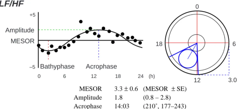
Fig. 2. An example of single cosinor analysis: Single cosinor analysis of LF/HF on a 22-year-old female patient with migraine without aura. Left: The base line is MESOR (3.3) and the points of +5 and –5 indicate MESOR ± 5. The hourly sequential data (J) are fitted to the cosinor curve. Right: Amplitude and acrophase are shown on the cosinor display by a vector on the coordinates. The outside circle indicates a 24-h period. Zero-amplitude test was performed. In a case where the 95% confidential circle, which is displayed by a small circle, doesn’t include a zero-point, the circadian rhythm is regarded as significant. HF, high frequency heart rate fluctuation; LF, low frequency heart rate fluctuation; MESOR, midline estimating statistic of rhythm.
MESOR 3.3 ± 0.6 (MESOR ± SE) Amplitude 1.8 (0.8 – 2.8) Acrophase 14:03 (210˚, 177–243) 12 18 6
LF/HF
0 0 6 12 18 24 (h) Bathyphase Acrophase Amplitude MESOR +5 –5 3.065.8 ± 14.1 ms, 46.0 ± 16.3 ms, 14.2 ± 9.0%, 2141 ± 1073 ms2, 495 ± 485 ms2 and 6.02 ± 2.78 in the controls, and 776 ± 80 ms, 61.7 ± 38.4 ms, 43.5 ± 18.3 ms, 15.2 ± 13.2%, 1683 ± 808 ms2, 582 ± 524 ms2 and 4.21 ± 2.07 in the migraine group, respectively. Averages of all parameters except for LF/HF were not sig-nificantly different between the migraine and the control group. LF/HF in the migraineurs was significantly smaller than that in the con-trols (unpaired t-test, P < 0.05).
Single cosinor analysis of MI, SD, RMSSD, %RR50, LF, HF and LF/HF
A typical example of single cosinor analysis is illustrated in Fig. 2. Nineteen out of 20 controls and all 20 migraineurs showed significant MI rhythms. The incidence of significant circadian rhythms in SD, RMSSD and %RR50 were 9, 15 and 17/20 in the controls and 10, 14 and 18/20 in the migraineurs, respectively. The incidence of significant circadian rhythms of LF, HF and
LF/HF were 10, 13 and 8/20 in the controls and 11, 9 and 11/20 in the migraineurs, respectively. The incidence of significant rhythms of these time domain and spectral parameters showed no significant difference between the migraine group and the controls (χ2-test).
Group mean cosinor analysis of MI, SD, RMSSD and %RR50
With the group mean cosinor method, signifi-cant circadian rhythms were determined in MI, SD, RMSSD and %RR50 in both the migraine group and the controls. In MI, there were no significant differences in the MESOR or the vector of amplitude-acrophase between the 2 groups. The amplitude-acrophase vectors of SD, RMSSD and %RR50 in the migraine group significantly differed from those in the controls (amplitude-acrophase test, P < 0.05). There were no significant differences in MESORs of SD, RMSSD and %RR50 between both groups (MESOR test, Fig. 3).
Fig. 3. Group mean cosinor analysis of MI, SD, RMSSD and %RR50. The amplitude-acrophase vector and MESOR of the MI showed no significant difference between groups. Amplitude-acrophase vectors of SD, RMSSD and %RR50 in the migraine group were significantly different from those in controls (amplitude-acrophase test, P < 0.05, small circles represent the 95% confidence area). MESORs of these parameters showed no significant difference between groups (MESOR test). MESOR, midline estimating statistic of rhythm; MI, mean interval of heart rate; RMSSD, root mean square successive SD; %RR50, the proportion of cycles during which the RR difference is > 50 ms.
RMSSD
*
*
*
0 12 18 6 20 (ms) 0 12 18 6 150(ms) 0 12 18 6 12 (%) 0 12 18 6 15 (ms) Control Migraine MESOR Control 44.5 ± 0.2 Migraine 40.4 ± 0.2 MESOR Control 819.1 ± 1.6 Migraine 781.6 ± 1.5 MESOR Control 15.4 ± 0.2 Migraine 14.3 ± 0.1 MESOR Control 87.5 ± 4.6 Migraine 79.7 ± 3.5 *P < 0.05; amplitude-acrophase testMI
SD
%RR50
Group mean cosinor analysis of LF, HF and LF/HF
With the group mean cosinor method, circadian rhythms existed in LF, HF and LF/HF in both the migraine group and the controls. The amplitude-acrophase vectors of LF, HF and LF/ HF were significantly different between both groups (amplitude-acrophase test, P < 0.05). There were significant differences in the MESORs of LF and LF/HF between the migraine group and the controls (MESOR test, P < 0.05, Fig. 4).
Discussion
In most earlier studies, the time domain statis-tical analysis and power spectral analysis of heart rate variability were examined in a condi-tion of rest, supine posicondi-tion for a short period,
and the examinations were performed at a cer-tain time of day to exclude the influence of cir-cadian variations. We carried out 48-h Holter ECG recordings under free activity in order to obtain data during normal daily activity includ-ing circadian rhythms. Two-day recordinclud-ing and an hourly averaging method over a 2-day period can minimize the influence of environmental, physical and emotional incidental factors and enable an effective analysis of autonomic nervous functions during normal daily activity (Takeshima et al., 1997). Other advantages of this method are: i) the procedures are non-invasive; ii) examination of circadian variations in combination with cosinor analysis or other rhythm analysis technologies is possible; and iii) comparison of groups utilizing group-mean cosinor analysis can be performed.
The time domain analysis parameters, i.e., SD, RMSSD and %RR50, are thought to reflect the activities of the parasympathetic nervous system. LF (0.02–0.15 Hz), which is one type Fig. 4. Group mean cosinor analysis of LF, HF and LF/HF. Amplitude-acrophase vectors of LF, HF and LF/ HF in the migraine group were significantly different from those in controls (amplitude-acrophase test, P < 0.05, small circles represent the 95% confidence area). There were significant differences in the MESORs of the LF and LF/HF ratio between the migraine group and the controls (MESOR test, P < 0.05). These results suggest the dysfunction of the sympathetic nervous system and the possibility of circadian rhythm generators. HF, high frequency heart rate fluctuation; LF, low frequency heart rate fluctuation; MESOR, midline estimating statistic of rhythm.
*
*
0 12 18 6 400 (ms2) 0 12 18 6 700 (ms2) 0 12 18 6 15 (ms) Control Migraine MESOR Control 9.3 ± 0.2 Migraine 5.6 ± 0.2** MESOR Control 2152.1 ± 18.1 Migraine 1584.2 ± 10.8** MESOR Control 503.9 ± 8.5Migraine 524.4 ± 6.3 *P < 0.05; amplitude-acrophase test **P < 0.05; MESOR test
LF
LF/HF
of spectral analysis parameter, is mediated by both sympathetic and parasympathetic contri-butions. Below 0.04 Hz relates to sympathetic and parasympathetic function, and near 0.10 Hz reflects sympathetic activity of the vasomotor reflex. HF is mediated by parasympathetic ner-vous control and is related to respiratory sinus arrhythmia (Akselrod et al., 1981 and 1985; Pomeranz et al., 1985). Therefore, the LF/HF ratio reportedly represents sympathetic nervous control. These parameters of spectral analysis vary with respiratory frequency or posture. On standing with eupnea, the LF component in-creases, which shows a relation to sympathetic nervous activity, while the HF component decreases in the same conditions, relating to parasympathetic nervous activity (Pomeranz et al., 1985; Zigelman et al., 1994). In the present study, we did not record respiration, so we can not tell how much influence it may have on sympathetic function, and still more on LF and HF. All of the subjects were free from any heart or respiratory disease, so we think respiration may not have had muc effect on the data of either group.
In this study, only 2 patients had migraine with aura. We also analyzed data excluding migraine patients with aura (data not shown); the overall results were essentially identical to the presented data. Within 3 days before the examinations, 3 migraineurs each had an attack. One patient, who had an attack 24 h before the Holter ECG recording, did not take any medica-tion. Regarding the possibility that migraine pain by itself might influence their autonomic function, we performed the recordings during a headache free period and long enough after the last attack, so that these influences were mini-mal even if they existed.
Using the rhythm analysis technique, we found significant differences of time domain and spectral analysis parameters of heart rate fluctuation during normal daily activity be-tween migraineurs and controls. The vectors of amplitude-acrophases of SD, RMSSD, %RR50, LF, HF and LF/HF in the migraineurs were sig-nificantly different from those in the controls. There were no significant differences in the MESORs of these parameters except for LF and
LF/HF between the migraine group and con-trols. The significant differences of vectors of amplitude-acrophases for SD, RMSSD, %RR50 and HF indicate parasympathetic dysfunction in migraineurs during headache free periods. The MESORs of LF and LF/HF in migraineurs were smaller, which suggests hypofunction of the sympathetic nervous system.
As for the coordinates, the time of acro-phases in SD, RMSSD and %RR50 seemed almost similar between both groups, while the length of vectors of the migraine group were shorter than those of controls (Fig. 3). The de-crease in amplitudes might contribute mainly to significant differences in the amplitude-acrophase test. The decrease in heart rate variability in mi-graineurs may be due to hypofunction of the parasympathetic nervous system.
In the case of a certain degree of disorder in the effector organs, not only the amplitude but also the MESOR should decrease (Takemiya, 1994). The decreased MESOR of LF/HF sug-gests that sympathetic hypofunction might be associated with a disorder at the level of the effector organs. While only amplitudes of SD, RMSSD and %RR50 were decreased, the oscil-lator of circadian rhythms may form weak rhythms in parasympathetic parameters.
In mammals, the circadian rhythm of the autonomic systems including heart rate fluctu-ations, blood pressure, body temperature and hormonal regulations are generated at the suprachiasmatic nuclei, which have a rich in-nervation of serotonergic neurons (Moore et al., 1978; Malmgren, 1990). Serotonergic abnormal-ities in migraineurs are well documented (Sicuteri et al., 1961; Curran et al., 1965; Takeshima et al., 1987b). Recently, serotonin agonists have suc-cessfully been applied to the treatment of mi-graine. Dysfunction of serotonergic systems may cause the change of the circadian rhythm of heart rate fluctuations in migraineurs.
Considering the presented data, we conclud-ed that migraineurs might have both cardiac parasympathetic and sympathetic hypofunction and might have some disorder in the rhythm generator. Also, the cosinor method seems like a very useful tool in evaluating cardiac auto-nomic dysfunction in migraineurs.
Acknowledgments: We wish to thank Prof. Kenji
Nakashima, Div. of Neurology, Inst. of Neurological Sciences, Prof. Takao Sasaki of the Third Dept. of Internal Medicine, and Prof. Takayuki Nose of the Dept. of Public Health, Faculty of Medicine, Tottori University for their kind advice, useful suggestions and critical readings of the manuscript.
References
1 Akselrod S, Gordon D, Madwed JB, Snidman NC, Shannon DC, Cohen RJ. Hemodynamic re-gulation: investigation by spectral analysis. Am J Physiol 1985;249:H867–H875.
2 Akselrod S, Gordon D, Ubel FA, Shannon DC, Barger AC, Cohen RJ. Power spectrum analysis of heart rate fluctuation: a quantitative probe of beat-to-beat cardiovascular control. Science 1981;213:220–222.
3 Appel S, Kuritzky A, Zahavi I, Zigelman M, Akselrod S. Evidence for instability of the auto-nomic nervous system in patients with migraine headache. Headache 1992;32:10–17.
4 Curran DA, Hinterberger H, Lance JW. Total plasma serotonin, 5-hydroxyindoleacetic acid and p-hydroxy-m-methoxymandelic acid excre-tion in normal and migrainous subjects. Brain 1965;88;997–1010.
5 Fanciullacci M. Iris adrenergic impairment in idiopathic headache. Headache 1979;19:8–13. 6 Furlan R, Guzzetti S, Crivellaro W, Dassi S,
Tinelli M, Baselli G, et al. Continuous 24-hour assessment of the neural regulation of systemic arterial pressure and RR variabilities in ambulant subjects. Circulation 1990;81:537–547. 7 Gotoh F, Komatsumoto S, Araki N, Gomi S.
Nor-adrenergic nervous activity in migraine. Arch Neurol 1984;41:951–955.
8 Havanka-Kanniainen H, Tolonen U, Myllyla VV. Autonomic dysfunction in adult migraineurs. Headache 1986;26:425–430.
9 Headache Classification Committee of the International Headache Society. Classification and diagnostic criteria for headache disorders, cranial neuralgias and facial pain. Cephalalgia 1988;8:1–96.
10 Herman P. The pupil and headaches. Headache 1983;23:102–105.
11 Malmgren R. The central serotoninergic system. Cephalalgia 1990;10:199–204.
12 Mikamo K, Takeshima T, Takahashi K.
Cardio-vascular sympathetic hypofunction in muscle contraction headache and migraine. Headache 1989;29:86–89.
13 Moore RY, Halaris AE, Jones BE. Serotonin neurons of the midbrain raphe: ascending projec-tions. J Comp Neurol 1978;180:417–438. 14 Nelson W, Tong YL, Lee JK, Halberg F.
Meth-ods for cosinor-rhythmometry. Chronobiologia 1979;6:305–323.
15 Pogacnik T, Sega S, Pecnik B, Kiauta T. Auto-nomic function testing in patients with migraine. Headache 1993;33:545–550.
16 Pomeranz B, Macaulay RJB, Caudill MA, Kutz I, Adam D, Gordon D, et al. Assessment of auto-nomic function in humans by heart rate spectral analysis. Am J Physiol 1985;248:H151–H153. 17 Rubin LS, Graham D, Pasker R, Calhoun W.
Autonomic nervous system dysfunction in com-mon migraine. Headache 1985;25:40–48. 18 Sicuteri F, Testi A, Anselmi B. Biochemical
in-vestigations in headache: increase in the hydroxy-indoleacetic acid excretion during migraine attacks. Int Arch Allergy 1961;19:55–58. 19 Takemiya T. Circadian rhythmicity in the
elder-ly. Jiritsu Shinkei 1994;31:400–407.
20 Takeshima T, Mishima K, Tabata M, Burioka N, Nakashima K. Acrophase amplitude of ambula-tory blood pressure decreases in migraineurs. Headache 1997;37:577–582.
21 Takeshima T, Shimomura T, Takahashi K. Platelet activation in muscle contraction head-ache and migraine. Cephalalgia 1987b;7:239– 243.
22 Takeshima T, Takao Y, Takahashi K. Pupillary sympathetic hypofunction and asymmetry in muscle contraction headache and migraine. Cephalalgia 1987a;7:257–262.
23 Takeshima T, Takao Y, Urakami K, Nishikawa S, Takahashi K. Muscle contraction headache and migraine. Platelet activation and plasma nor-epinephrine during the cold pressor test. Cephal-algia 1989;9:7–13.
24 Thomsen LL, Iversen HK, Boesen F, Olesen J. Transcranial doppler and cardiovascular respons-es during cardiovascular autonomic trespons-ests in mi-graineurs during and outside attacks. Brain 1995; 118:1319–1327.
25 Zigelman M, Appel S, Davidovitch S, Kuritzky A, Zahavi I, Akselrod S. The effect of verapamil calcium antagonist on autonomic imbalance in migraine: evaluation by spectral analysis of beat-to-beat heart rate fluctuations. Headache 1994; 34:569–577.