New Strategy for Overcoming Resistance to Chemotherapy of Ovarian Cancer
Junzo KigawaTottori University Hospital Cancer Center, Yonago 683-8504, Japan
The incidence of ovarian cancer in 2008 was projected to be 225,500 new cases and 140,200 deaths worldwide, representing 3.7% of all female cancers and 4.2% of all cancer deaths in women.1 Ovarian cancer, one of the
major causes of death from cancer in women, is com-monly diagnosed at advanced stage. Cytoreductive surgery followed by platinum and taxane-based combi-nation chemotherapy is currently the standard treatment for ovarian cancer.2 Ovarian cancer is one of the most
sensitive solid tumors, with objective responses ranging from 60 to 80% even in patients with advanced stage.3
However, most patients ultimately recur and develop chemo-resistance.
An international study, GOG 182-ICON 5, sought to improve the efficacy of standard platinum-taxane thera-py by incorporating newer cytotoxic agents (gemcitabine, pegylated liposomal doxorubicin and topotecan).4
Un-fortunately, the combination of these agents used in standard therapy has not improved overall survival (OS). Furthermore, the most effective regimen for second line chemotherapy has not yet been determined. The second line chemotherapies have achieved objective responses in about 10 to 30% of patients and even lower percent-age of complete response.5 As a result, the survival rate
for patients with ovarian cancer has not improved over the past 20 years.6 A new strategy is needed to improve
the prognosis of patients with ovarian cancer.
Resistance to chemotherapy presents a major ob-stacle to attempt to improve the prognosis of patients with ovarian cancer. Accordingly, it is important for the management of ovarian cancer to elucidate the mecha-nisms of chemoresistance and to get over the resistance. Recently, the biological characteristics of ovarian cancer have been clear and molecular-targeted agents including signal-transduction inhibitors and anti-angiogenesis have developed. Here, a new strategy for treatment of ovarian cancer is discussed from the viewpoint of the conquest of chemoresistance based on our data.
CHEMORESISTANCE MECHANISMS AND SECOND LINE CHEMOTHERAPY
Anticancer drugs interfere indiscriminately with DNA synthesis, DNA repair systems and mitosis. Many mech-anisms have been postulated to explain chemoresistance, including decreased drug accumulation inside tumor ABSTRACT
Background Ovarian cancer is one of the most sen-sitive solid tumors, with objective responses ranging from 60 to 80% even in patients with advanced stage. However, most patients ultimately recur and develop resistance to chemotherapy. As a result, the survival rate for patients with ovarian cancer has not improved over the past 20 years. Resistance to chemotherapy presents a major obstacle to attempt to improve the prognosis of patients with ovarian cancer. A new strategy is neces-sary to improve the prognosis of patients with ovarian cancer.
Methods The mechanism of chemoresistance was reviewed to get over the resistance. Additionally, the bio-logical characteristics of ovarian cancer and molecular-targeted agents including signal-transduction inhibitors and anti-angiogenesis were discussed.
Results Genetic diagnosis for chemosensitivity with drug-resistance genes may be a useful predictor. Un-fortunately, molecular-targeted therapy alone has been insufficient to improve the prognosis for patients with advanced ovarian cancer. Molecular molecular-targeted therapy should be carried out together with conventional cytotoxic agents. On the occasion of the use of the mo-lecular targeted-agents, care of the appearance of the unexpected adverse effect should be important.
Conclusion The future research in this field will en-able to develop an effective strategy for conquest of che-moresistance in ovarian cancer.
Key words chemotherapy; molecular targeted agent; ovarian cancer; resistance
Corresponding author: Junzo Kigawa, MD [email protected]
Received 2012 December 10 Accepted 2013 March 22
Abbreviations: CDDP, cisplatin; CPT-11, 7-ethyl-10-[4-(1-piperidino)-1-piperidino] carbonyloxycamptothecin; ERK, ex-tracellular signal-regulated kinase kinase; FIGO, International Federation of Gynecology and Obstetrics; GOG, Gynecologic Oncology Group; ICON, International Collaborative Group for Ovarian Neoplasia; KFr, cisplatin-resistant KF; MDR-1, multidrug-resistance-1; MEK, mitogen-activated protein kinase kinase; MRP, multidrug resistance-associated protein; OS, overall survival; PDGF, platelet-derived growth factor; PFS, progression-free survival; PI3K, phosphatidylinositol 3'-kinase; PMA, phorbol 12-myristate 13-acetate; PTX, paclitaxel; topo, topoisomerase; VEGF, endothelium growth factor
cells, increased cellular detoxification and increased DNA repair activity.7, 8, 9 Apoptosis and survival signal
pathway also contribute to sensitivity to anti-cancer agents10, 11 (Fig. 1). Platinum and taxane agents are key
drugs for first line chemotherapy of ovarian cancer.3
Second line chemotherapy should be chosen in consider-ation of chemoresistance mechanisms.
Platinum agent such as cisplatin (CDDP) is one of the most active anticancer agents now used. The length of the progression free interval following the first plati-num-based chemotherapy is the most important predic-tive factor of response to second line chemotherapy.12, 13, 14, 15 The patients who relapse after over 24 months have
the highest chance of responding to the first platinum-based chemotherapy. In contrast, patients who recur within 6 months show low response rate.13
Despite its broad clinical applications, however, in-trinsic or acquired resistance to CDDP often culminates in chemotherapeutic failure. Of the several approaches used to overcome the resistance to CDDP, the use of other drugs given in combination seems to be essential. Experimental studies on the combined effects of CDDP and other anticancer agents can provide information on the most appropriate sequence.
Multidrug resistance-associated protein (MRP) gene functions as an ATP-dependent pump for cytotoxic drugs and MRP plays the significant role for platinum resistance in ovarian cancer.8 Interestingly,
topoisom-erase (topo) I enzymatic activity was 2- to 4-fold higher in the CDDP-resistant cell lines compared with their respective parent cell lines. Additionally, topo II protein contents in resistant ovarian cancer cells were signifi-cantly greater compared with those of their parent cell lines.11 It is reported that the expression levels of
topoi-somerase were associated with sensitivity to topoisomer-ase inhibitors.16, 17
DNA topo inhibitors are of interest as targets for cancer chemotherapy.18
7-Ethyl-10-[4-(1-piperidino)-1-piperidino] carbonyloxycamptothecin (CPT-11) inhib-its topo I through the formation of stable topo I-DNA cleavable complexes.19 CPT-11, a semisynthetic
water-soluble derivative of camptothecin, has a potent antitu-mor activities.19 CPT-11 is known as an effective against
pleiotropic drug-resistant tumors in vitro and in vivo and shows some lack of cross resistance with CDDP.20, 21, 22, 23 Furthermore, the synergism between CDDP and
CPT-11 has been demonstrated and clinical studies showed that the CPT-11/CDDP combination was more effica-cious than CPT-11 alone.24
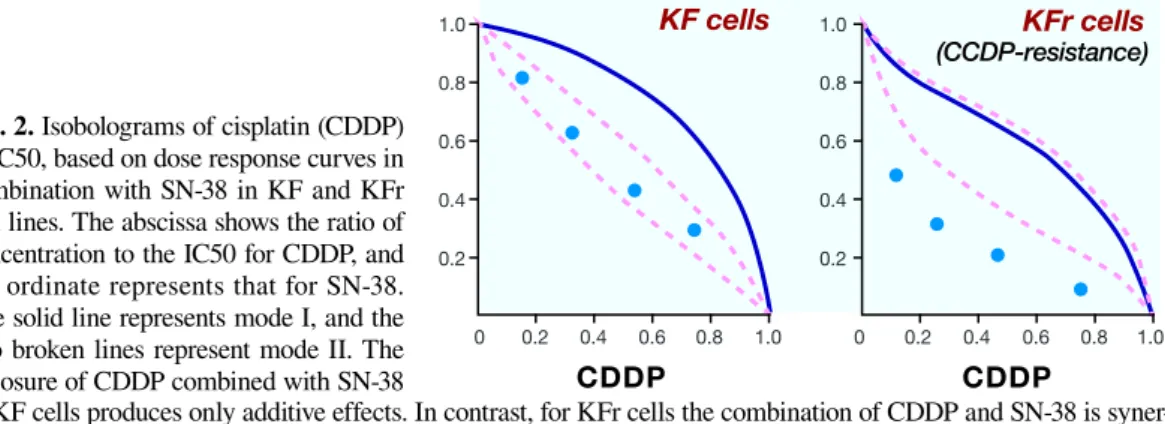
KF cells, epithelial ovarian carcinoma cell line and a CDDP resistant subline, KFr were used as an acquired CDDP-resistant model. The effects of CDDP in com-bination with SN-38, an active metabolite of CPT-11, on KF and KFr cells were analyzed using an improved isobologram method (Fig. 2). For KF cells, the com-bined data points fell within an envelope surrounded by three lines, indicating an additive effect. In contrast, for KFr, CDDP-resistant cell line, the data points were below the envelopes, indicating a supra-additive (syner-gistic) effect. These results suggest that the combination of CDDP and SN-38 may be more effective in CDDP-resistant cells than in the parent cells. To determine the biological factors involved in the sensitivity to the com-bination of CDDP and SN-38, we compared topo I cata-lytic activity between KF and KFr cells (Fig. 3). Topo I activity in KFr was greater than in KF cells; the increase was 4-fold for KFr cells versus KF cells. A potential mechanism to explain the synergism between CDDP and topo I inhibitors has been proposed; an increased DNA interstrand cross-linking by the simultaneous exposure to CDDP and topo I inhibitor and an enhance-ment of the activity of topo I inhibitors by high doses of CDDP.25 Although these findings do not explain why the
enhancement of CDDP cytotoxicity by a topo I inhibitor was observed only in the CDDP-resistant cells, the en-hanced topo I activity could, at least in part, contribute
Fig. 1. Many mechanisms of anticancer agents have been proposed, including decreased drug accumulation, increased cellar detoxification by proteins such as glutathione, decreased the induction of apoptosis and increased DNA repair. GCS, glutamylcysteine synthetase; GSH, glutathione; MDR-l, multidrug-1; MRP, multidrug resistance-associated protein.
to the enhancement of CDDP cytotoxicity in CDDP-re-sistant cells more effectively than in the parent cells. Ad-ditionally, a significant correlation between the sensitiv-ity for SN-38 and topo I activsensitiv-ity was observed. Fukuda et al. demonstrated that the sensitivity of tumor cells to topo I inhibitors was associated with intracellular topo I level.26 Topo I expression reflects sensitivity to topo I
inhibitor.26, 27 Hochster et al.27 found a negative
correla-tion of topo I protein level with area under the curve of topo I inhibitor, and Subramanian et al.28 demonstrated
that topo I activity measured by ICE bioassay related to response to topo I inhibitor. Those findings including our study mentioned above suggest topo I activity may be a useful predictor of resistance or sensitivity to CPT-11 based chemotherapy.
Next, we examined the relationship between topo I activity and the sensitivity to chemotherapy consisting of CDDP and CPT-11 in patients with relapsed ovarian can-cer.29 A total of 30 subjects were selected from patients
for recurrent epithelial ovarian cancer with measurable lesions. All patients had initially undergone platinum-based chemotherapy. All patients underwent surgery to remove recurrent tumor before second line chemothera-py consisting of CPT-11 and CDDP, but did not receive complete resection. Tumor samples were obtained at the surgery. All patients received 50 to 60 mg/m2 CPT-11
intravenously over a 90-min period on days 1, 8 and 15.
Additionally, 60 mg/m2 CDDP was infused
intravenous-ly over a 120- min period on day 1. Of the 30 patients, 18 responded to CPT-11 and CDDP (complete response = 2; partial response = 16) and 12 did not (no change = 8; progressive disease = 4). There were no significant differences in patient characteristics and performance status between the responders and the nonresponders to the second line chemotherapy. The minimum amount of extracted nuclear protein after platinum-based chemo-therapy showing complete DNA relaxation ranged from 15 to 388 ng. The amount of nuclear protein required for complete DNA relaxation was significantly greater in responders than in nonresponders (201.7 ± 92.5 versus 124.1 ± 59.4 ng, P = 0.0164; Fig. 4). This result reflects our in vitro finding that topo I activity was enhanced by exposure to CDDP and related to chemosensitivity. Therefore, the combination chemotherapy consisting of CPT-11 and CDDP may be an effective second line che-motherapy for CDDP resistant ovarian cancer.
GENETIC DIAGNOSIS fOR CHEMOSENSITIv-ITY wITH DRuG-RESISTANCE GENES
In previous chemosensitivity assays, cancer cells were isolated from the tumor, and these cultured cells were exposed to chemotherapeutic agents for evaluating the effectiveness of agents. The assays are complex and
dif-Fig. 3. Topo I catalytic activity in KF and KFr cells. Topo I catalytic activity in KFr cells is 4-fold of that in KF cells. KFr, cisplatin-resistant KF; topo, topoisomerase.
Fig. 2. Isobolograms of cisplatin (CDDP) at IC50, based on dose response curves in combination with SN-38 in KF and KFr cell lines. The abscissa shows the ratio of concentration to the IC50 for CDDP, and the ordinate represents that for SN-38. The solid line represents mode I, and the two broken lines represent mode II. The exposure of CDDP combined with SN-38
to KF cells produces only additive effects. In contrast, for KFr cells the combination of CDDP and SN-38 is syner-gistic. IC50, half maximal inhibitory concentration; KFr, cisplatin-resistant KF.
ficult. In contrast, genetic diagnosis for chemosensitivity is more convenient because cell culture is unnecessary and the transportation of specimen is easy for genetic diagnosis. cDNA microarray analysis of non-small cell lung cancers after laser-capture microdissection of can-cer cells from primary tumors, showing a significant as-sociation between expression levels of genes and chemo-sensitivity.30 The reference of the gene, which is peculiar
to each anticancer agent, may be useful in determining sensitivity.
Although chemoresistance is a multifactorial phe-nomenon, on the occasion of the clinical application, several candidate agents must be tested and validated on small specimens of the patient’s tumor to select effective agents for that individual. Accordingly, it is important to limit the number of examined genes for the cost benefit on the occasion of the clinical application. Anticancer agents against epithelial ovarian cancer are still limited to a few drugs, such as platinum agents, taxane com-pounds and DNA topoisomerase inhibitors.
The expression of multidrug-resistance-1 (MDR-1) gene encoding P-glycoprotein gene was significantly higher in non-responders of taxane and carboplatin therapy.31 Due to the hydrophilic nature of taxane
com-pound, resistance to taxane agent is associated with MDR-1. MDR-1 gene expression is related to sensitivity to taxane compounds in ovarian cancer.32 In contrast,
there were no differences in the level of MRP between responders and non-responders prior to chemotherapy.8
After chemotherapy, however, MRP gene expression was significantly higher in non-responders than in re-sponders. MRP gene expression, at least before chemo-therapy, may not relate to chemoresponse. MDR-1 gene might be a useful predictor for combination taxane and carboplatin therapy.
Copy number of topo I gene inversely correlated with sensitivity to SN38. Additionally, the expression levels of topo I were lower in CPT-resistant cell lines,
and the expression levels of topo IIalpha were lower in etoposide-resistant cell lines than in their parent cell lines that contained fewer copies than their parent cells. The expression of topo I was significantly higher in CPT-11/CDDP responders and topo IIalpha was also higher in etoposide responders, suggesting that the expression level of topoisomerase contributes to sensitivity to topoi-somerase inhibitor.31
As a result, the accuracy of the present genetic diag-nosis for chemosensitivity was 85.7 % for taxane/carbo-platin therapy, 77.8% for etoposide /cistaxane/carbo-platin and 100.0% for CPT-11/CDDP therapy.
MOLECuLAR-TARGETED THERAPY
Recent advances in our understanding of the molecular biology of cancer and carcinogenesis have led to a vari-ety of targeted agents. Targeted agents affect tumor cells, tumor stroma, tumor vasculature and cellular signaling mechanisms that are aberrant in tumor tissue. They are now entering clinical trials and are expected to become attractive treatment options for malignancies.33, 34 Now,
molecular-targeted therapies for ovarian cancer includ-ing signal-transduction inhibitors and anti-angiogenesis have developed and several clinical trials have been performed for ovarian cancer.33, 35, 36 Unfortunately,
molecular-targeted agent alone has been insufficient to improve the prognosis for advanced ovarian cancer. Therefore, molecular-targeted therapy should be carried out together with conventional cytotoxic agents.
Angiogenesis is a process critical for continuous tumor growth, invasion and metastasis. The new blood vessels created by angiogenesis from existing vascula-ture (neovascularization) is a complex process regulated by a number of growth factors and cytokines, such as vascular endothelium growth factors (VEGFs), fibroblast growth factors, angiopoietin, platelet-derived growth factors (PDGFs), tumor necrosis factor-alpha and inter-leukin 6 and 8 receptor pathways. The VEGF family and its receptors (VEGFRs) are one of the major pathways involved in tumor neovascularization. In ovarian cancer, overexpression of VEGF is often observed and has been associated with increased poor prognosis.37
Bevacizumab is a recombinant, humanized, mono-clonal immunoglobulin G1 antibody targeted at VEGF-A. This antibody binds to and neutralizes all biologi-cally active forms of VEGF-A (e.g., VEGF-A165) which suppresses growth of tumors and inhibits progression of metastatic disease.38, 39 Additionally, anti-VEGF drugs
are thought to enhance the effects of chemotherapeutic agents by improving the structure and function (nor-malization) of tumor vessels.40 Two phase III studies,
Fig. 4. The amount of the extraction is sig-nificantly greater in nonresponders than in responders (201.7 ± 92.5 versus 124.1 ± 59.4 ng).
the Gynecologic Oncology Group (GOG) 218 and the International Collaborative Group for Ovarian Neoplasia (ICON) 7, examined the combined effects with bevaci-zumab and standard chemotherapy [paclitaxel (PTX) + carboplatin] in a first-line/adjuvant chemotherapy set-ting.41, 42 The GOG218 trial was a 3-arm, double-blind,
placebo-controlled, randomized, phase III study of 1873 patients with macroscopic residual stage III or any stage IV ovarian, fallopian tube and peritoneal cancer.41
Pa-tients who received bevacizumab (15 mg/kg) plus che-motherapy with 16 cycles of maintenance bevacizumab had significantly longer progression-free survival (PFS) than those who received chemotherapy alone (median PFS, 14.1 versus 10.3 months). However, no significant difference in OS was observed. The rate of hypertension requiring medical therapy was higher in the chemother-apy plus bevacizumab arm compared to chemotherchemother-apy alone, though gastrointestinal events above grade 2 were observed in less than 3 % of patients. ICON7 had a similar design as a double-armed phase III study that enrolled 1528 patients with newly diagnosed high-risk stage I/IIA and stage III/IV ovarian, fallopian tube and peritoneal cancer, who were assigned randomly to che-motherapy alone or cheche-motherapy plus bevacizumab (7.5 mg/kg) with 12 cycles of maintenance bevacizumab.42
Although the dose of bevacizumab used in this trial was half of that used in GOG218, it significantly prolonged PFS (median PFS, 17.4 versus 19.8 months). Hyperten-sion of grade 2 or higher was observed more often in the bevacizumab arm (18 versus 2 % with chemotherapy alone). Among patients at high risk for progression (i.e., FIGO stage IV disease or stage III disease and > 1.0 cm of residual disease after debulking surgery), the median PFS (15.9 versus 10.5 months) and median OS (28.8 ver-sus 36.6 months) were significantly longer in the bevaci-zumab arm.
The Ovarian Cancer Education Awareness Network trial evaluated the efficacy of bevacizumab in com-bination with carboplatin and gemcitabine in patients
with recurrent, platinum-sensitive ovarian, primary peritoneal, or fallopian tube cancers after front-line, platinum-based therapy.43 A taotal of 484 patients were
assigned randomly to a group administered 6 cycles of chemotherapy combined with bevacizumab (15 mg/kg) or to placebo followed by maintenance bevacizumab or placebo until their disease progressed or unaccept-able toxicity developed. PFS for the bevacizumab arm was significantly longer than for the placebo arm (me-dian PFS, 12.4 versus 8.4 months). Grade 3 or higher hypertension and proteinuria occurred more frequently in the bevacizumab arm. Another study, GOG213, is enrolling a targeted 660 patients with platinum-sensitive recurrent ovarian, primary peritoneal, or fallopian tube cancer. This randomized, phase III trial is comparing chemotherapy (PTX + carboplatin) every 3 weeks for 6 to 8 cycles and chemotherapy plus bevacizumab (15 mg/ kg) with maintenance bevacizumab every 3 weeks. The surgical candidates among these patients will undergo secondary randomization to surgery or no surgery.
Chemotherapy-induced activation of cell-survival pathways is observed frequently when conventional anticancer drugs are used, and targeting these survival signals will be invaluable in designing rational and novel combination therapies. A number of agents designed to inhibit the kinase components of those signaling pathways, including mitogen-activated protein kinase kinase (MEK)-extracellular signal-regulated kinase ki-nase (ERK) and phosphatidylinositol 3'-kiki-nase (PI3K)/ Akt, have been developed and clinically tested in cancer treatment, such as lung, breast cancer and melanoma.44, 45 PTX functions by binding to intracellular
beta-tubu-lin, thus stabilizing microtubules. This action blocks normal cell-cycle progress when the mitotic metaphase and anaphase merge, preventing chromosome segrega-tion and tumor-cell death. PTX also has the potential to modulate various signaling pathways, such as the MEK/ ERK signaling cascade and a PI3K–Akt pathway, which are proliferation and cell-survival pathways.46, 47, 48
Ac-Fig. 5. The receiver operat-ing characteristic (ROC) curve according to chemo-response for TC (A), EP (B) and CPT-11/CDDP therapy (C). The optimal cut points are 80 for MDR-1, 90 for topo IIalpha and 200 for topo I by selecting the point on the ROC curve that
maxi-mized both sensitivity and specificity. CDDP, cisplatin; CPT-11, 7-ethyl-10-[4-(1-piperidino)-1-piperidino] carbonyloxycamptoth-ecin; EP, combination with etoposide and CDDP; MDR-1, multidrug-resistance-1; TC, combination with paclitaxel and carboplatin; topo, topoisomerase.
tivating these pathways leads to phosphorylated Bc12 antagonist of cell death and B-cell leukemia/lymphoma (Bcl)-2, and inhibits apoptosis. Amplified PI3K and activated Akt have been found in 30 to 40% of ovarian carcinomas and could represent the mechanism of drug resistance in patients with ovarian cancer.49
Kawaguchi et al. reported that the additive effect of MEK inhibitor combined with PTX in ovarian cancer cell lines tested with a range of pMEK protein expression levels after PTX treatment. Enhanced PTX-induced cell death in the presence of MEK inhibitor may relate to sup-pression of the survival-signaling function of the MEK– ERK pathway, although the exact mechanism is unclear50
(Fig. 5). The additive effect of PI3K inhibitor and PTX in all ovarian cancer cell lines was studied, regardless of the pAkt expression levels. Interestingly, upregulation of pAkt protein expression levels was seen after treating the cells with MEK inhibitor and PTX. Overexpression of activated Akt may decrease the sensitivity to the combi-nation of MEK inhibitor and PTX. Inhibiting MEK and ERK upregulates Akt kinase activity.51 Inhibiting MEK
pharmacologically does not only activate Akt, but also prolongs cell survival in normal cells. These findings suggest that simultaneously suppressing the MEK–ERK and PI3K–Akt pathways could dramatically intensify PTX-induced apoptosis in ovarian cancer cells.
Targeting these MEK–ERK and PI3K–Akt path-ways may be important in treating ovarian cancer. Nonaka et al. treated ovarian cancer cell lines with CDDP and any of a PI3K inhibitor (LY294002), a MEK inhibitor (PD98059), or a MEK/ERK activator [phorbol 12-myristate 13-acetate (PMA)], and assessed cell via-bility, expression of MEK/ERK and PI3K/Akt, cell cycle distribution and apoptosis.52 CDDP and LY294002 had
an additive effect on inhibiting cell growth, and CDDP and PD98059 had and antagonistic effect on cell growth in all cell lines (Fig. 6). In CDDP-resistant cells, CDDP and PMA dramatically suppressed the cell growth,
up-regulated the expression of phosphorylated ERK and cleaved caspase-9, down-regulated the expression of checkpoint kinases, and increased the proportion of cells in the synthesis-phase fraction and apoptotic cells. Those findings indicate that further study is warranted to determine the effectiveness of combination treatment with anticancer agents and molecular targeting therapy for platinum-resistant ovarian carcinoma.
On the occasion of the use of the molecular target-ed-agents, we must take care of the appearance of the unexpected adverse effect.53 Additionally, the
cost-ef-fectiveness should be an important issue. Because tailor made treatment based on the characteristic of the cancer cell is expected, translational research for biomarker is necessary.
The author declares no conflict of interest.
REfERENCES
1 Naniwa J, Itamochi H, Kigawa J. Implication of clear cell and mucinous histology [Internet]. In: Díaz-Padilla I, editor. Ovar-ian cancer – a clinical and translational update. Chapter 5. New York: InTech; c2004-2013. Available from: http://www- cancerorg/Research/CancerFactsFigures/GlobalCancerFacts-Figures/global-facts-figures-2nd-ed.
2 Heintz AP, Odicino F, Maisonneuve P, Quinn MA, Benedet JL, Creasman WT, et al. Carcinoma of the ovary. FIGO 26th Annual Report on the Results of Treatment in Gynecological Cancer. Int J Gynecology Obstet. 2006;95 Suppl 1:S161-92. PMID: 17161157.
3 Japan Society of Gynecologic Oncology, The, editor. [Ovar-ian cancer treatment guideline 2010]. Tokyo: Kanehara; 2010. Japanese.
4 Bookman MA, Brady MF, McGuire WP, Harper PG, Alberts DS, Friedlander M, et al. Evaluation of new platinum-based treatment regimens in advanced-stage ovarian cancer: a Phase III Trial of the Gynecologic Cancer Intergroup. J Clin Oncol. 2009;27:1419-25. PMID: 19224846.
5 Williams LL. Secondary cytoreduction of ovarian malignan-cies. In: Markman M, Hoskins WJ, editors. Cancer of the ovary. New York: Raven Press; 1993. p. 187-203.
Fig. 6. The effects of paclitaxel (PTX) combined with mitogen-acti-vated protein kinase kinase (MEK) and phosphatidylinositol 3'-kinase (PI3K) inhibitors on ovarian cancer cells. Cell proliferation was signifi-cantly suppressed by PTX combined with MEK and PI3K inhibitors compared with other treatment con-ditions. LY294002, PI3K inhibitor; PD98059, MEK inhibitor.
6 Thigpen JT, Bertelsen K, Eisenhauer EA, Hacker NF, Lund B, Sessa C. Long-term follow-up of patients with advanced ovarian carcinoma treated with chemotherapy. Ann Oncol. 1993;4:35-40. PMID: 8312199.
7 Andrews PA, Velury S, Mann SC. Cis-Diamminedichloro-platinum (II) accumulation in sensitive and resistant human ovarian carcinoma cells. Cancer Res. 1988;48:68-73. PMID: 3335000.
8 Kigawa J, Minagawa Y, Cheng X, Terakawa N. Gamma-glu-tamyl cysteine synthetase up-regulates glutathione and multi-drug resistance-associated protein in patients with chemore-sistant epithelial ovarian cancer. Clin Cancer Res 1998;4:1737-41. PMID: 9676849.
9 Lautier D, Canitrot Y, Deeley RG, Cole SP. Multidrug resis-tance mediated by the multidrug resisresis-tance protein (MRP) gene. Biochem Pharmacol. 1996;52:967-77. PMID: 8831715. 10 Kigawa J, Terakawa N. Adenovirus-mediated transfer of a p53
gene in ovarian cancer. Adv Exp Med Biol. 2000;465:207-14. PMID: 10810628.
11 Minagawa Y, Kigawa J, Itamochi H, Kanamori Y, Shimada M, Takahashi M, et al. Cisplatin-resistant HeLa cells are resistant to apoptosis via p53-dependent and -independent pathways. Jpn J Cancer Res. 1999;90:1373-9. PMID: 10665656.
12 Eisenhauer EA, Vermorken JB, van Glabbeke M. Predictors of response to subsequent chemotherapy in platinum pretreat-ed ovarian cancer: a multivariate analysis of 704 patients. Ann Oncol. 1997;8:963-8. PMID: 9402168.
13 Markman M, Rothman R, Hakes T, Reichman B, Hoskins W, Rubin S, et al. Second-line platinum therapy in patients with ovarian cancer previously treated with cisplatin. J Clin Oncol. 1991;9:389-93. PMID: 1999708.
14 Thigpen JT, Vance RB, Khansur T. Second-line chemothera-py for recurrent carcinoma of the ovary. Cancer. 1993;71:1559-64. PMID: 8094320.
15 Thigpen JT, Blessing JA, Ball H, Hummel SJ, Barrett RJ. Phase II trial of paclitaxel in patients with progressive ovarian carcinoma after platinum-based chemotherapy: a Gynecologic Oncology Group study. J Clin Oncol. 1994;12:1748-53. PMID: 7916038.
16 Braybrooke JP, Levitt NC, Joel S, Davis T, Madhusudan S, Turley H, et al. Pharmacokinetic study of cisplatin and infu-sional etoposide phosphate in advanced breast cancer with correlation of response to topoisomerase IIalpha expression. Clin Cancer Res. 2003;9:4682-8. PMID: 14581337.
17 Yasui K, Mihara S, Zhao C, Okamoto H, Saito-Ohara F, Tomida A, et al. Alteration in copy numbers of genes as a mechanism for acquired drug resistance. Cancer Res. 2004;64:1403-10. PMID: 14973057.
18 Drlica K, Franco RJ. Inhibitors of DNA topoisomerases. Bio-chemistry. 1988;27:2253-9. PMID: 2838070.
19 Hertzberg RP, Caranfa MJ, Hecht SM. On the mechanism of topoisomerase I inhibition by camptothecin: evidence for binding to an enzyme-DNA complex. Biochemistry. 1989;28:4629-38. PMID: 2548584.
20 Kunimoto T, Nitta K, Tanaka T, Uehara N, Baba H, Takeuchi M, et al. Antitumor activity of 7-ethyl-10-[4-(1-piperidino)-1-piperidino]carbonyloxycamptothecin, a novel water-soluble derivative of camptothecin, against murine tumors. Cancer Res. 1987;47:5944-7. PMID: 3664496.
21 Masuda N, Fukuoka M, Kusunoki Y, Matsui K, Takifuji N, Kudoh S, et al. CPT-11: a new derivative of camptothecin for the treatment of refractory or relapsed small-cell lung cancer. J Clin Oncol. 1992;10:1225-9. PMID: 1321891.
22 Mathijssen RH, van Alphen RJ, Verweij J, Loos WJ, Nooter K, Stoter G, et al. Clinical pharmacokinetics and metabolism of irinotecan (cpt CPT-11). Clin Cancer Res. 2001;7:2182-94. PMID: 11489791.
23 Tsuruno T, Matsuzaki T, Matsushita M, Fukuoka M, Kusuno-ki Y, Saito H, Yokokura T. Antitumor effect of CPT-11, a new derivative of camptothecin, against pleiotropic drug-resistant tumors in vitro and in vivo. Cancer Chemother Pharmacol. 1988;21:71-4. PMID: 3342468.
24 Kano Y, Suzuki K, Akutsu M, Suda K, Inoue Y, Yoshida M, et al. Effects of CPT-11 in combination with other anti-cancer agents in culture. Int J Cancer. 1992;50:604-10. PMID: 1537625.
25 Minagawa Y, Kigawa J, Irie T, Kanamori Y, Itamochi H, Cheng X, et al. Enhanced topoisomerase I activity and topoi-somerase II alpha content in cisplatin-resistant cancer cell lines. Jpn J Cancer Res. 1997;88:1218-23. PMID: 9473741. 26 Fukuda M, Nishio K, Kanzawa F, Ogasawara H, Ishida T,
Arioka H, et al. Synergism between cisplatin and topoisomer-ase I inhibitors, NB-506 and SN-38, in human small cell lung cancer cells. Cancer Res. 1996;56:789-93. PMID: 8631015. 27 Hochster H, Liebes L, Speyer J, Sorich J, Taubes B, Oratz R,
et al. Effect of prolonged topotecan infusion on topoisomerase 1 levels: a phase I and pharmacodynamic study. Clin Cancer Res. 1997;3:1245-52. PMID: 9815806.
28 Subramanian D, Kraut E, Staubus A, Young DC, Muller MT. Analysis of topoisomerase I/DNA complexes in patients ad-ministered topotecan. Cancer Res. 1995;55:2097-103. PMID: 7743509.
29 Kigawa J, Takahashi M, Minagawa Y, Oishi T, Sugiyama T, Yakushiji M, et al. Topoisomerase-I activity and response to second-line chemotherapy consisting of camptothecin-11and cisplatin in patients with ovarian cancer. Int J Cancer. 1999;84:521-4. PMID: 10502731.
30 Kikuchi T, Daigo Y, Katagiri T, Tsunoda T, Okada K, Kakiu-chi S, et al. Expression profiles of non-small cell lung cancers on cDNA microarrays: identification of genes for prediction of lymph-node metastasis and sensitivity to anti-cancer drugs. Oncogene. 2003;22:2192-205. PMID: 12687021.
31 Naniwa J, Kigawa J, Kanamori Y, Itamochi H, Oishi T, Shi-mada M, et al. Genetic diagnosis for chemosensitivity with drug-resistance genes in epithelial ovarian cancer. Int J Gyne-col Cancer. 2007;17:76-82. PMID: 17291235.
32 Kamazawa S, Kigawa J, Kanamori Y, Itamochi H, Sato S, Iba T, et al. Multidrug resistance gene-1 is a useful predictor of paclitaxel-based chemotherapy for patients with ovarian can-cer. Gynecol Oncol. 2002;86:171-6. PMID: 12144824. 33 Itamochi H. Targeted therapies in epithelial ovarian
can-cer: Molecular mechanisms of action. World J Biolo Chem. 2010;1:209-20. PMID: 21537476.
34 Ma WW, Adjei AA. Novel agents on the horizon for cancer therapy. CA Cancer J Clin. 2009;59:111-37. PMID: 19278961. 35 Kigawa J. Releveance of genetic and epigenetic changes to
treatment. Chemotherapeutic strategies for gynecologic can-cers. In: Kikuchi Y, editor. Chemotherapeutic strategies for gynecological cancers. Kerala: Transworld Research Network; 2011. p. 5-19.
36 Kigawa J. Molecular-targeted therapy for ovarian cancer. (re-view) Int J Clin Oncol. 2012;17:423. PMID: 22926639. 37 Shimogai R, Kigawa J, Itamochi H, Iba T, Kanamori Y, Oishi
T, et al. Expression of hypoxia-inducible factor 1alpha gene affects the outcome in patients with ovarian cancer. Int J Gy-necol Cancer. 2008;18:499-505. PMID: 18476949.
induced angiogenesis suppresses tumour growth in vivo. Nature. 1993;362;841-4. PMID: 7683111.
39 Jain RK. Normalization of tumor vasculature: an emerging concept in antiangiogenic therapy. Science. 2005;307;58-62. PMID: 15637262.
40 Micha JP, Goldsten BH, Rettenmainer MA, Genesen M, Gra-ham C, Bader K, et al. A phase II study of outpatient first-line paclitaxel, carboplatin, and bevacizumab for advanced-stage epithelial ovarian, peritoneal, and fallopian tube cancer. Int J Gynecol Cancer. 2007;17:771-6. PMID: 17343605.
41 Burger RA, Brady MF, Bookman MA, Fleming GF, Monk BJ, Huang H. et al. Incorporation of bevacizumab in the primary treatment of ovarian cancer. N Engl J Med. 2011;365:2473-83. PMID: 22204724.
42 Perren TJ, Swart AM, Pfisterer J, Ledermann JA, Pujade-La-uraine E, Kristensen G, et al. A Phase 3 trial of bevacizumab in ovarian cancer. N Engl J Med. 2011;365:2484-96. PMID: 22204725.
43 Aghajanian C, Blank SV, Goff BA, Judson PL, Teneriello MG, Husain A, et al. OCEANS: a randomized, double-blind, placebo-controlled phase III trial of chemotherapy with or without bevacizumab in patients with platinum-sensitive recurrent epithelial ovarian, primary peritoneal, or fallopian tube cancer. J Clin Oncol. 2012;30:2039-45. PMID: 22529265. 44 Downward J. Cancer biology: signatures guide drug choice.
Nature. 2006;19:274-5. PMID: 16421553.
45 Powis G, Ihle N, Kirkpatrick DL. Practicalities of drugging the phosphatidylinositol-3-kinase/Akt cell survival signaling pathway. Clin Cancer Res. 2006;12:2964-6. PMID: 16707590. 46 Mabuchi S, Ohmichi M, Kimura A, Hisamoto K, Hayakawa J,
Nishio Y, et al. Inhibition of phosphorylation of BAD and
Raf-Biol Chem. 2002;277:33490-500. PMID: 12087097.
47 Qiu L, Di W, Jiang Q, Scheffler E, Derby S, Yang J, et al. Targeted inhibition of transient activation of the EGFR-mediated cell survival pathway enhances paclitaxel-induced ovarian cancer cell death. Int J Oncol. 2005;27:1441-8. PMID: 16211241.
48 Sunters A, Madureira PA, Pomeranz KM, Aubert M, Brosens JJ, Cook SJ, et al. Paclitaxel-induced nuclear translocation of FOXO3a in breast cancer cells is mediated by c-Jun NH2-terminal kinase and Akt. Cancer Res. 2006;66:212-20. PMID: 16397234.
49 Johnstone RW, Ruefli AA, Lowe SW. Apoptosis: a link be-tween cancer genetics and chemotherapy. Cell. 2002;108:153-64. PMID: 11832206.
50 Kawaguchi W, Itamochi H, Kigawa J, Kanamori Y, Oishi T, Shimada M, et al. Simultaneous inhibition of the mitogen-activated protein kinase kinase and phosphatidylinositol 3'-ki-nase pathways enhances sensitivity to paclitaxel in ovarian carcinoma. Cancer Sci. 2007;98:2002-8. PMID: 17900261. 51 Sinha D, Bannergee S, Schwartz JH, Lieberthal W, Levine
JS. Inhibition of ligand-independent ERK1/2 activity in kid-ney proximal tubular cells deprived of soluble survival fac-tors up-regulates Akt and prevents apoptosis. J Biol Chem. 2004;279:10962-72. PMID: 14701865.
52 Nonaka M, Itamochi H, Kawaguchi W, Kudoh A, Sato S, Uegaki K, et al. Activation of the mitogen-activated protein kinase kinase/extracellular signal-regulated kinase pathway overcomes cisplatin resistance in ovarian carcinoma cells. Int J Gynecol Cancer. 2012;22:922-9. PMID: 22672985.
53 Punt CJ, Mol L, Koopman M. Bevacizumab and cancer treatment-related mortality. JAMA. 2011;305:2292. PMID: 21642679.