Morphology of Stomach Ossicles in Brachyura
著者
NAKAMURA Kaworu, TAKEMOTO Takayuki
journal or
publication title
鹿児島大学水産学部紀要=Memoirs of Faculty of
Fisheries Kagoshima University
volume
35
number
1
page range
7-15
別言語のタイトル
短尾類の胃骨形態に関する研究
Vol. 35, No. 1, pp. 7-15 (1986)
Morphology of Stomach Ossicles in Brachyura
Kaworu Nakamura* and Takayuki Takemoto*
Abstract
For understanding of the mechanism of gastric mill function, medial and lateral teeth were morphologically investigated with their articulated ossicles and muscular connections.
In 15 brachyuran species of 5 families, the fundamental structure of the cardiac stomach was the same without remarkable difference among species. Based on positional relations, 13 ossicles and 9 muscles of the cardiac stomach were identified. High movability of the articulation suggested that the propyloric ossicles, linked at each end with the urocardiac or pyloric ossicles, would be a principal part of the gastric mill for manifesting the medial tooth movement. Finally, transcribed illustrations of the medial and lateral teeth of the species examined were presented.
Brachyura is a very specialized organization morphologically in decapod. For example, its digestive tracts diverge, spreading diverticula over the internal space, and the intestine
is extremely short compared to other decapod because of degenerated abdomen1'. These
differentiations have been considered to be derived from its geological adaptability. One of the essential organs for the nutrimental ingestion is the stomach which consists of cardiac, pyloric and gastric mill parts. The gastric mill possesses two typed teeth named medial and lateral teeth, and has been investigated in the crayfish, crabs, lobsters and shrimps
morphologically2"7' and in the crabs and lobsters physiologically8"12', since early times. In
this report, investigation of the teeth morphology is conducted in about 15 brachyuran
species of 5 families, as well as a functional examination of teeth movements for a
preliminary basis of physiological approach.
Materials and Methods
Samplings were carried out from May to the middle of August, at each estuary of Inari, Isaku and Koutsuki rivers as well as the seashore of Hajima in Kagoshima Prefecture.
Following species were collected as materials ' family Grapsidae; Chiromantes dehaani, C.
haematocheir, Eriocheir japonicus, Gaeticedepressus, Helice tridens, Hemigrapsus penicillatus, H.
sanguineus, Parasesarma erythrodactylum, Perisesarma bidens, Sesarmops intermedium, family
Ocypodidae; Ilyoplaxpusilla, Macrophthalmus {Mareotis) japonicus. family Xanthidae; Eriphia
smithii. In addition, Geothlphusa dehaani (family Potamidae) and Charybdis (Charybdis) * Lab. of Propagation Physiology, Fac. of Fisheries, Kagoshima University, 50-20 Shimoarata 4,
Mem. Fac. Fish., Kagoshima Univ. Vol. 35, No. 1 (1986)
feriata (family Portunidae) were collected at Imuretani of Ijuhin City in Kagoshima Pref.
and Nishinoomote Island of Okinawa Pref., respectively.
After identifications of species, materials were supplied to two different experiments as
follows ! I ) Investigation of mechanical relations of stomach ossicles and muscles with biting and grinding functions of the gastric mill. Articular movement of ossicles connected with the gastric mill composed of medial and lateral teeth was especially traced under a vivisectional condition, and their movability was examined. II) Morphological observation and description of medial and lateral teeth of the gastric mill. After fixation with 10 %
formalin, medial and lateral teeth with their related ossicles were extirpated to illustrate
their shapes and recognize a specific difference of medial tooth or a component number of
lateral teeth. The nomenclature followed Maynard and Dandcs article7'.Results and Discussion
Fundamental structure of cardiac stomach (Fig. 1 and 2)
Ossicles and muscles distributed on the cardiac stomach were symmetrically arranged.
The median ossicles of the dorsal wall were arc-shaped mesocardiac ossicle (MCO),
spatula-shaped urocardiac os. (UCO) which was connected to MCO along the latter length
and its ventro-posterior transformed to a medial tooth, and propyloric os. (PPO) with
inferior part articulated at an obtuse angle with the posterior end of UCO. Apart from the
midline, each paired ossicle of pterocardiac os. (PCO) connected to MCO at the end,
zygocardiac os. «(ZCO) which was a slender triangle distorted slightly and posteriorly its
end being opposite to another pair as lateral teeth, exopyloric os. (EPO) and pyloric os.
(PYO) was situated.For the extrinsic muscle, anterior gastric muscle (AGM) at the frontal wall, each of the
endo-lateral posterior gastric m. (ENLPGM),
exo-lateral posterior gastric m.
(EXLPGM) and mesial posterior gastric m. (MPGM) at the posterior wall, were
recognized dorsally. AGM and MPGM had a thick bundle. As for the intrinsic muscle of thedorsal wall, mesial cardio-pyloric m. (MCPM) and lateral cardio-pyloric m. (LCPM) were distinguished. The latter showed superior to and larger than the former.
Connections of these muscles with previously mentioned ossicles were as follows '. AGM
attached itself to MCO and PCO. EXLPGM attached itself to EPO. ENLPGM and MPGM attached to PYO. LCPM connected MCO to PYO. MCPM connected MCO to
PPO.
The median ossicles of the ventral wall was cardio-pyloric valve (CPV). Paired
prepectineal os. (PRPO) articulated to PCO showed its slender body as a beam on the
ventro-lateral part of the cardiac stomach. PRPO connected pectineal os. (PEO) at the posterior end, and the latter linked to postpectineal os. (POPO). PEO was an oval plate
possessing many setae at its edge toward the cavity. POPO was a component of the anterior
appendants of CPV. At the level of PEO, a rod-like subdentate os. (SDO) was situated. Dorsal tip of SDO showed a hook-shape connected to the ventral hollow of the lateral teeth, and the other end articulated with a protrusion of the inferior lateral cardiac os. (ILCO)
EXLPGM
ENLI
MPGM
MCO
Fig. 1. Dorsal and ventral views of ossicles and muscles of the cardiac stomach in brachyura. Upper, dorsal; lower, ventral. Abbrev., oeso, oesophagus; pyl, pylorus: Ossicles; CPV, cardio-pyloric valve; EPO, exopyloric ossicle; ILC0, inferior lateral cardiac ossicle; MCO, mesocardiac ossicle; PCO, pterocardiac
ossicle; PEO, pectineal ossicle; PLCP, postero-lateral cardiac plate; POPO,
postpectineal ossicle; PPO, propyloric ossicle; PRPO, prepectineal ossicle; PYO, pyloric ossicle; SDO, subdentate ossicle; UCO, urocardiac ossicle; ZCO, zygocardiac ossicle: Muscles; AGM, anterior gastric muscle; ALCM, ante
rolateral cardiac muscle; ENLPGM, endo-lateral posterior gastric muscle;
EXLPGM, exo-lateral posterior gastric muscle; LCPM, lateral cardio-pyloric muscle; MCPM, mesial cardio-pyloric muscle; MPGM, mesial posterior gastric muscle; PLCM, postero-lateral cardiac muscle.
10 Mem. Fac. Fish., Kagoshima Univ. Vol. 35, No. 1 (1986) PPO PCO CPV PLCP PRPO ILCO
Fig:. 2. Structural relation of medial and lateral teeth provided to
urocardiac or zygocardiac ossicle, and their connection to other ossicles. Abbrev., CPV, cardio-pyloric valve; EPO,
exopylor-ic ossexopylor-icle; ILCO, inferior lateral cardiac ossexopylor-icle; MCO,
mesocar-diac ossicle; PCO, pterocarmesocar-diac ossicle; PEO, pectineal ossicle; PLCP, postero-lateral cardiac plate; POPO, postpectineal ossicle; PPO, propyloric ossicle; PRPO, prepectineal ossicle:
PYO, pyloric ossicle; SDO, subdentate ossicle; UCO, urocar diac ossicle; ZCO, zygocardiac ossicle.
which formed, with POPO and posterio-lateral cardiac plate (PLCP), the anterior
appendant of CPV.
As for ventral muscles, extrinsic ventro-lateral dilator (VLD) and two intrinsic muscles
as antero-lateral and postero-lateral cardiac m. (ALCM and PLCM) were easily
recognized among other muscles.
Connections of these muscles with ossicles were as follows '. VLD attached to POPO. ALCM, connected ILCO with the ventro-anterior area of ZCO. PLCM connected PLCP
with the ventro-posterior area of ZCO.
Movability of the articulation was different at each jointing site. Its high movability was
observed at two connecting sites of PPO with UCO and PYO. It suggested a dynamic movement of the medial tooth provided to UCO during a grinding procedure.
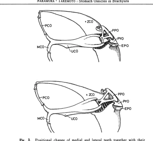
Mechanical consideration of medial and lateral teeth movements (Fig. 3)
Gastric mill function could be divided into two movements, biting and grinding. These movements were conducted by specific muscles related with medial and lateral teeth. At closing procedure of biting, the lateral teeth came in contact with another, rising upward, as
a result of coordinated movements of muscular contraction and relaxation. Due to
examination of articular movability during biting, ZCO provided with lateral teeth was
MCO
Fig. 3. Positional change of medial and lateral teeth together with their related ossicles. Upper, opening of lateral teeth; lower, closing of the teeth. Abbev., EPO, exopyloric ossicle; MCO, mesocardiac ossicle; PCO, pterocardiac ossicle; PPO, propyloric ossicle; PYO, pyloric ossicle; UCO, urocardiac ossicle; ZCO, zygocardiac ossicle.
muscles such as AGM, ALCM, ENLPGM, EXLPGM and PLCM would contract, and
conversely MPGM seemed to be relaxed at closing movement. As for the medial tooth, its
bearer UCO was moved upward or downward to bite or grind together with lateral teeth situated inferiorly. This movement of the medial tooth was conducted mainly by functions
of a few UCO-connected or related muscles as AGM, LCPM and MCPM. As mentioned
previously, UCO had an articular connection with PPO. The latter had further a
fulcrum-linkage to PYO. Powerful and effective movement of UCO would be yielded by an
assurance of the high movability of PPO. The latter seemed therefore to be a principal part of the gastric mill ossicles.
Morphology of medial and lateral teeth of brachyura
Some of medial and lateral teeth examined were drawn in Plates I -M. In same species, the size of both teeth and number of lateral teeth had some variance according to individual
age and sexuality. However, their fundamental structure seemed to be equal not only in species but also in genus or family. As a result, it was difficult to show a phylogenic interpretation concerning teeth morphology of brachyura. Brachyura had been considered to
12 Mem. Fac. Fish., Kagoshima Univ. Vol. 35, No. 1 (1986)
be an extremely differentiated tribe in decapod. It was possible, therefore, to indicate that the characteristic constancy of brachyuran teeth had resulted from a convergence of
functional rationality of the gastric mill.
References
Smith, R. I. (1978) I The midgut caeca and the limits of the hindgut of Brachyura I A
clarification. Crustaceana, 35, 195-205.
HUXLEY, T. H. (1880) '. The Crayfish, an introduction to the study of zoology, Paul, London,
1-371.
PEARSON, J. (1908) : Cancer (the edible crab), L. M. B. C. Mem., 16, Liverpool Univ.
Press, Liverpool, 1-209.
YONGE, C. M. (1924) ! Studies on the comparative physiology of digestion. II- The mechanism
of feeding, digestion, and assimilation in Nephrops norvegicus. J. Exp. Biol., 1, 343-389.
YOUNG, J. H. (1959) '. Morphology of the white shrimp Penaeus setiferus (Linnaeus 1758). Fish.
Bull, 59, 1-168.
Dall, W. (1967) '. The functional anatomy of the digestive tract of a shrimp, Metapenaeus bennettae RACER and DALL (Crustacea ! Decapoda '. Penaeidae). Austr. J. Zool., 15, 699-714. Maynard, D. M. andM. R. Dando (1974) I The structure of the stomatogastric neuromuscular system in Callinectessapidus, Homarus americanus and Panulirus argus(Decapoda Crustacea). Phil.
Trans. R. Soc. Lond., 268 B, 161-220.
SELVERSTON, A. I. and B. Mulloney (1974) : Organization of the stomatogastric ganglion of
the spiny lobster. A. Neurons driving the medial tooth. J. Comp. Physiol., 91, 33-51.
Govind, C. K., H. L. Atwood and D. M. Maynard (1975) : Innervation and neuromuscular
physiology of intrinsic foregut muscles in the blue crab and spiny lobster. J. Comp. Physiol., 96,
185-204.
Hartline, D. K. andD. M. Maynard (1975) : Motor patterns in the stomatogastric ganglion of
the lobster Panulirus argus. J. Exp. Biol., 62, 405-420.
Maynard, D. M. and A. I. Selverston (1975) : Organization of the stomatogastric ganglion of
the spiny lobster. IV. The pyloric system. J. Comp. Physiol., 100, 161-182.
VEDEL, J. P. and M. MOULINS (1977) I Functional properties of interganglionic motor neurons
Family Grapsldae
Chiromantes dehaani Carapace length 28.9mm
No.of lateral teeth 14
Chiromantes haematocheir Carapace length 30.2ma
No.of lateral teeth 14
:r1oche1rJaponicus No.of iateral teeth 18
Family Grapsldae
Gaetice depressus Carapace length 18.2mt>
No.of lateral teeth IS
Helice tHdens No.of lateral teeth 15
Hemigrapsus penicillatus Carapace length 2S.8o»
No.oflateral teeth 13
Plate I. Illustrations of the medial and. lateral teeth of Chiromantes dehaani, Chiromantes
haematocheir, Eriocheir japonicus, Gaetice depressus, Helice tridens and Hemigrapsus penicillatus ( family Grapsidae ).
14 Mem. Fac. Fish., Kagoshima Univ. Vol. 35, No. 1 (1986)
Family Grapsldae
Hemigrapsus sanguineus Carapace length 20.1mm
No.oflateral teeth 13
Parasesarma erythrodactylum Carapace length 16.7mm
No.of lateral teeth IS
Perisesarma bidens No.of lateral teeth 16
Family Grapsldae
Sesarmops Intermedium Carapace length 22.8mm
No.oflateral teeth16
Family Ocypodidae
Ilyoplax pallia, Carapace length 7.2mm
No.of lateral teeth 10
Hacroohthalmus (Mareotis) Japonicus Carapace length 24.5mm
No.of lateral teeth 14
Plate n . Illustrations of the medial and lateral teeth of Hemigrapsus sanguineus, Parasesarma
erythrodactylum, Perisesarma bidens, Sesarmops intermedium ( family Grapsidae ), and two
Family Portunidae
Charybdis (Charybdis) feriata Carapace length 49.8mm
No.of lateral teeth 8
Family Potamidae
Geothelphusa dehaani No.of lateral teeth 15
Family Xanthidae
Eriphia smith 11 Carapace length 20.8mm No.of lateral teeth 8
Plate 11. Illustrations of the medial and lateral teeth of Charybdis ( Charybdis ) feriata ( family Portunidae ), Geothelphusa dehaani ( family Potamidae ) and Eriphia smithii (family