Studies on the Proteases of Marine Bacteria
III : Purification and Some Enzymatic
Properties of Proteases
著者
SAKATA Taizo, SUMIYOSHI Kazuro, KAKIMOTO
Daiichi
journal or
publication title
鹿児島大学水産学部紀要=Memoirs of Faculty of
Fisheries Kagoshima University
volume
26
page range
71-78
別言語のタイトル
海洋細菌のプロテアーゼに関する研究 III : プロ
テアーゼの精製と酵素学的性質について
Mem. Fac. Fish., Kagoshima Univ. Vol.26 pp. 71-78(1977)
Studies on the Proteases of Marine Bacteria—III Purification and Some Enzymatic Properties
of Proteases
Taizo Sakata, Kazuro Sumiyoshi and Daiichi Kakimoto*
Abstract
Four components with casein hydrolysing activity were found in extracellular protease pro duced by a marine Pseudomonas sp. 1-6. Each protease component was fractionated by use of DEAE-Sephadex A-50, Sephadex G-100 and polyacrylamide gel electrophoresis. Protease activity was determined by the digestion of casein in the reaction mixture for fractions obtained on column chromatography and on a casein agar plate for polyacrylamide gels, respectively. The method of a casein agar plate was found to be convenient to detect the protease activity
on the gel.
Protease fractions were alkaline proteases of which optimal pHs were from pH 10.0 to pH 11.0. Proteases F-IIa and F-IIb were very low in activity without Ca++ ion but reactivated with the addition of Ca++ ion remarkably, whereas the activities of F-Ia and F-Ib were un affected or slightly affected by Ca++ ion. Proteases of this strain were stabilized to heat by Ca++ ion but no substantial decrease in activity was observed during 6 hr incubation at 25 C
regardless of the absence of Ca++ ion.
Numerous proteases of terrestrial and marine bacteria have been purified and
characterized1""3^. Kato et al4) showed that a marine bacterium Pseudomonas sp.
No. 548 produced at least four kinds of extracellular protease and that all four en
zymes were inactivated by EDTA and three enzymes were stabilized by Ca++ ion.
In a preceding paper5) it was shown that the supplement of Ca++ ion to a basal suspending medium was indispensable to the production of extracellular protease in
marine isolates.
To determine the effect of cations on enzyme proteins an investigation on purifica tion and some enzymatic properties of a marine isolate Pseudomonas sp. 1-6 has now been undertaken.
Materials and Methods
Organism. A marine isolate, Pseudomonas sp. 1-6 was used in this work.
Medium and cultural conditions. A modification of the medium of ZoBell
2216E contained polypeptone (0.5%) yeast extract (0.1%) and Herbst's artificial sea
water (ASW). Cultures (250 ml of medium in 500 ml flasks) were incubated on a
72 Mem. Fac. Fish., Kagoshima Univ. Vol. 26 (1977)
shaker at 25 C until early stationary phase (24 hr) and harvested by centrifugation.
Enzyme assay. The assay method of protease activity was the same as described previously5^.
Protein concentration. The protein was determined spectrophotometrically by
measuring its absorbance at 280 nm.
Acrylamide gel electrophoresis. Gel electrophoresis was performed by the method of Davis (pH 9.4 gel)6). After electrophoresis of enzyme preparations the
gel was divided lengthwise to two parts with a razor knife. One of them was stained with amino black. The other was put on a casein agar plate and incubated for 6-12 hr at 30 C. A casein agar plate was made from 0.6% casein (Hammarsten) and
1.0% agar mixture in 0.05 sodium carbonate buffer (pH 10.0) and dried at 37 C
overnight.
Results
Purification of proteases
Step 1. Acetone precipitation. Cold acetone was added to culture superna
tant (about 1 1) to 70% (v/v). After standing in a cold room for one hour the pre
cipitate was collected by filtration on a Buchner funnel and dissolved in 0.05 M
Tris-HC1 buffer (pH 7.6, about 30 ml) containing 2 mM CaCl2. Insoluble materials
were removed by filtration and the filtrate was dialyzed for 24 hr against the same
buffer using cellulose tubing (Visking co. type 18/32).
Step 2. DEAE-Sephadex A-50 column chromatography. The dialyzed solu
tion was applied to a DEAE-Sephadex A-50 column (1.5x25 cm) which had been
equilibrated with 0.05 M Tris-HCl (pH 7.6) containing 2 mM CaCl2. The pro teases adsorbed were eluted with a linear gradient increase in sodium chloride con centration at a flow rate of 20 ml per hr and 5 ml fractions were collected. The
reservoir contained 250 ml of 0.05 M Tris-HCl buffer containing 0.7 M sodium chlo
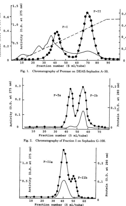
ride and 2 mM CaCl2 and the mixing chamber 250 ml of the same buffer without sodium chloride. A typical chromatogram is shown in Fig. 1. Two active peaks appeared in which the front and rear peaks were designated as F-I and F-II, respec
tively. The active fractions were combined and precipitated by the addition of solid
ammonium sulfate to a final concentration of 0.80 saturation. The precipitate was collected by centrifugation, dissoleved in the same buffer and dialyzed overnight a-gainst the buffer.
Step 3. Sephadex G-100 filtration. The dialyzed solutions were then passed through a column of Sephadex G-100 (2.5 X75 cm) and the buffer was allowed to
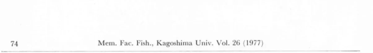
flow at a rate of 16 ml/hr. The elution patterns on Sephadex G-100 of Fractions I and II are illustrated in Figs. 2 and 3, respectively. Enzyme preparations of both Fractions I and II were separated to two fractions with protease activity on a column
pro-a 6 e •H 0) +> O U
Sakata-Sumiyoshi-Kakimoto: Studies on the Proteases of Marine Bacteria—III 73
•1.5 i n CM F - I I „ 0 . 6 -•1.0 Q F-I 7 ^ 0.4 -"0.5 O •P > •H +) 0.2 -n < • • • ^-" • • 1 1 1 1 *"•' # --P d -P •P O 10 20 30 40 50 60 70
Fraction number (5 ml/tube)
80 90
Fig. 1. Chromatography of Protease on DEAE-Sephadex A-50.
0.3
0.2
-0 . 1
-10 20 30 40 50 60 70
Fraction number (5 ml/tube)
Fig. 2. Chromatography of Fraction I on Sephadex G-100.
" 0.2 0.1 a 6 Q) •p o u 10 20 30 40 50 60 °
Fraction number (5 ml/tube)
Fig. 3. Chromatography of Fraction II on Sephadex G-100.
0.8 0.6 0.4 g o u 0.2 3 is
74 Mem. Fac. Fish.. Kagoshima Univ. Vol. 26 (1977)
tease activity of this strain is fractionated to at least four components, which are des ignated as F-Ia, F-Ib, F-IIa and F-IIb.
Acrylamice gel electrophoresis. Acrylamide gel electrophoresis was per formed with each enzyme preparation and protease activity was detected on a casein-agar plate according to materials and methods. As shown in Fig. 4, crude enzyme preparation had four components and each enzyme preparation gave a mixture com
posed of a major band and one or two minor bands with protease activity.
Unfor-F - I I a
Fig. 4. Polyacrylamide Gel Electrophoresis of Enzyme Preparations. The gels were put
on casein agar plates and incubated at 30 C.
Sakata-Sumiyoshi-Kakimoto: Studies on the Proteases of Marine Bacteria—III 75
tunately, the enzyme preparations which were completely pure were not obtained by
the present procedure of purification.
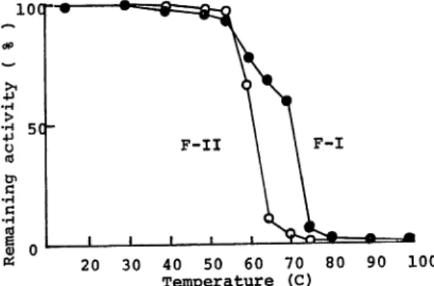
Thermal stability of enzyme activity
The enzyme solutions in 0.05 M Tris-HCl buffer (pH 7.6) with or without the
supplement of Ca++ ion were incubated at various temperatures for 10 min followed
by rapid cooling. The remaining activities were assayed under standard conditions. As shown in Fig. 5, F-I preparation was somewhat more stable than F-II to tem perature. Almost complete inactivation of F-II was observed at 65 C, while most of F-I activity remained at this temperature after ten min, and thermal stability of F-I increased by the addition of Ca++ ion. The result was shown on thermal sta bility of F-II with Ca++ and was not without Ca++ ion because in the absence of
Ca++ ion the activity of F-II decreased markedly.
i o c t a +> o c 5C-<d 6 0) 40 50 60 Temperature 100
Fig. 5. Effect of Temperature on the Stability of Proteases. Enzyme solutions in 0.05 M
Tris-HCl buffer (pH 7.6) with 0.002 M CaCl2 were incubated at the temperature
indicated for 10 min. The remaining activity was determined.
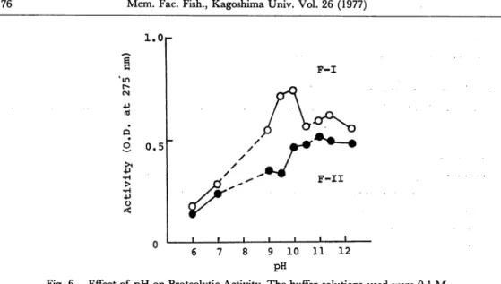
Effect of pH on enzyme activity
Fig. 6 illustrates the effect of pH on the activity of F-I and F-II preparations. Both enzyme preparations showed pH optima at the alkaline side. Optimal activi ties of enzyme preparations F-I and F-II were at pH 10.0 and pH 11.0, respectively.
Effect of cations and some reagents
Tables 1 and 2 summarize the effect of various cations, EDTA and DFP on the
activities of enzyme preparations. The enzyme preparations used in this experiment had been dialyzed against 0.05 M Tris buffer for 24 hr, which was changed one time or three times during dialysis. After enzyme preparations were incubated at 30 C for 1 hr with the addition of various cations (final cone. 1 mM), the remaining activities were determined. Among cations tested heavy metal ions, such as Zn++, Cu++ and Hg++, were inhibitory to the enzyme activities. DFP (diisopropylfluorophosphate,
76 Mem. Fac. Fish., Kagoshima Univ. Vol. 26 (1977) 1.0r 2 0,5 •H > -P O < 6 8 9 10 pH
Fig. 6. Effect of pH on Proteolytic Activity. The buffer solutions used were 0.1 M
Na2HP04-KH2P04 (pH 6.0-7.0), 0.1 M NaHC08-Na2C03 (pH 9.0-10.5)
and 0.1 M glycine-NaOH (pH 11.0-12.5).
F-I
11 12
Table 1. Effect of Chemicals on Protease Activity. The enzyme
preparations were used after dialyzed for 24 hr. The outer fluid was not changed during dialysis.
Chemical Cone. Remaining activity (%)
F-I F-II None 100 100 KC1 1 mM 101 89 LiCl 99 92 AgN03 76 69 MgCl2 93 52 MnCl2 95 89 ZnCl2 40 26 CuSO< 56 27 HgCl2 23 11 CaCl2 85 306 EDTA 78 85 / / 10 40 19 DFP 1 43 11 / / 10 24 12
serine enzyme inhibitor) and EDTA (ethylenediaminetetraacetate, metal chelating agent) inactivated both enzyme preparations F-I and F-II and more inhibitory ef fect was observed on F-II than on F-I. When the dialysis solution was changed 3 times, enzyme preparations F-IIa and F-Ilb lost protease activities almost complete ly and reactivated markedly with the addition of Ca++ ion in contrast to the case of
Sakata-Sumiyoshi-Kakimoto: Studies on the Proteases of Marine Bacteria—III 77
Table 2. Effect of Chemicals on Protease Activity. In this experiment
the enzyme preparations were used after the outer fluid of dialysis was changed three times.
Chemical Cone. Remaining activity (O. D. at 275 nm)
F-Ia F-Ib F-IIa F-IIb
None 0.68 0.36 0.19 0.03 CaCl2 1 mM 0.64 0.39 1.08 0.60 EDTA 1 0.70 — — — / / 10 0.70 0.21 — — DFP 1 0.07 0.16 — — / / 10 0.01 0.17 — —
F-Ia and F-Ib which decreased less in protease activity after exhaustive dialysis.
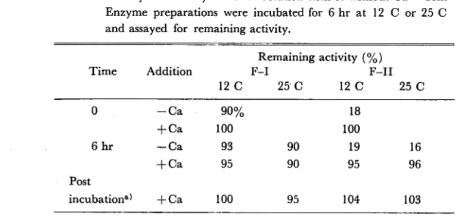
Enzyme stability
In a previous paper it was demonstrated that protease activity of this strain was not observed after incubation for 6 hr in a medium without the supplement of Ca++ ion. To approach the possibilities that enzyme proteins were not released at all into the medium or that enzyme proteins were unstable and lost activity rapidly in the
absence of Ca++ ion, stabilities of enzyme activities during 6 hr incubation period
were examined. After being dialysed thoroughly against 0.05 M Tris-HCl buffer, enzyme preparations F-I and F-II were incubated for 6 hr at 12 C or 25 C with or
without the addition of Ca++ ion (final cone. 5 mM) and assayed for remaining ac
tivity. When remaining activity was estimated, Ca++ ion was added to enzyme solu tions to which Ca++ ion had not been added during incubation. As shown in Table 3, no substantial decrease in enzyme activity was observed after 6 hr incubation both
Table 3. Enzyme Stability in Buffer Solution with or without Ca++ Ion. Enzyme preparations were incubated for 6 hr at 12 C or 25 C and assayed for remaining activity.
Remaining activity (%)
Time Addition F-I F-II
12 C 25 C 12 C 25 C 0 - C a 90% 18 + Ca 100 100 6hr — Ca 93 90 19 16 H-Ca 95 90 95 96 Post incubation*} + Ca 100 95 104 103
a) Enzyme preparations were incubated in 0.05 M Tris-HCl
buffer (pH 7.6) without Ca++ ion and supplemented with CaCl2 (5 mM) after incubation.
78 Mem. Fac. Fish., Kagoshima Univ. Vol. 26 (1977)
at 12 C and 25 C regardless of the absence of Ca++ ion. In the case of F-II, protease activity was very low without Ca++ ion but the addition of Ca++ ion after 6 hr incu bation reactivated the enzyme activity.
Discussion
Four components with casein hydrolysing activity were demonstrated in superna tant medium from a marine Pseudomonas sp. 1-6, by use of two steps of column chro matography with DEAE-Sephadex A-50 and Sephadex G-100 or acrylamide gel-electrophoreses. To examine the purity of enzyme preparations samples of each step in purification were subjected to electrophoresis in polyacrylamide gel and incubated on a casein agar plate to detect protease activities. The method of casein agar plates used in this experiment was found be very convenient to detect protease activity and check the purity of the samples. This method may be able to be applied to various enzymes which hydrolyse macromolecular substances.
The protease fractions obtained were alkaline proteases of which the optimum pHs
were from pH 10.0 to pH 11.0. Fractions F-IIa and F-IIb were reactivated mark edly and became more stable to heat by the addition of Ca++ ion. On the other
hand, Fraction F-Ia was not affected and F-Ib was slightly affected by the presence of Ca++ ion. Fractions F-I and F-II obtained on DEAE-Sephadex A-50 column
chromatography did not decrease in activity substantially during. 6 hr incubation in
buffer solution at 25 C. F-II was especially low in activity without Ca++ but the addition of Ca++ ion after incubation reactivated its activity remarkably. These
results suggest the possibility that proteases were not produced by the cells suspended
in the medium without Ca++ rather than the possibility that proteases produced into the medium lost their activities in the absence of Ca++ ion during 6 hr incubation.
Namely Ca++ ion is suggested to be an indispensable factor which controls the syn
thesis and secretion of extracellular proteases of this strain.
References
1) Hagihara, B. (1958): "The Enzyme", vol. 4, p. 193. Academic Press Inc., New York. 2) Tsuru, D., T. Yamamoto and J. Fukumoto (1966): Studies on bacterial protease-XIII. Agr.
Biol Chem., 30, 651-658.
3) Prestidge, L., V. Gage and J. Spizizen (1971): Protease activities during sporulation in Bacil
lus subtilis. J. BacterioL, 107, 815-823.
4) Kato, N., T. Nagasawa, S. Adachi, Y. Tani and K. Ogata (1972): Purification and proper
ties of proteases from a marine-psychrophilic bacterium. Agr. Biol Chem., 36, 1185-1192.
5) Sakata, T., K. Ueda and D. Kakimoto (1977): Studies on the proteases of marine bacteria
(II). Mem. Fac. Fish. Kagoshima Univ., to be published.
6) Davis, B. J. (1964): Disc gel electrophoresis. II. Method and application on human serum pro