—Reviews—
ホスホジエステラーゼ7A(PDE7A)阻害薬の
免疫および皮膚反応における作用
後 藤 恵
*, 門 嶋 - 山 岡 久 美 子
Effect of Phosphodiesterase 7A Inhibitor on Immune and Skin Reaction
Megumi G
OTO*and Kumiko K
ADOSHIMA-Y
AMAOKAAsubio Pharma Co., Ltd., 6-4-3 Minatojima-Minamimachi, Chuo-ku, Kobe 650-0047, Japan (Received October 29, 2010; Accepted November 30, 2010)
Phosphodiesterase 7A (PDE7A) has been suggested to be involved in activation of T lymphocytes. In the present study, a possible involvement of PDE7A in function of immune cells and skin reaction was investigated using ASB16165, an inhibitor for PDE7A. ASB16165, which has an IC50 value of 15 nM for human PDE7A, suppressed immune responses, cytokine production by T lymphoblasts, cytotoxic T lymphocyte (CTL) induction, NKT cell proliferation and cytokine production. Also, ASB16165 suppressed keratinocyte proliferation and cytokine production in vitro. In phorbol ester 12-O-tetradecanoylphorbol-13-acetate (TPA) induced skin inflammation model, ASB16165 showed inhibitory effect on skin edema and TNF-α production in skin, and proliferation of skin epidermal keratinocyte in vivo. These indicate that PDE7A inhibitors such as ASB16165 will be beneficial for the patients with immunological disorders and skin disorders with TNF-α production or keratinocyte hyperproliferation (e.g. psoriasis).
Key words——phosphodiesterase 7A (PDE7A), cAMP, CTL, NKT, keratinocytes; formononetin
はじめに
ホスホジエステラーゼ(PDE)は,細胞内セカ ンドメッセンジャーである cAMP および cGMP を それぞれ 5'-AMP 及び 5'-GMP に加水分解するこ とにより,細胞内のシグナル伝達を調節する酵素 である.PDE は,現在までに 21 種の遺伝子がク ローニングされ,これらはアミノ酸配列の相同性, 生化学的特性及び阻害薬に対する感受性の違いか ら 11 種類のファミリー(PDE1~PDE11)に分類 されている1, 2).PDE7 は cAMP を特異的に分解する PDE ファミリー分子であり,同じく cAMP を基質 とする PDE4 の阻害薬であるロリプラムに非感受 性の PDE として 1993 年に同定された3).PDE7 に は PDE7A 及び PDE7B の 2 つの遺伝子が存在し, 各々少なくとも 3 種のスプライスバリアントの存 在が報告されている.PDE7A は,脾臓やリンパ節, 血中リンパ球といった免疫系の細胞に特異的に発 現し,PDE7B はリンパ球では検出されず,脳や肝 臓,心臓,甲状腺や骨格筋といった非リンパ組織 に多く発現が認められている4, 5). 免疫応答と PDE7 との関係に関する知見として は,PDE7 は T 細胞などの免疫細胞に発現してい ることが報告されており,さらに PDE7A のアン *アスビオファーマ株式会社,〒 650-0047 神戸市中央区港島南町 6 丁目 4 番 3 号, e-mail: [email protected] 本論考は,後藤 恵,山岡 久美子の博士論文をもとに再構成したものである.チセンスオリゴヌクレオチドが T 細胞の増殖と IL-2 産生を強く抑制することが報告されている6). これらの結果は PDE7 が T 細胞の活性化において 重要な役割を担っていることを示唆している.一 方で,PDE7 阻害薬単独では T 細胞の活性化抑制 には不十分であり PDE4 阻害薬との併用によって のみ顕著な抑制作用を示すという報告7)や,PDE7A のノックアウトマウスで T 細胞機能に異常がない という報告8)もあり,T 細胞における PDE7 の役割 について明確な結論は得られていない.一方,皮 膚における PDE7 の役割についての報告はない. 我々は,PDE7 の生理学的な役割を解析する目的 で,PDE7A 選択的な阻害薬 ASB16165 を使用し 検討を行った.
1. PDE 7A 阻害薬 ASB16165
ASB16165(1-Cyclohexyl-N-[6-(4-hydroxy-1-piperidinyl)-3-pyridinyl]-3-methyl-1H-thieno[2,3-c] pyrazole-5-carboxamide monohydrate (Fig.1) の PDE7 阻害活性及びその他の PDE に対する特異 性を明らかにするため各種リコンビナントヒト PDE を用いて酵素阻害活性を測定した.その結 果,ASB16165 は PDE7A に対して強力な抑制作 用を示し,その IC50は 15 nM であった.一方, PDE4 に対する阻害作用は弱く,IC50は 2.1 μM で, PDE7A に対する IC50よりも約 140 倍高値であっ た.その他の PDE に対する IC50は 10 μM 程度も しくはそれ以上であり,ASB16165 は選択的な PDE7A 阻害薬であることが確認された (Table 1).
2. 免疫細胞における PDE7A 阻害薬の作用
1) T 細胞活性化刺激における PDE7A 阻害薬の
作用
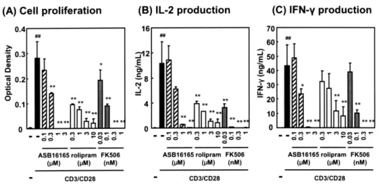
T 細 胞 の 活 性 化 に 対 す る 新 規 PDE7A 阻 害 薬 ASB16165 の作用の有無を明らかにする目的で, マウス脾臓由来の T 細胞を用いて抗 CD3/CD28 抗 体刺激における ASB16165 の効果を検討した.そ の結果,ASB16165 は T 細胞の増殖及び培養上清 中のサイトカイン(IL-2,IFN-γ)産生を濃度依存 的かつ有意に抑制した (Fig.2).PDE4 阻害薬ロリ プラムやカルシニューリン阻害薬 FK506 も同様 の抑制作用を示した. 次に既に活性化した T 細胞に対する ASB16165 の作用を明らかにするため,マウス T 細胞を抗 CD3 抗体刺激と IL-2 培養により作製した活性化 T 細胞(T リンフォブラスト)を用い,IL-12 刺 激で誘導される IFN-γ 産生に対する ASB16165 の 効果を検討した.その結果,ASB16165 は T リン フォブラストからの IFN-γ 産生を濃度依存的かつ 有意に抑制した(Fig.3).PDE4 阻害薬ロリプラム も抑制作用を示したが,カルシニューリン阻害薬 FK506 は抑制作用を示さなかった.一方,細胞内 cAMP 上昇薬であるフォルスコリン及び db-cAMP が抑制作用を示したことから,本試験系におい て細胞内 cAMP 濃度の上昇が IL-12 刺激による IFN-γ 産生に対して抑制的に働くことが示唆され た.また本試験系において,ASB16165 の効果に 対するプロテインキナーゼ A(PKA)阻害薬の作 用を検討した結果,PKA 阻害薬により ASB16165 による抑制作用の一部が解除された(Data not shown)ことから,ASB16165 の効果の少なくと も一部が,PDE7 阻害による cAMP 上昇と,それ による PKA の活性化を介していることが示唆され た9). 以 上 よ り,PDE7 選 択 的 阻 害 薬 で あ るFigure 2 Effect of ASB16165 on proliferation and cytokine production by T cells activated by CD3/CD28
costimulation.
Resting T cells were prepared from C57BL/6N mouse splenocytes using mouse T cell Enrichment Column kit. The cells (4×10⁵ cells/mL) were cultured on anti-CD3 mAb-coated dishes with CD28 (1 μg/mL) in the presence of various
concentration of ASB16165, rolipram or FK506. CD3 mAb-coated dishes has been prepared by incubating with anti-CD3 mAb (10 μg/mL in PBS) for 3 h at 37˚C and washed with PBS before use. (A) Cell proliferation was assessed at 72 h using Cell Counting kit. (B) IL-2 and (C) IFN-γ protein levels in the supernatant were measured using ELISA at 72 h. The data are expressed as mean ± SD of triplicate cultures. ##P < 0.01, compared with the group in which T cells were not activated with
CD3/CD28 costimulation (Student’s t-test). *P <0.05, **P < 0.01, compared with the group in which T cells were activated and
no test compound was added (Dunnett’s test)
Figure 1 Molecular structure of ASB16165
PDE activity, the hydrolysis of cAMP or cGMP to 5’-AMP or 5’-GMP, was determined by a modification of the two-step reaction in which 5’-nucleotidase was used to convert 5’AMP or 5’GMP to adenosine or guanosine in combination with PDE. The reaction mixture (50 μL final volume) contained 20 mM Tris (pH 7.5), 1 mM MgCl2, 0.1 mM EDTA, 330 μg/mL metal-free
BSA, 2 units/mL 5’-nucleotidase, 0.05 μM [3H]-cAMP or 0.15 μM [3H]-cGMP and one of PDE isozymes in the presence of various concentration of ASB16165 and 1% DMSO. The substrate was [3H]-cAMP for PDE7A1, PDE1A3, PDE2A3, PDE3cat, PDE4cat, PDE8A1 and PDE10A1 or [3H]-cGMP for PDE5cat and PDE9A2. In the case of PDE1A3 and PDE2A3 assay, 50 units/mL calmodulin plus 3 mM CaCl2 and 1 μM cGMP was added to the reaction mixture, respectively. The concentration of
each PDE in the reaction mixture was set to hydrolyze about 20% of substrate added to the reaction mixture.
The reaction was initiated by adding the substrate, carried out for 2 h at 25˚C and terminated by adding 100 μL of 50% slurry of QAE-sephadex A-25 in 10 mM HEPES pH 7.0. After settling for about 2 min, 50 μL of supernatants were transferred to another plate containing 100 μL of 50% slurry of QAE-sephadex A-25 in each well and mixed. After settling for about 2 min, 20 μL of supernatants were transferred to a LumaPlateTM (PerkinElmer).
The radioactivity of each well in the LumaPlateTM was counted using the TopCount® NTX microplate scintillation and
luminescence counter (PerkinElmer).
ASB16165 が T 細胞の活性化に伴う増殖とサイ トカイン産生及び活性化 T 細胞からのサイトカイ ン産生を抑制すること,その作用が cAMP 上昇 を介していることが示された.これらの知見は, PDE7A が T 細胞の増殖と機能に重要な役割を担っ ていることを示唆する.
2) 細胞傷害性 T 細胞における PDE7A 阻害薬の
作用
細胞傷害性 T 細胞(CTL)は,CD8+T 細胞が分 化し細胞傷害活性を獲得したエフェクター細胞で あり,自己免疫疾患の発症と病態形成に重要な 役割を担っていると予想される.CD8+T 細胞に 対する ASB16165 の効果を明らかにする目的で, CD8+T 細胞の CTL への分化に対する ASB16165 の 効 果 を 検 討 し た. 異 な る 主 要 組 織 適 合 抗 原 (MHC,マウスでは H-2 抗原)を有する 2 系統の マウスのリンパ球を用いて一方向混合リンパ球反 応(MLR)を行い,同種異系(アロ)特異的な CTL を誘導し,P815 ターゲット細胞に対する傷 害活性を測定したところ,ASB16165 は濃度依存 的かつ有意に傷害活性の誘導を抑制した (Fig.4). 一方,PDE4 阻害薬ロリプラムの効果は部分的で, 最大効果は ASB16165 と比較して弱かった.カル シニューリン阻害薬 FK506 は傷害活性に対して 非常に強い抑制作用を示した. 以上より,PDE7A が CTL の分化において重 要な役割を担っていることが示唆され,CTL が エフェクターとして機能するような種々の自己 免 疫 疾 患 の 治 療 に お い て,ASB16165 な ど の PDE7A 阻害薬が治療薬として有用である可能性 を示唆する10).3) NKT 細胞における PDE7A 阻害薬の作用
Natural Killer T(NKT)細胞は,T細胞マーカー(e.g. T 細胞抗原レセプター /T cell receptor, TCR) 及び NK 細胞マーカー(e.g. NK1.1)の両方を有す るT細胞サブセットであり,活性化された後速や かに Th1/Th2/Th17 サイトカインを産生する.ま た,NK 細胞と同様に癌細胞などの標的細胞に対 して細胞傷害作用を有するが,加えて自己免疫疾 患などの様々な疾患(e.g. I 型糖尿病,多発性硬化 症,強皮症,肝炎)においても重要な役割を担う ことが示唆されている.
Figure 3 Effect of ASB16165 on IL-12-induced IFN-
γ production by T lymphoblasts.
T lymphoblasts were generated by culturing T cells on anti-CD3 mAb-coated dishes for 1 day and subsequent incubation with IL-2 (50 U/mL) for 2 days. CD3 mAb-coated dishes has been prepared by incubating with anti-CD3 mAb (10 μg/mL in PBS) for 3 h at 37˚C and washed with PBS before use. lymphoblasts (1.2×10⁶ cells/mL) were activated by restimulation with
IL-12 (100 ng/mL) for 24 h at 37˚C, and IFN-γ protein level in the supernatant was measured by ELISA. Test compounds (ASB16165, rolipram, FK506, forskolin or db-cAMP) were added to the assays 30 min prior to the activation. The data are expressed as mean ± SD of triplicate cultures. ##P < 0.01, compared with the group in which T lymphoblasts were cultured
without IL-12 for 24 h (Student’s t-test) *P < 0.05, **P < 0.01, compared with the group in which no test compound was added
and T lymphoblasts were cultured with IL-12 for 24 h (Dunnett’s test).
Figure 4 Effect of PDE7 inhibitor ASB16165 on induction of CTL activity in MLR.
Spleen cells from C57BL/6N mice (responder, H-2Db) and MMC-treated spleen cells from BALB/c mice (stimulator, H-2Db)
were cocultured in the presence of various concentration of ASB16165, rolipram or FK506 for 4 days, and cytotoxicity of MLR-induced CTL was assessed by Eu release assay using Eu-labeled P815 cells as target. In brief, cytotoxicity assay was performed by incubating the effector cells with the Eu-labeled P815 target cells (2 x 10⁴ cells) in 96-well round bottom plate
for 4 h at 37˚C. The number of effector cells was dependent on the effector/target cell ratio (E/T ratio). To determine the spontaneous release and maximum release of Eu, 0.1 mL of Eu release assay medium and 1% TritonX-100 were added instead of effector cells, respectively. After 4 h incubation, the supernatant was transferred to 96-well fluorescence plate and mixed with 4-5 times volume of Enhancement solution (PerkinElmer). The Eu release was assessed by time-resolved fluorescence using EnVisionTM 2102 Multilabel Reader (PerkinElmer). The percentage of released Eu of each sample was calculated as
(experimental release - spontaneous release) / (maximum release - spontaneous release) x 100. (A) Effect of ASB16165, rolipram and FK506 on CTL activity induction (E/T ratio=50). (B) Effect of ASB16165 on CTL activity induction at various E/T ratios. ■, 0 μM (i.e. 0.1% of DMSO); ○, 0.1 μM; ×, 0.3 μM; △, 1 μM; ◇, 3 μM of ASB16165 and ◆, MLR was performed without stimulator cells. The data are expressed as mean ± SD of triplicate assays. ##P < 0.01, compared with the group in
which MLR was performed without stimulator cells (Student’s t-test). **P < 0.01, compared with the control group, in which
NKT 細胞における PDE7A の役割を解析する目 的で,マウスより単離した NKT 細胞を用い,抗 CD3 抗体と IL-2 で刺激した際の増殖及びサイト カイン産生に対する ASB16165 の効果を検討し た.その結果,ASB16165 は NKT 細胞の増殖及 びサイトカイン産生を有意に抑制することが明ら かとなった(Fig. 5).同様の効果が細胞内 cAMP 上昇薬によっても認められたことから (Data not shown),NKT 細胞の細胞増殖およびサイトカイ ン産生に cAMP が寄与し,ASB16165 は PDE7A 阻害作用を介して細胞内の cAMP 量を上昇させ ることにより抑制作用を示した可能性が示唆され た.本結果は,NKT 細胞の増殖及び機能における PDE7A と細胞内 cAMP の役割を示す新たな知見 である11).
3. 皮膚における PDE7A 阻害薬の作用
1) ヒト表皮ケラチノサイトにおける PDE7A 阻
害薬の作用
皮膚における PDE7A の役割を明らかにする目 的で,ヒト表皮ケラチノサイトを用い,細胞機能 及び細胞増殖における PDE7A 阻害薬 ASB16165 の作用を検討した.in vitro
においてヒト表皮ケラチノサイトを TPA 及び A23187 で刺激したところ,TNF-α が産生さ れ,この TNF-α 産生を ASB16165 は有意に抑制 した (Fig.6).次に ASB16165 の細胞増殖に対する 作用を検討したところ,ASB16165 による有意な 増殖抑制作用が認められた (Fig.7).PDE4 阻害薬 のロリプラムは,TNF-α 産生及び細胞増殖のいずFigure 7 Effect of ASB16165 on cell proliferation of human epidermal keratinocytes in vitro.
Human keratinocytes were incubated with various concentrations of ASB16165, rolipram, db-cAMP, betamethasone (BD) and vitamin D3 in 96-well collagen I-coated flat bottom plate. After incubation for 48 h, cell proliferation was analyzed with BrdU incorporation assay. The data are expressed as mean ± SD of triplicate cultures. **P<0.01, *P<0.05, compared with the control
group in which no test compound was added (Dunnett’s test).
Figure 6 Effect of ASB16165 on TNF-
α production by human epidermal keratinocytes stimulated with TPA and
calcium ionophore.
Human keratinocytes were incubated with various concentration of ASB16165, rolipram, forskolin and db-cAMP for 30 min in 96-well collagen I-coated flat bottom plate, and TPA (100 ng/ml) and A23187 (1 μg/ml) were added to the culture. After incubation for additional 8 hr, the concentration of TNF-α in the culture supernatant was determined by ELISA. The data are expressed as mean ± SD of triplicate cultures. ##P < 0.01, compared with the group in which keratinocytes were not activated with
TPA plus calcium ionophore (Student’s t-test). **P < 0.01, compared with the group in which keratinocytes were activated and no
test compound was added (Dunnett’s test).
Figure 5 Effects of ASB16165, rolipram and FK506 on cell proliferation and production of IFN-
γ by isolated
murine NKT cells.
For purification of NKT cells, spleen cells from C57BL/6N mice were first labeled with biotin-conjugated anti-NK1.1 mAb, and NK1.1+ cells were subsequently collected by positive selection using anti-biotin microbeads and MACS Systems (Miltenyi Biotech). The enriched NK1.1+ cells were then labeled with FITC-conjugated anti-TCRβ mAb and PE-conjugated streptavidin. NKT cells (TCRβ+/NK1.1+ cells) were purified by sorting with FACS Aria (BD). NKT cells (5 x 10⁴ cells/well, the purity of
the cells was 90.3%) were stimulated with IL-2 (100 U/mL) for 96 h in the presence or absence of various concentration of ASB16165, rolipram, or FK506 in the anti-CD3 mAb-coated plates that has been prepared by incubating with anti-CD3 mAb (10 μg/mL in PBS) for 3 h at 37˚C and washed with PBS before use. (A) Cell proliferation was assessed at 96 h using Cell Counting kit. (B) IFN-γ protein levels in the supernatant were measured using ELISA at 96 h. The data are expressed as mean ± SD of triplicate assays. ##P<0.01, compared with the control group in which cells were not stimulated (Student’s t-test). *P<0.05, **P<0.01, compared with the control group in which cells were stimulated and no compound was added (Dunnett’s test).
れに対しても殆ど抑制作用を示さなかった.一方, これらの試験系で細胞内 cAMP 上昇薬であるフォ ルスコリン或いは db-cAMP が有意な抑制作用を 示したことから,これらの試験系において cAMP が細胞内のセカンドメッセンジャーとして寄与し ている可能性が示唆された. 以上の結果より,ヒト表皮ケラチノサイトにお いて,ASB16165 が細胞機能及び増殖を抑制した ことから,皮膚ケラチノサイトにおいて PDE7A が重要な役割を担っていることが示唆された.
2)
TPA(12-O-tetradecanoylphorbol-13-acetate)誘発耳浮腫モデルにおける PDE7A
阻害薬の作用
TPA 誘発耳浮腫モデルは,強力な発癌プロモー ターとして知られるホルボールエステル(TPA) を皮膚に塗布することにより惹起される皮膚炎モ デルであり,初期の浮腫にケラチノサイトから の TNF-α 産生が関与する可能性が示唆されてい る.また我々は,PDE7A が正常マウスの耳介組織 の表皮層に発現することを確認している12).そこで TPA 誘発皮膚炎モデルを用い,皮膚炎症における PDE7A の役割を明らかにするため ASB16165 の 作用について検討を行ったところ,ASB16165 が TPA 誘発皮膚炎モデルの初期の浮腫反応を用量依 存的に抑制することが明らかとなった (Fig. 8).ま た,本モデルにおける耳介組織中の TNF-α 産生量 を測定したところ,ASB16165 が TNF-α 産生に対 して抑制作用を示した(Fig.9). ASB16165 がin vitro
においてヒト表皮ケラチ ノサイトからの TNF-α 産生を抑制すること,TPA 誘発皮膚炎モデルの初期の浮腫にケラチノサイト からの TNF-α 産生が関与している可能性を考え合 わせると,TPA 耳浮腫モデルにおける ASB16165 の耳介浮腫抑制作用の少なくとも一部は,表皮ケ ラチノサイトからの TNF-α 産生抑制に基づくもの である可能性が示唆される.Figure 8 Effect of ASB16165 on TPA-induced ear edema in mice.
20 μl of TPA (50 μg/ml) containing indicated concentration of ASB16165 or betamethasone dipropionate was applied topically to the both sides of the right ear of BALB/c mice. Ear thickness was measured using a thickness gauge at various time points. The increase in ear thickness (edema) was determined by subtracting ear thickness before painting from that of each time point. (A) The increase of ear thickness at each time points. (B) Area under the curve from 0 hr to 6 hr shown in A. The data are expressed as mean ± SD (N=7). **P < 0.01, compared with vehicle (Dunnett’s test). $$P < 0.01, compared with vehicle
(Student’s t-test)
Figure 9 Effect of ASB16165 on TPA-induced TNF-
α production in the application site of the skin.
20 μl of TPA (50 μg/ml) containing indicated concentration of ASB16165 or betamethasone dipropionate was applied topically to the both sides of the right ear of BALB/c mice, and ear biopsy samples were taken using a punch at 3 hr after TPA application. The biopsies were homogenized vigorously in 50 mM Tris-HCl buffer (pH 7.5) with 150 mM NaCl, 1 mM EDTA and protease inhibitor cocktail, and the homogenate was incubated on ice for 15 min in the presence of 0.1% Triton X-100. The homogenate was centrifuged at 14,000 rpm for 10 min at 4˚C. After the centrifugation, TNF-α in the supernatants were measured using ELISA kit. (A), Effects of 1% ASB16165 and 0.1% betamethasone dipropionate (time course). (B), Effects of various concentrations of ASB16165 at 3 hr after TPA application. The data are expressed as mean ± SD (N = 5 for a, N = 7 for b). $$P < 0.01, compared with vehicle (Student’s t-test). ##P < 0.01, compared with normal (Student’s t-test). **P < 0.01,
3) TPA 反復塗皮膚炎モデルにおける PDE7A 阻
害薬の作用
角化細胞の増殖は種々の増殖因子によって制御 されている.角化細胞の過増殖は自己免疫性の皮 膚疾患である乾癬に特徴的な病理像であり,古く からこの疾患における有望な創薬ターゲットの一 つと考えられてきた.乾癬に類似した動物モデル としては,TPA を皮膚に繰り返し塗布すること で表皮の過剰増殖が観察される皮膚炎モデルがあ る.マウス TPA 反復塗布皮膚炎モデルを用い,in
vivo
での表皮ケラチノサイトの細胞増殖における PDE7A の役割について解析を行った.その結果, ASB16165 は耳介厚の肥厚を抑制し(Fig.10A), さらに耳介組織中の表皮厚を測定したところ,表 皮厚の増加に対しても用量依存的かつ有意な抑制 作用を示した(Fig.10B, C).in vivo
においても表 皮ケラチノサイトの増殖に対し ASB16165 が抑制 作用を示したこと,また本モデルが乾癬モデルと して用いられていることから,ASB16165 は乾癬 のような表皮の過増殖を伴う疾患に有用であると 考えられた13).4. 結語
PDE7A 選択的な阻害薬である ASB16165 を用 いた検討より,免疫細胞では T 細胞の活性化によ る増殖やサイトカイン産生及び,活性化 T 細胞か らのサイトカイン産生,細胞傷害性 T 細胞の誘導, NKT 細胞の増殖及びサイトカイン産生において, PDE7A が関与している可能性が示唆された.ま た,皮膚においては表皮角化細胞の増殖及び機能, 或いは皮膚炎症において ASB16165 が抑制作用を 示したことから,皮膚炎症や表皮の増殖に PDE7A が関与している可能性が示唆された. 以上より,PDE7A は免疫担当細胞及び皮膚にお いて重要な役割を担っていることが示された.こ れらの知見より,PDE7A 阻害薬は活性化 T 細胞や CTL,NKT 細胞の関与する免疫異常,あるいは皮 膚炎症,皮膚ケラチノサイトの過増殖を伴う乾癬 のような疾患の治療に有用である可能性が示唆さ れた.Figure 10 Effect of ASB16165 on increase of ear and epidermal thickness induced by multiple topical
application of TPA.
(Figure 10 caption).
20 μl of TPA (10 μg/ml) containing indicated concentration of ASB16165 or betamethasone dipropionate was applied topically to the both sides of the right ear of BALB/c mice on days 0, 2, 4, 7 and 9. On the days except those of the TPA application, each compound was dissolved in methanol and applied. Ear thickness was measured everyday except for days 5 and 6 using a thickness gauge. The increase in ear thickness was determined by subtracting ear thickness before painting from that of each time point. Ear samples were excised on day 10 and fixed in 10% (v/v) buffered formalin, embedded in paraffin, and sectioned at 4 μm. The sections were then stained with hematoxylin and eosin. The thickness of the epidermis (from the basal layer to stratum corneum) was measured at 30 random sites along the length of the specimen (approximately 1 mm) using Lumina Vision version 2. 5. 0.15 (Mitani Corp.). A) The increase of ear thickness at each time points. (B) Representative vertical sections stained with hematoxylin and eosin (×200). (C) Quantitative analysis of the effect of ASB16165 on the increase of epidermal thickness.
The data are expressed as mean ± SD (N=10). Control, solvent for TPA was applied instead of TPA solution. Vehicle, TPA but not any compound was applied. ##P < 0.01, compared with control (Student’s t-test). *P < 0.05, **P < 0.01, compared with
謝辞 終始御親切な御指導と御助言を賜りま した,大阪薬科大学生体防御学研究室 天野富美 夫教授に慎んで感謝の意を表します.また本研究 の遂行にあたり,御指導と御鞭撻を賜りました北 海道大学遺伝子病制御研究所 免疫制御分野 西 村孝司教授に謹んで感謝の意を表します.本研究 の遂行にあたり多大なる御尽力を賜りましたアス ビオファーマ株式会社の皆様に,心より御礼申し 上げます.
REFERENCES
1) Mehats, C.; Andersen, C. B.; Filopanti, M.; Jin, S. L.; Conti, M., Trends. Endocrinol. Metab., 13, 29-35 (2002).
2) Conti, M.; Beavo, J., Annu. Rev. Biochem., 76, 481-511 (2007).
3) Michaeli, T.; Bloom, T. J.; Martins, T.; Loughney, K.; Ferguson, K.; Riggs, M.; Rodgers, L.; Beavo, J. A.; Wigler, M., J. Biol. Chem., 268, 12925-32 (1993). 4) 4) Bloom, T. J.; Beavo, J. A., Proc. Natl. Acad. Sci.
U.S.A., 93, 14188-92 (1996).
5) Han, P.; Zhu, X.; Michaeli, T., J. Biol. Chem., 272, 16152-7 (1997).
6) Li, L.; Yee, C.; Beavo, J. A., Science., 283, 848-51 (1999). 7) Smith, S. J.; Cieslinski, L. B.; Newton, R.; Donnelly,
L. E.; Fenwick, P. S.; Nicholson, A. G.; Barnes, P. J.; Barnette, M. S.; Giembycz, M. A., Mol. Pharmacol., 66, 1679-89 (2004).
8) Yang, G.; McIntyre, K. W.; Townsend, R. M.; Shen, H. H.; Pitts, W. J.; Dodd, J. H.; Nadler, S. G.; McKinnon, M.; Watson, A. J., J. Immunol., 171, 6414-20 (2003). 9) Kadoshima-Yamaoka, K.; Murakawa, M.; Goto, M.;
Tanaka, Y.; Inoue, H.; Murafuji, H.; Nagahira, A.; Hayashi, Y.; Nagahira, K.; Miura, K.; Nakatsuka, T.; Chamoto, K.; Fukuda, Y.; Nishimura, T., Immunol.Lett., 122, 193-7 (2009).
10) Kadoshima-Yamaoka, K.; Murakawa, M.; Goto, M.; Tanaka, Y.; Inoue, H.; Murafuji, H.; Hayashi, Y.;
Nagahira, K.; Miura, K.; Nakatsuka, T.; Chamoto, K.; Fukuda, Y.; Nishimura, T., Int. Immunopharmacol., 9, 97-102 (2009).
11) Goto, M.; Murakawa, M.; Kadoshima-Yamaoka, K.; Tanaka, Y.; Inoue, H.; Murafuji, H.; Hayashi, Y.; Miura, K.; Nakatsuka, T.; Nagahira, K.; Chamoto, K.; Fukuda, Y.; Nishimura, T., Cell. Immunol., 258, 147-51 (2009). 12) Kadoshima-Yamaoka, K.; Goto, M.; Murakawa, M.;
Yoshioka, R.; Tanaka, Y.; Inoue, H.; Murafuji, H.; Kanki, S.; Hayashi, Y.; Nagahira, K.; Ogata, A.; Nakatsuka, T.; Fukuda, Y., Eur. J. Pharmacol., 613, 163-6 (2009).
13) Goto, M.; Kadoshima-Yamaoka, K.; Murakawa, M.; Yoshioka, R.; Tanaka, Y.; Inoue, H.; Murafuji, H.; Kanki, S.; Hayashi, Y.; Nagahira, K.; Ogata, A.; Nakatsuka, T.; Fukuda, Y., Eur. J. Pharmacol., 633, 93-7 (2010).